Chromosomal aberrations refer to changes in chromosome structure or number. The document discusses various types of structural aberrations like deletions, duplications, inversions, and translocations, and numerical aberrations involving changes in ploidy. Karyotype analysis involves staining and arranging chromosomes to identify aberrations. Abnormal karyotypes can lead to diseases like Down syndrome, Patau syndrome, and Cri du chat syndrome. The normal human karyotype contains 22 pairs of autosomes and an XX or XY sex chromosome complement.


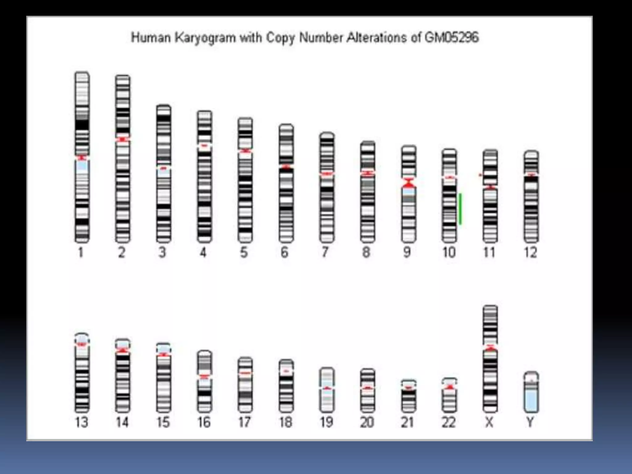


![ The normal human karyotypes contain 22 pairs of
autosomal chromosomes and one pair of sex chromosomes
[allosomes].
Normal karyotype for females contain two X
chromosomes[XX]
Normal karyotype for male contain one X and Y
chromosome[XY]
Any variation from the standard karyotype may lead to
developmental abnormalities .
NORMALHUMANKARYOTYPE](https://image.slidesharecdn.com/2016-1-180406045157/75/HUMAN-CHROMOSOMAL-ABERRATIONS-AND-KARYOTYPE-ANALYSIS-25-2048.jpg)