Downloaded 704 times


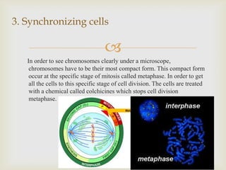
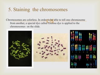
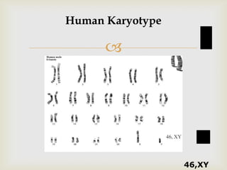


Karyotyping is a technique used in cytogenetics to examine chromosomes and identify genetic abnormalities that can cause disorders or disease. It involves collecting a cell sample, treating the cells to synchronize them in metaphase, staining the chromosomes, and analyzing the number, size, shape, and banding pattern of chromosomes to create a karyotype. Abnormal karyotypes can provide information about genetic conditions like Down syndrome, Klinefelter syndrome, and Turner syndrome. The main purpose is to detect changes in chromosome number or structure that can help diagnose these genetic disorders.