Downloaded 606 times
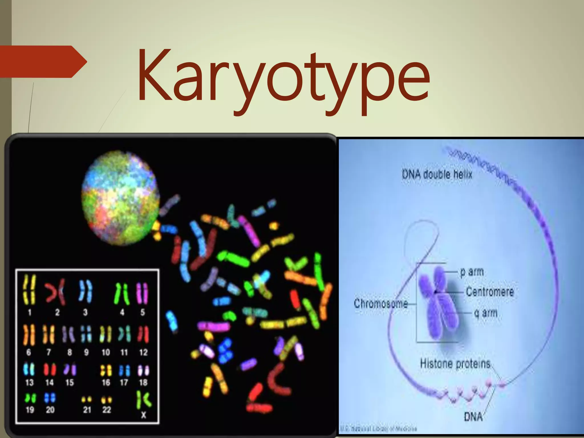


A karyotype is the number and appearance of chromosomes in a cell. It depicts the complete set of chromosomes and can detect abnormalities. The study of whole chromosome sets is called karyology. Chromosomes are arranged in a standard format called a karyogram or idiogram. A karyotype is prepared by culturing cells to induce cell division, arresting mitosis, staining the chromosomes, and analyzing their number, size, shape, and banding pattern under a microscope. This allows detection of chromosomal abnormalities that can indicate genetic disorders.
Introduction to karyotype, its definition, and function, along with details about chromosome structure.
Definitions of karyology and idiogram; characteristics of chromosomes depicted in ordered pairs.
Structure and function of chromosomes, with species-specific chromosome counts.
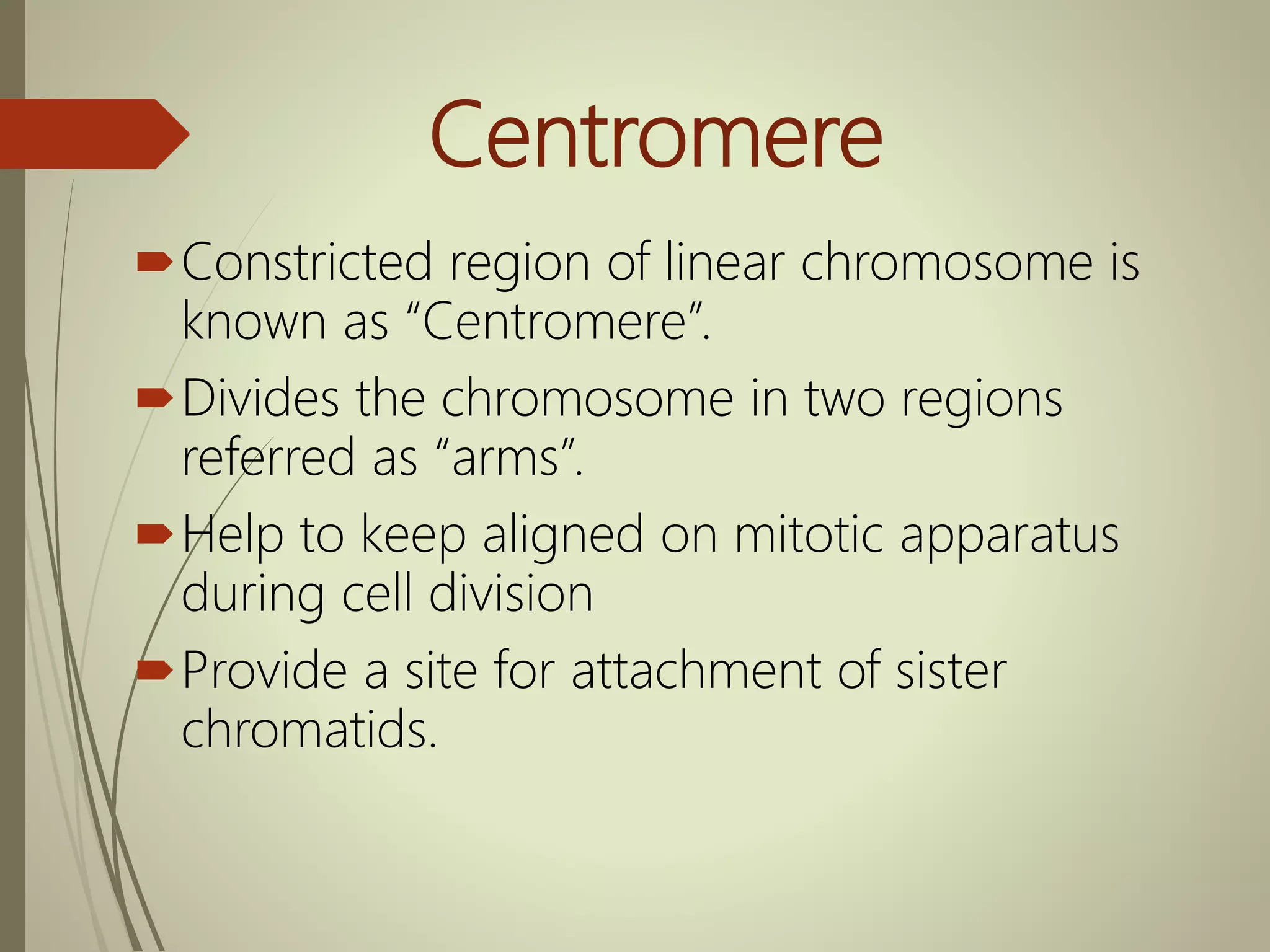
Definition and significance of centromeres in chromosome alignment during division.
Classification of chromosomes based on centromere positions: metacentric, submetacentric, acrocentric, and telocentric.
Detailed description of human chromosomes' organization and types in an ideogram.
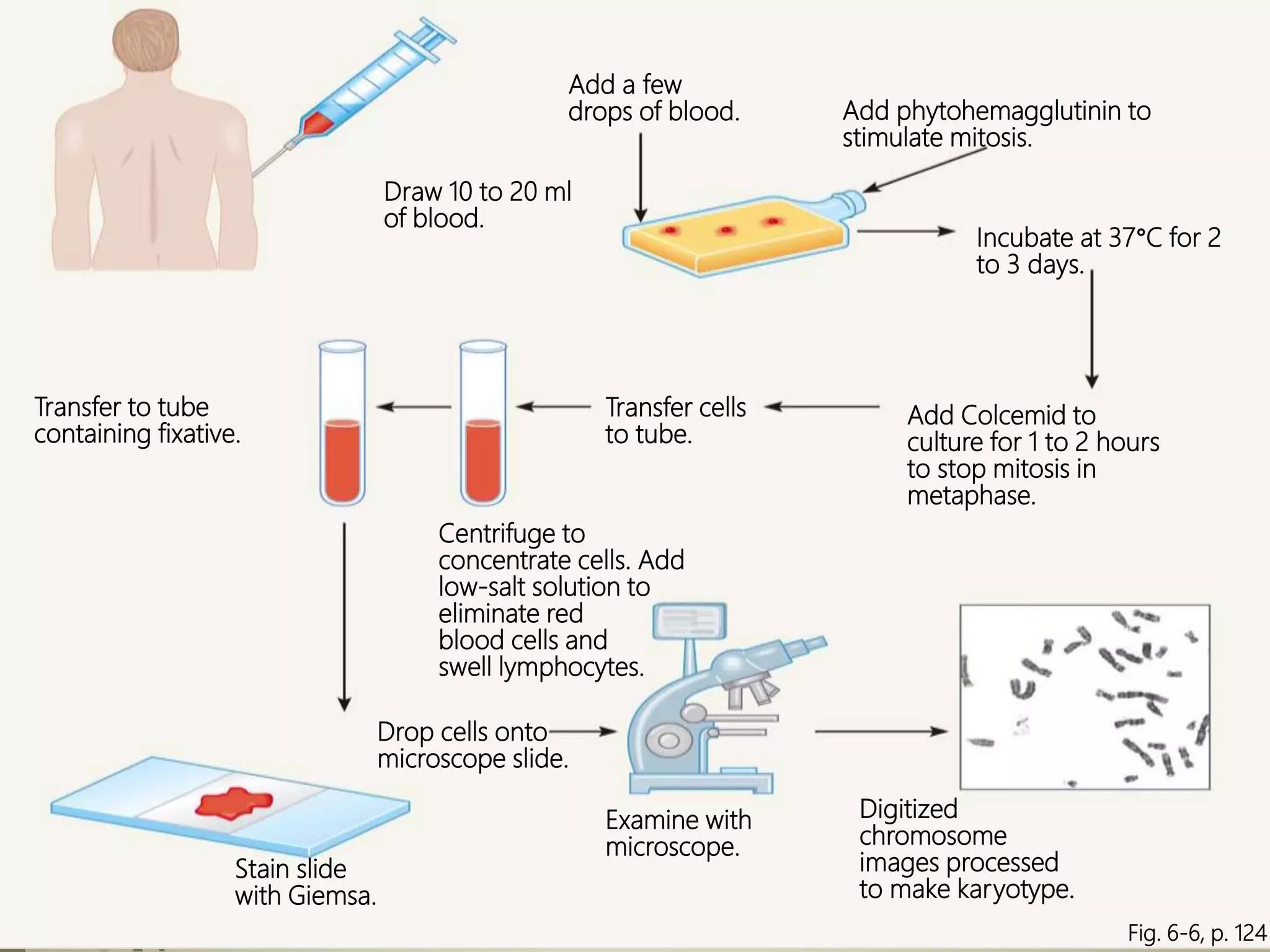
Step-by-step process of karyotyping and analysis techniques used to detect abnormalities.
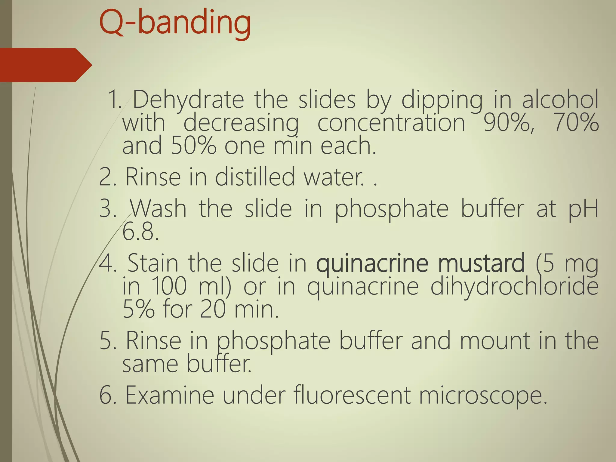
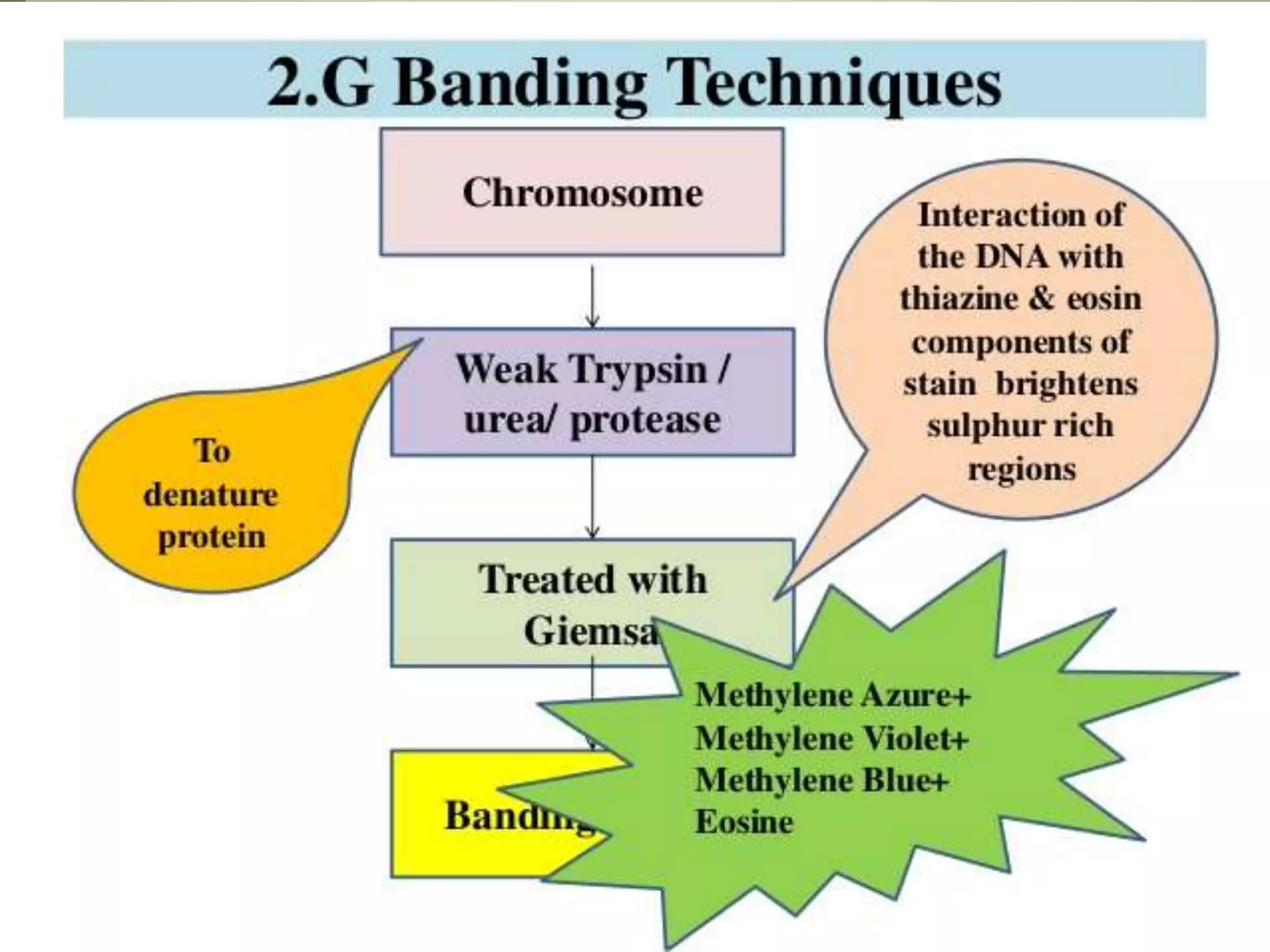
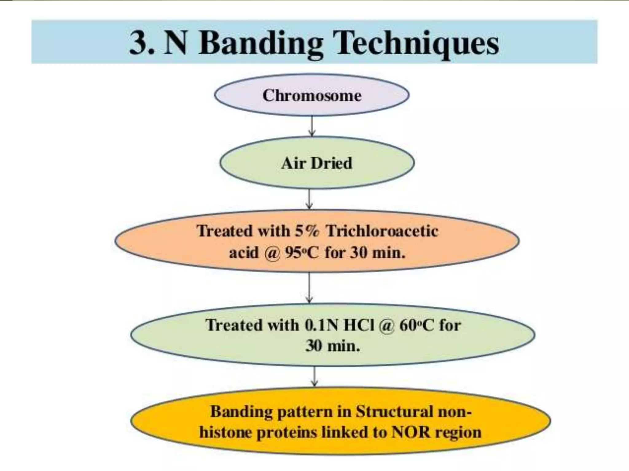
Various chromosome banding techniques like G-banding, Q-banding, N-banding, C-banding.
Various chromosome banding techniques like G-banding, Q-banding, N-banding, C-banding.
Various chromosome banding techniques like G-banding, Q-banding, N-banding, C-banding.
Various chromosome banding techniques like G-banding, Q-banding, N-banding, C-banding.
Various chromosome banding techniques like G-banding, Q-banding, N-banding, C-banding.
Overview of chromosome painting technology for visualizing chromosomal structures.
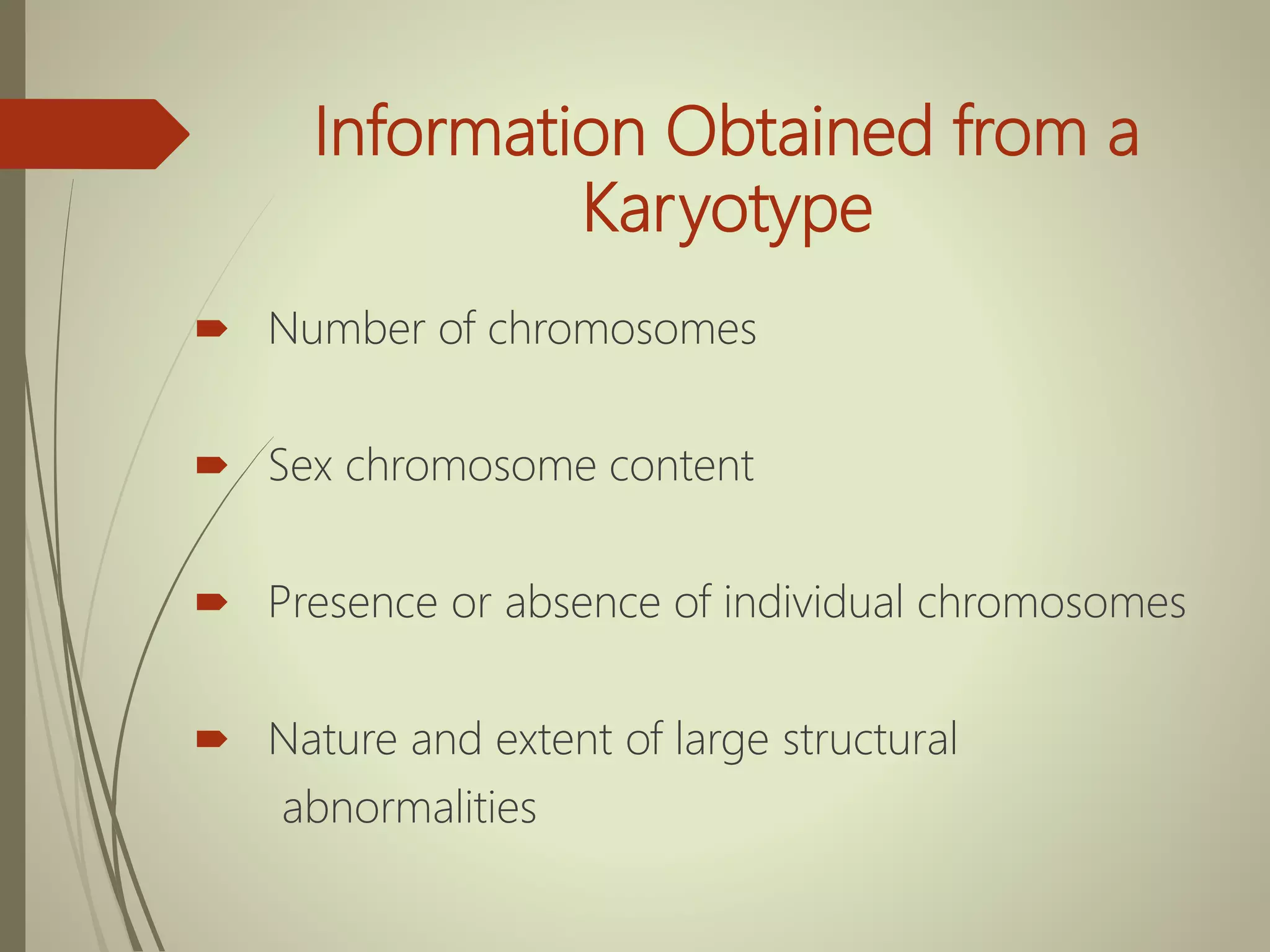
Information deduced from karyotyping, including chromosome number and abnormalities.
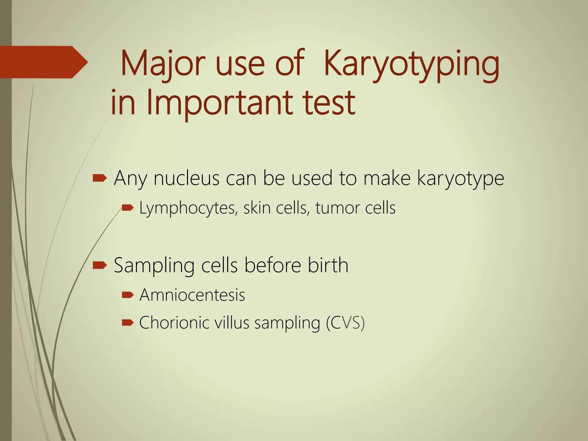
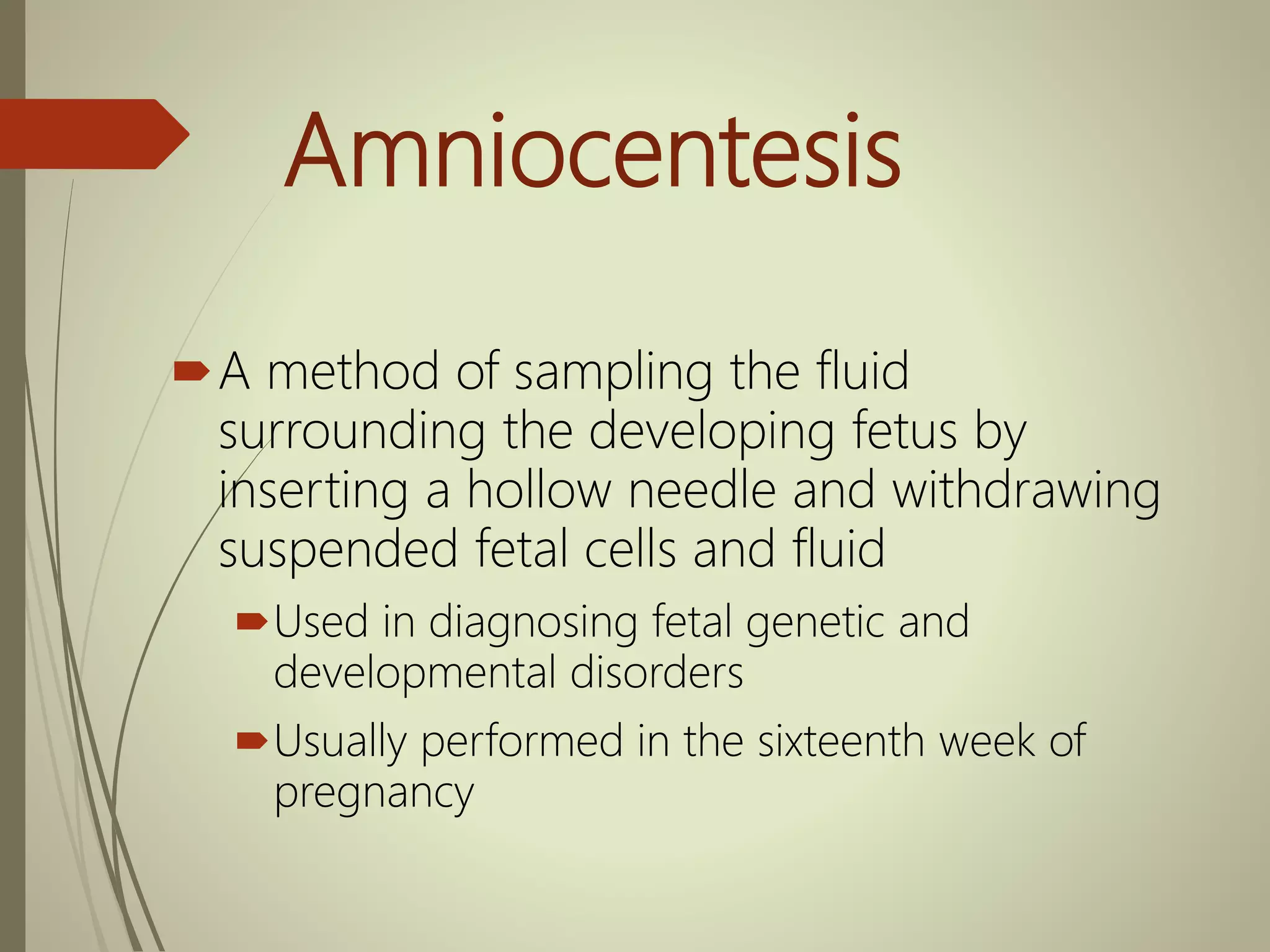
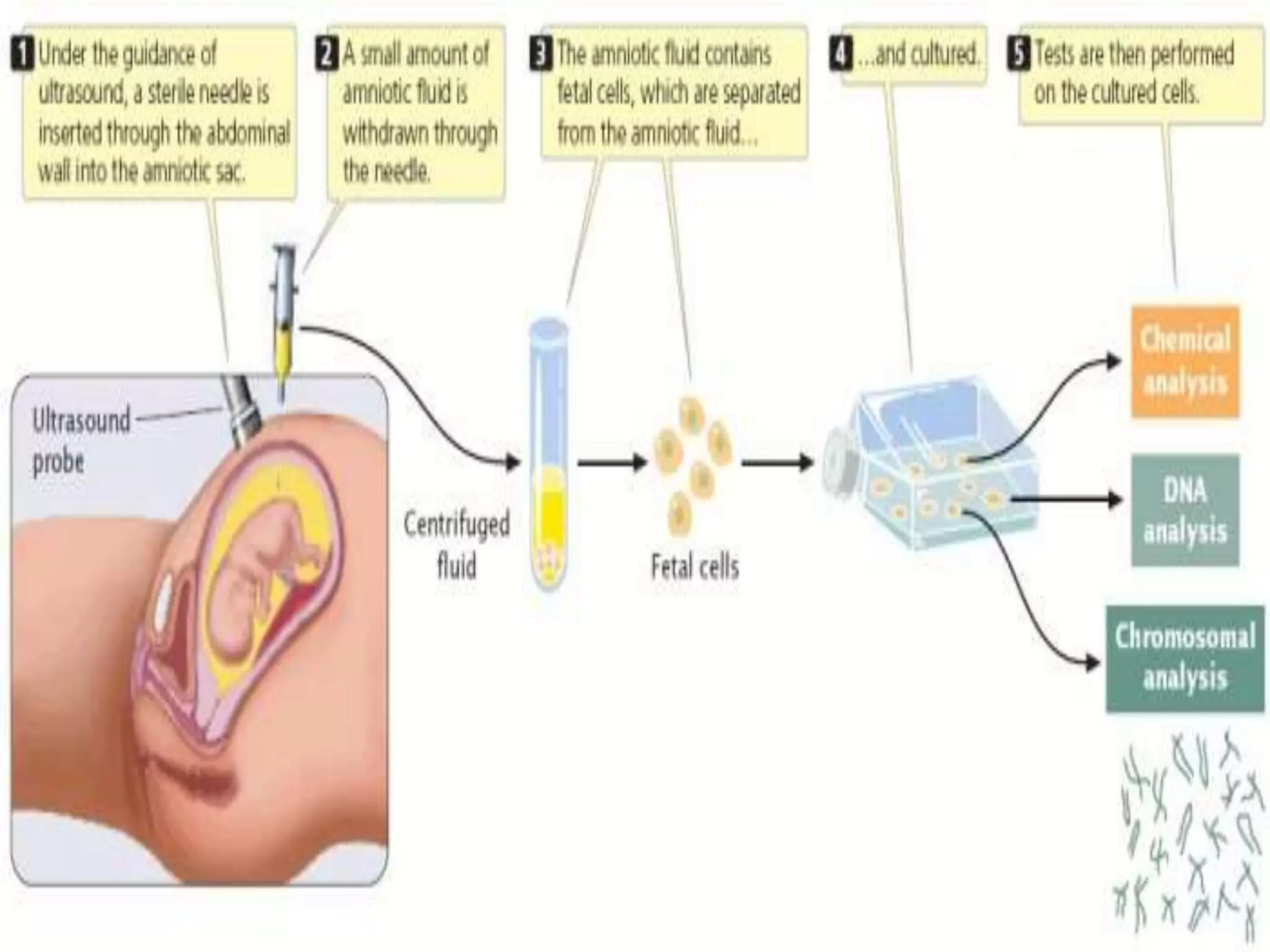
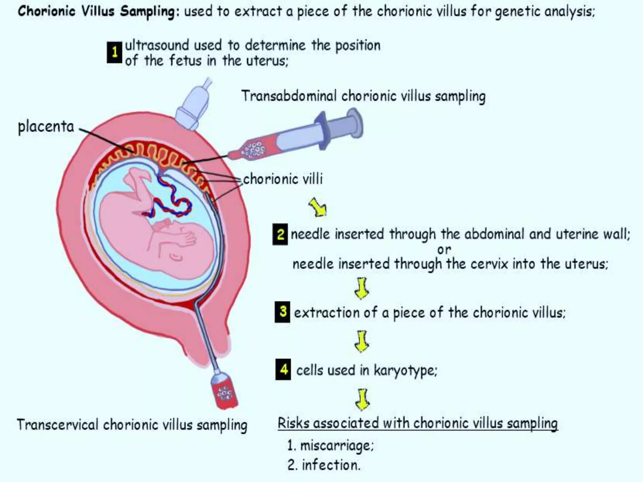
Uses of karyotyping in genetic diagnostics, including amniocentesis and CVS procedures.
Uses of karyotyping in genetic diagnostics, including amniocentesis and CVS procedures.
Uses of karyotyping in genetic diagnostics, including amniocentesis and CVS procedures.
Benefits and limitations of karyotyping including detection powers and risks of procedures.