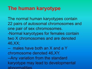
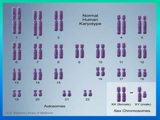
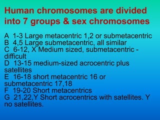
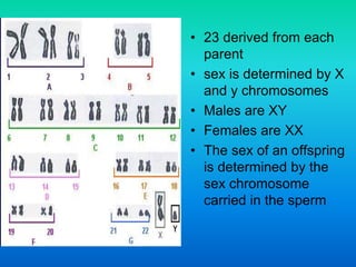
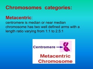
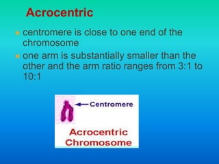
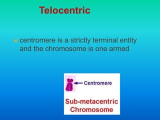
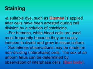
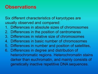
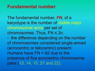
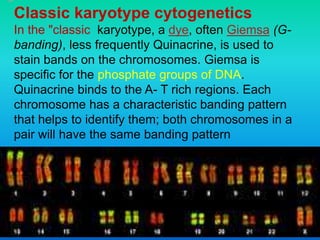
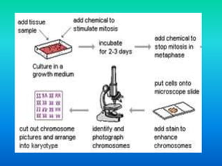
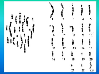
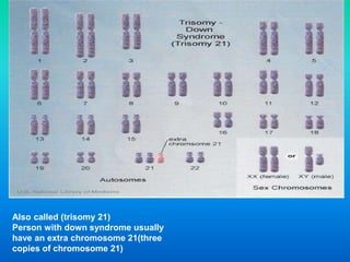
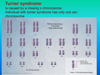
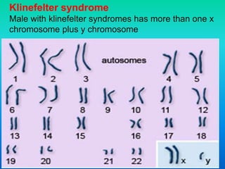
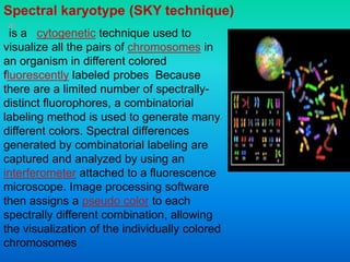
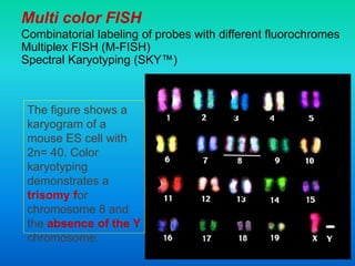
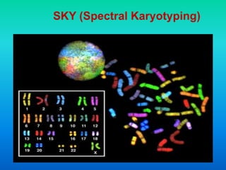
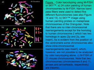
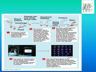
Karyotyping involves staining and analyzing chromosomes to identify any abnormalities. A normal human karyotype contains 23 chromosome pairs, including 22 autosomal and 1 sex chromosome pair. Chromosomes are categorized by centromere position and size. Karyotyping is used to diagnose conditions like Down syndrome that involve extra or missing chromosomes. New techniques like spectral karyotyping allow full chromosome visualization with color-coded labeling for detailed analysis.
Chromosomes are bundles of tightly coiled DNA located within the nucleus of almost every cell in our body. A chromosome is a DNA molecule with part or all of the genetic material (genome) of an organism. Chromosomes are normally visible under a light microscope only when the cell is undergoing the metaphase of cell division. Before this happens, every chromosome is copied once (S phase), and the copy is joined to the original by a centromere, resulting in an X-shaped structure. The original chromosome and the copy are now called sister chromatids. During metaphase, when a chromosome is in its most condensed state, the X-shape structure is called a metaphase chromosome.
Definition
Centromere Particular chromosome complement of an individual or a related group of individuals, as defined by the chromosome size, morphology, and number –Karyotype.
Karyotype
CLASSIFICATION OF CHROMOSOMES FORKARYOTYPING
Types of karyotype
Asymmetric Karyotype
• Show larger difference
between smaller and
larger chromosome in a
set.
• Have more acrocentric
chromosomes.
• Have relatively
advanced feature.
Symmetric Karyotype
Show lesser difference
between smaller and
larger chromosome in a
set.
• Have more metacentric
chromosomes.
• Have no relatively
advanced feature
Procedure of karyotyping
SPECIMENS USED
Types of banding
G-banding
R-banding
c-banding
Q-banding
T-banding
Karyotype Detects Various Chromosome Abnormalities
Aneuploidy
Deletions
Duplications
Translocations
Idiogram
Advantages of Karyotyping
Disadvantages:
This is a lecture presented by Dr.Omer Yahia Describing the first step of in the Role of molecular diagnostics through out the life. Give a brief shading out on the procedures for sample collection and types of diseases and syndromes undergone such tests .
The advances likes Next Generation Sequencing is more advanced than Microarray Compatability Genomic hybridization and it is 100% of sensitivity and specificity regarding aneuploidy sequencing from all biological samples.
Title: Sense of Taste
Presenter: Dr. Faiza, Assistant Professor of Physiology
Qualifications:
MBBS (Best Graduate, AIMC Lahore)
FCPS Physiology
ICMT, CHPE, DHPE (STMU)
MPH (GC University, Faisalabad)
MBA (Virtual University of Pakistan)
Learning Objectives:
Describe the structure and function of taste buds.
Describe the relationship between the taste threshold and taste index of common substances.
Explain the chemical basis and signal transduction of taste perception for each type of primary taste sensation.
Recognize different abnormalities of taste perception and their causes.
Key Topics:
Significance of Taste Sensation:
Differentiation between pleasant and harmful food
Influence on behavior
Selection of food based on metabolic needs
Receptors of Taste:
Taste buds on the tongue
Influence of sense of smell, texture of food, and pain stimulation (e.g., by pepper)
Primary and Secondary Taste Sensations:
Primary taste sensations: Sweet, Sour, Salty, Bitter, Umami
Chemical basis and signal transduction mechanisms for each taste
Taste Threshold and Index:
Taste threshold values for Sweet (sucrose), Salty (NaCl), Sour (HCl), and Bitter (Quinine)
Taste index relationship: Inversely proportional to taste threshold
Taste Blindness:
Inability to taste certain substances, particularly thiourea compounds
Example: Phenylthiocarbamide
Structure and Function of Taste Buds:
Composition: Epithelial cells, Sustentacular/Supporting cells, Taste cells, Basal cells
Features: Taste pores, Taste hairs/microvilli, and Taste nerve fibers
Location of Taste Buds:
Found in papillae of the tongue (Fungiform, Circumvallate, Foliate)
Also present on the palate, tonsillar pillars, epiglottis, and proximal esophagus
Mechanism of Taste Stimulation:
Interaction of taste substances with receptors on microvilli
Signal transduction pathways for Umami, Sweet, Bitter, Sour, and Salty tastes
Taste Sensitivity and Adaptation:
Decrease in sensitivity with age
Rapid adaptation of taste sensation
Role of Saliva in Taste:
Dissolution of tastants to reach receptors
Washing away the stimulus
Taste Preferences and Aversions:
Mechanisms behind taste preference and aversion
Influence of receptors and neural pathways
Impact of Sensory Nerve Damage:
Degeneration of taste buds if the sensory nerve fiber is cut
Abnormalities of Taste Detection:
Conditions: Ageusia, Hypogeusia, Dysgeusia (parageusia)
Causes: Nerve damage, neurological disorders, infections, poor oral hygiene, adverse drug effects, deficiencies, aging, tobacco use, altered neurotransmitter levels
Neurotransmitters and Taste Threshold:
Effects of serotonin (5-HT) and norepinephrine (NE) on taste sensitivity
Supertasters:
25% of the population with heightened sensitivity to taste, especially bitterness
Increased number of fungiform papillae
The prostate is an exocrine gland of the male mammalian reproductive system
It is a walnut-sized gland that forms part of the male reproductive system and is located in front of the rectum and just below the urinary bladder
Function is to store and secrete a clear, slightly alkaline fluid that constitutes 10-30% of the volume of the seminal fluid that along with the spermatozoa, constitutes semen
A healthy human prostate measures (4cm-vertical, by 3cm-horizontal, 2cm ant-post ).
It surrounds the urethra just below the urinary bladder. It has anterior, median, posterior and two lateral lobes
It’s work is regulated by androgens which are responsible for male sex characteristics
Generalised disease of the prostate due to hormonal derangement which leads to non malignant enlargement of the gland (increase in the number of epithelial cells and stromal tissue)to cause compression of the urethra leading to symptoms (LUTS
Anti ulcer drugs and their Advance pharmacology ||
Anti-ulcer drugs are medications used to prevent and treat ulcers in the stomach and upper part of the small intestine (duodenal ulcers). These ulcers are often caused by an imbalance between stomach acid and the mucosal lining, which protects the stomach lining.
||Scope: Overview of various classes of anti-ulcer drugs, their mechanisms of action, indications, side effects, and clinical considerations.
ARTIFICIAL INTELLIGENCE IN HEALTHCARE.pdfAnujkumaranit
Artificial intelligence (AI) refers to the simulation of human intelligence processes by machines, especially computer systems. It encompasses tasks such as learning, reasoning, problem-solving, perception, and language understanding. AI technologies are revolutionizing various fields, from healthcare to finance, by enabling machines to perform tasks that typically require human intelligence.
MANAGEMENT OF ATRIOVENTRICULAR CONDUCTION BLOCK.pdfJim Jacob Roy
Cardiac conduction defects can occur due to various causes.
Atrioventricular conduction blocks ( AV blocks ) are classified into 3 types.
This document describes the acute management of AV block.
Pulmonary Thromboembolism - etilogy, types, medical- Surgical and nursing man...VarunMahajani
Disruption of blood supply to lung alveoli due to blockage of one or more pulmonary blood vessels is called as Pulmonary thromboembolism. In this presentation we will discuss its causes, types and its management in depth.
Title: Sense of Smell
Presenter: Dr. Faiza, Assistant Professor of Physiology
Qualifications:
MBBS (Best Graduate, AIMC Lahore)
FCPS Physiology
ICMT, CHPE, DHPE (STMU)
MPH (GC University, Faisalabad)
MBA (Virtual University of Pakistan)
Learning Objectives:
Describe the primary categories of smells and the concept of odor blindness.
Explain the structure and location of the olfactory membrane and mucosa, including the types and roles of cells involved in olfaction.
Describe the pathway and mechanisms of olfactory signal transmission from the olfactory receptors to the brain.
Illustrate the biochemical cascade triggered by odorant binding to olfactory receptors, including the role of G-proteins and second messengers in generating an action potential.
Identify different types of olfactory disorders such as anosmia, hyposmia, hyperosmia, and dysosmia, including their potential causes.
Key Topics:
Olfactory Genes:
3% of the human genome accounts for olfactory genes.
400 genes for odorant receptors.
Olfactory Membrane:
Located in the superior part of the nasal cavity.
Medially: Folds downward along the superior septum.
Laterally: Folds over the superior turbinate and upper surface of the middle turbinate.
Total surface area: 5-10 square centimeters.
Olfactory Mucosa:
Olfactory Cells: Bipolar nerve cells derived from the CNS (100 million), with 4-25 olfactory cilia per cell.
Sustentacular Cells: Produce mucus and maintain ionic and molecular environment.
Basal Cells: Replace worn-out olfactory cells with an average lifespan of 1-2 months.
Bowman’s Gland: Secretes mucus.
Stimulation of Olfactory Cells:
Odorant dissolves in mucus and attaches to receptors on olfactory cilia.
Involves a cascade effect through G-proteins and second messengers, leading to depolarization and action potential generation in the olfactory nerve.
Quality of a Good Odorant:
Small (3-20 Carbon atoms), volatile, water-soluble, and lipid-soluble.
Facilitated by odorant-binding proteins in mucus.
Membrane Potential and Action Potential:
Resting membrane potential: -55mV.
Action potential frequency in the olfactory nerve increases with odorant strength.
Adaptation Towards the Sense of Smell:
Rapid adaptation within the first second, with further slow adaptation.
Psychological adaptation greater than receptor adaptation, involving feedback inhibition from the central nervous system.
Primary Sensations of Smell:
Camphoraceous, Musky, Floral, Pepperminty, Ethereal, Pungent, Putrid.
Odor Detection Threshold:
Examples: Hydrogen sulfide (0.0005 ppm), Methyl-mercaptan (0.002 ppm).
Some toxic substances are odorless at lethal concentrations.
Characteristics of Smell:
Odor blindness for single substances due to lack of appropriate receptor protein.
Behavioral and emotional influences of smell.
Transmission of Olfactory Signals:
From olfactory cells to glomeruli in the olfactory bulb, involving lateral inhibition.
Primitive, less old, and new olfactory systems with different path
Prix Galien International 2024 Forum ProgramLevi Shapiro
June 20, 2024, Prix Galien International and Jerusalem Ethics Forum in ROME. Detailed agenda including panels:
- ADVANCES IN CARDIOLOGY: A NEW PARADIGM IS COMING
- WOMEN’S HEALTH: FERTILITY PRESERVATION
- WHAT’S NEW IN THE TREATMENT OF INFECTIOUS,
ONCOLOGICAL AND INFLAMMATORY SKIN DISEASES?
- ARTIFICIAL INTELLIGENCE AND ETHICS
- GENE THERAPY
- BEYOND BORDERS: GLOBAL INITIATIVES FOR DEMOCRATIZING LIFE SCIENCE TECHNOLOGIES AND PROMOTING ACCESS TO HEALTHCARE
- ETHICAL CHALLENGES IN LIFE SCIENCES
- Prix Galien International Awards Ceremony
Explore natural remedies for syphilis treatment in Singapore. Discover alternative therapies, herbal remedies, and lifestyle changes that may complement conventional treatments. Learn about holistic approaches to managing syphilis symptoms and supporting overall health.
Are There Any Natural Remedies To Treat Syphilis.pdf
Introduction to karyotyping examination.
1. Professor Dr. Najat A. Hasan (MB ChB, MSc, PhD in Clinical
Biochemistry, College of Medicine -Alnahrain University, Baghdad.
Iraq)
2. A karyotype is the characteristic chromosome
complement of an eukaryote species. The
preparation and study of karyotypes is part of
cytogenetics.
- The chromosomes are depicted (by rearranging a
microphotograph) in a standard format known as a
karyogram or idiogram: in pairs, ordered by size
and position of centromere for chromosomes of
the same size.
3. The human karyotype
The normal human karyotypes contain
22 pairs of autosomal chromosomes and
one pair of sex chromosomes.
-Normal karyotypes for females contain
two X chromosomes and are denoted
46,XX;
-- males have both an X and a Y
chromosome denoted 46,XY.
--Any variation from the standard
karyotype may lead to developmental
4.
5. - Karyotypes are arranged with the short
arm of the chromosome on top, and the long
arm on the bottom. Some karyotypes call
the short and long arms p and q,
respectively. In addition, the differently
stained regions and sub-regions are given
numerical designations from proximal to
distal on the chromosome arms. For
example, Cri du chat syndrome involves a
deletion on the short arm of chromosome 5.
It is written as 46,XX,5p-. The critical region
for this syndrome is deletion of 15.2, which
Chromosomes are always arranged
6. Human chromosomes are divided
into 7 groups & sex chromosomes
A 1-3 Large metacentric 1,2 or submetacentric
B 4,5 Large submetacentric, all similar
C 6-12, X Medium sized, submetacentric -
difficult
D 13-15 medium-sized acrocentric plus
satellites
E 16-18 short metacentric 16 or
submetacentric 17,18
F 19-20 Short metacentrics
G 21,22,Y Short acrocentrics with satellites. Y
no satellites.
7. • 23 derived from each
parent
• sex is determined by X
and y chromosomes
• Males are XY
• Females are XX
• The sex of an offspring
is determined by the
sex chromosome
carried in the sperm
9. centromere is close to one end of the
chromosome
one arm is substantially smaller than the
other and the arm ratio ranges from 3:1 to
10:1
Acrocentric
10. centromere is a strictly terminal entity
and the chromosome is one armed
Telocentric
11. Staining
-a suitable dye, such as Giemsa is applied
after cells have been arrested during cell
division by a solution of colchicine.
- -For humans, white blood cells are used
most frequently because they are easily
induced to divide and grow in tissue culture.
- Sometimes observations may be made on
non-dividing (interphase) cells. The sex of an
unborn fetus can be determined by
observation of interphase cells (Barr body).
12. Observations
Six different characteristics of karyotypes are
usually observed and compared:
1. Differences in absolute sizes of chromosomes
2. Differences in the position of centromeres
3. Differences in relative size of chromosomes
4. Differences in basic number of chromosomes
5. Differences in number and position of satellites,
6. Differences in degree and distribution of
heterochromatic regions. Heterochromatin stains
darker than euchromatin, and mainly consists of
genetically inactive repetitive DNA sequences.
13. Fundamental number
The fundamental number, FN, of a
karyotype is the number of visible major
chromosomal arms per set of
chromosomes .Thus, FN ≤ 2n.
- the difference depending on the number
of chromosomes considered single-armed
(acrocentric or telocentric) present.
Humans have FN = 82 due to the
presence of five acrocentric chromosome
pairs (13, 14, 15, 21 and 22).
14. Ploidy: the number of sets in a karyotype
1. Polyploidy, where there are more
than two sets of homologous
chromosomes in the cells, occurs
mainly in plants. Polyploidy in
animals is much less common.
2. Haplo-diploidy, where one sex is
diploid, and the other haploid. It is a
common arrangement in the
Hymenoptera.
15. 3. Endopolyploidy
occurs when in adult differentiated tissues
the cells have ceased to divide by mitosis,
but the nuclei contain more than the
original somatic number of chromosomes.
Aneuploidy
the condition in which the chromosome
number in the cells is not the typical number
for the species. This would give rise to a
chromosome abnormality such as an extra
chromosome or one or more chromosomes
lost. Abnormalities in chromosome number
usually cause a defect in development. Down
syndrome and Turner syndrome .
16. Classic karyotype cytogenetics
In the "classic karyotype, a dye, often Giemsa (G-
banding), less frequently Quinacrine, is used to
stain bands on the chromosomes. Giemsa is
specific for the phosphate groups of DNA.
Quinacrine binds to the A- T rich regions. Each
chromosome has a characteristic banding pattern
that helps to identify them; both chromosomes in a
pair will have the same banding pattern
17. Types of banding
G-banding is obtained with Giemsa stain following
digestion of chromosomes with trypsin. It yields a series of
lightly and darkly stained bands
the dark regions tend to be heterochromatic, late-replicating
and AT rich.
The light regions tend to be euchromatic, early-replicating
and GC rich. There is300-400 bands in a normal, human
genome.
R-banding is the reverse of G-banding (the R stands for
"reverse"). The dark regions are euchromatic (guanine-
cytosine rich regions) and the bright regions are
heterochromatic (thymine-adenine rich regions).
C-banding: Giemsa binds to constitutive heterochromatin,
so it stains centromeres.
18. Q-banding
Is a fluorescent pattern obtained using
quinacrine for staining. The pattern of
bands is very similar to that seen in G-
banding.
T-banding: visualize telomeres.
Silver staining: Silver nitrate stains the
nucleolar organization region-associated
protein. This yields a dark region where the
silver is deposited, denoting the activity of
rRNA genes within the NOR.
19. Chromosome abnormalities
1. numerical, as in the presence of extra or missing
chromosomes,
2. structural, as in derivative chromosome, translocations,
inversions, large-scale deletions or duplications.
Numerical abnormalities, also known as aneuploidy, often occur
as a result of nondisjunction during meiosis in the formation
of a gamete; trisomies, in which three copies of a
chromosome are present instead of the usual two,
Structural abnormalities often arise from errors in homologous
recombination.
Both types of abnormalities can occur in gametes and therefore
will be present in all cells of an affected person's body, or
they can occur during mitosis and give rise to a genetic
mosaic individual who has some normal and some abnormal
cells.
20. Chromosomal abnormalities that lead to
disease in humans include:
Turner syndrome results from a single X chromosome
(45, X or 45, X0).
Klinefelter syndrome, the most common male
chromosomal disease, otherwise known as 47, XXY is
caused by an extra X chromosome.
Edwards syndrome is caused by trisomy (three copies) of
chromosome 18.
Down syndrome, a common chromosomal disease, is
caused by trisomy of chromosome 21.
lymphoblastic leukemia. Patau syndrome is caused by
trisomy of chromosome 13.
Also documented are trisomy 8, trisomy 9 and trisomy 16,
although they generally do not survive to birth.
21. Some disorders arise from loss of just a piece of one
chromosome, including
Cri du chat (cry of the cat), from a truncated short arm on
chromosome 5. The name comes from the babies' distinctive cry,
caused by abnormal formation of the larynx.
1p36 Deletion syndrome, from the loss of part of the short arm of
chromosome 1.
Angelman syndrome – 50% of cases have a segment of the long
arm of chromosome 15 missing; a deletion of the maternal genes,
example of imprinting disorder.
Prader-Willi syndrome – 50% of cases have a segment of the
long arm of chromosome 15 missing; a deletion of the paternal
genes, example of imprinting disorder.
Chromosomal abnormalities can also occur in cancerous
cells of an otherwise genetically normal individual; one well-
documented example is the Philadelphia chromosome, a
translocation mutation commonly associated with chronic
myelogenous leukemia and less often with acute
22. Source of cell sample
any cell in the body , except red blood cell , which
lack a nucleus , can be a source of chromosomes
for karyotyping.
In adult
Can use white blood cells separated from a blood
sampling
In fetuses
Cells can be obtained by either
Amniocentesis
Chorionic vill sampling (cvs)
23. Amniocentesis is not usually performed until
about the 14th-17th week of pregnancy .
Karyotyping the chromosomes may be
delayed as long as four weeks so that the
cell can be cultured to increase their number
.
B)Chorionic villi sampling(cvs):
This procedure can be done as early as the fifth
week of pregnancy .
The cell do not have to be cultured , and
karyotyping can be done immediately .
24.
25. Results of a Karyotype test
*There are 46 chromosomes that can be
grouped as 22 matching pairs and 1 pair of
sex chromosomes (XX for a female and XY
for a male)
*The size, shape, and structure are
normal for each chromosome .
Normal
*There are more than or less
than 46 chromosomes.
*The shape or size of one or
more chromosomes is abnormal.
*A chromosome pair may be
broken or incorrectly separated
Abnormal
26.
27. Also called (trisomy 21)
Person with down syndrome usually
have an extra chromosome 21(three
copies of chromosome 21)
28. Turner syndrome
Is caused by a missing x chromosome .
Individual with turner syndrome has only one sex
chromosome
30. is a cytogenetic technique used to
visualize all the pairs of chromosomes in
an organism in different colored
fluorescently labeled probes Because
there are a limited number of spectrally-
distinct fluorophores, a combinatorial
labeling method is used to generate many
different colors. Spectral differences
generated by combinatorial labeling are
captured and analyzed by using an
interferometer attached to a fluorescence
microscope. Image processing software
then assigns a pseudo color to each
spectrally different combination, allowing
the visualization of the individually colored
chromosomes
Spectral karyotype (SKY technique)
31. Multi color FISH
Combinatorial labeling of probes with different fluorochromes
Multiplex FISH (M-FISH)
Spectral Karyotyping (SKY™)
The figure shows a
karyogram of a
mouse ES cell with
2n= 40. Color
karyotyping
demonstrates a
trisomy for
chromosome 8 and
the absence of the Y
chromosome.
33. Figure : Color karyotyping using M-FISH
or SKY™. a) 24 color painting of human
chromosomes by M-FISH. Narrow band
pass filters were used to detect five
different fluorochromes (see also Figure
14 and 15). b) SKY™ image using
human painting probes on metaphase
chromosomes of the Orangutan. Note
that there are no inter-chromosomal
rearrangements except for the homolog
to human chromosome 2 which has two
homologs in apes (2p and 2q, see
insert). As is evident from the position of
the centromere some chromosomes also
show intra-chromosomal
rearrangements (see insert), which,
however, are not possible to be analyzed
by color karyotyping alone. c) M-FISH of
21 mouse chromosomes. Note that two
chromosomes (chromosomes 8 and 11,
arrows and arrowheads, respectively)
are trisomic.