Download as PDF, PPTX
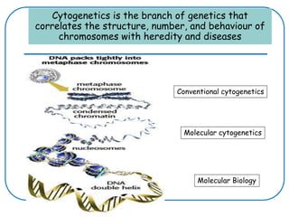
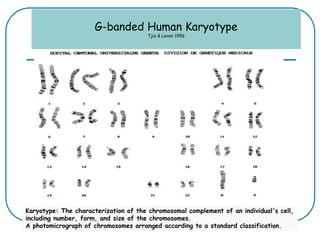
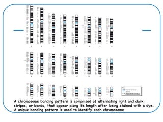
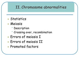
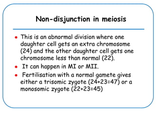
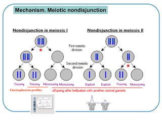
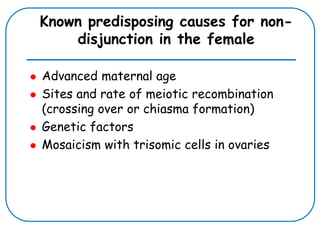
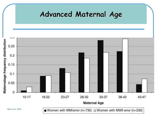
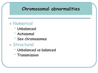
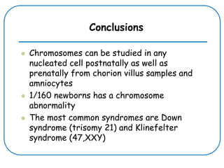

Cytogenetics is the study of chromosomes and chromosome abnormalities in relation to heredity and disease. It involves analyzing chromosomes through karyotyping and molecular cytogenetics techniques. Chromosome banding patterns allow chromosomes to be identified. Non-disjunction during meiosis can result in aneuploidies like Down syndrome. Advanced maternal age is a risk factor for non-disjunction. Both numerical and structural chromosome abnormalities can occur.