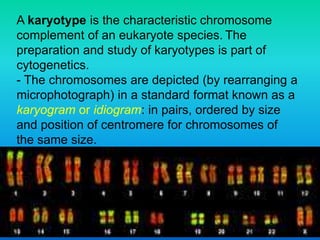
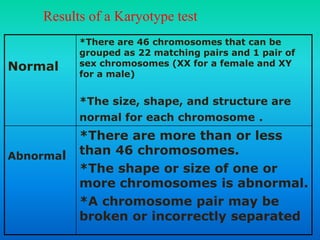
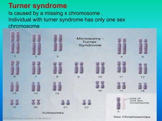
1. Professor Dr. Najat A. Hasan (MB ChB, MSc, PhD in Clinical
Biochemistry, College of Medicine -Alnahrain University, Baghdad.
Iraq)
2. A karyotype is the characteristic chromosome
complement of an eukaryote species. The
preparation and study of karyotypes is part of
cytogenetics.
- The chromosomes are depicted (by rearranging a
microphotograph) in a standard format known as a
karyogram or idiogram: in pairs, ordered by size
and position of centromere for chromosomes of
the same size.
3. The human karyotype
The normal human karyotypes contain
22 pairs of autosomal chromosomes and
one pair of sex chromosomes.
-Normal karyotypes for females contain
two X chromosomes and are denoted
46,XX;
-- males have both an X and a Y
chromosome denoted 46,XY.
--Any variation from the standard
karyotype may lead to developmental
4.
5. - Karyotypes are arranged with the short
arm of the chromosome on top, and the long
arm on the bottom. Some karyotypes call
the short and long arms p and q,
respectively. In addition, the differently
stained regions and sub-regions are given
numerical designations from proximal to
distal on the chromosome arms. For
example, Cri du chat syndrome involves a
deletion on the short arm of chromosome 5.
It is written as 46,XX,5p-. The critical region
for this syndrome is deletion of 15.2, which
Chromosomes are always arranged
6. Human chromosomes are divided
into 7 groups & sex chromosomes
A 1-3 Large metacentric 1,2 or submetacentric
B 4,5 Large submetacentric, all similar
C 6-12, X Medium sized, submetacentric -
difficult
D 13-15 medium-sized acrocentric plus
satellites
E 16-18 short metacentric 16 or
submetacentric 17,18
F 19-20 Short metacentrics
G 21,22,Y Short acrocentrics with satellites. Y
no satellites.
7. • 23 derived from each
parent
• sex is determined by X
and y chromosomes
• Males are XY
• Females are XX
• The sex of an offspring
is determined by the
sex chromosome
carried in the sperm
9. centromere is close to one end of the
chromosome
one arm is substantially smaller than the
other and the arm ratio ranges from 3:1 to
10:1
Acrocentric
10. centromere is a strictly terminal entity
and the chromosome is one armed
Telocentric
11. Staining
-a suitable dye, such as Giemsa is applied
after cells have been arrested during cell
division by a solution of colchicine.
- -For humans, white blood cells are used
most frequently because they are easily
induced to divide and grow in tissue culture.
- Sometimes observations may be made on
non-dividing (interphase) cells. The sex of an
unborn fetus can be determined by
observation of interphase cells (Barr body).
12. Observations
Six different characteristics of karyotypes are
usually observed and compared:
1. Differences in absolute sizes of chromosomes
2. Differences in the position of centromeres
3. Differences in relative size of chromosomes
4. Differences in basic number of chromosomes
5. Differences in number and position of satellites,
6. Differences in degree and distribution of
heterochromatic regions. Heterochromatin stains
darker than euchromatin, and mainly consists of
genetically inactive repetitive DNA sequences.
13. Fundamental number
The fundamental number, FN, of a
karyotype is the number of visible major
chromosomal arms per set of
chromosomes .Thus, FN ≤ 2n.
- the difference depending on the number
of chromosomes considered single-armed
(acrocentric or telocentric) present.
Humans have FN = 82 due to the
presence of five acrocentric chromosome
pairs (13, 14, 15, 21 and 22).
14. Ploidy: the number of sets in a karyotype
1. Polyploidy, where there are more
than two sets of homologous
chromosomes in the cells, occurs
mainly in plants. Polyploidy in
animals is much less common.
2. Haplo-diploidy, where one sex is
diploid, and the other haploid. It is a
common arrangement in the
Hymenoptera.
15. 3. Endopolyploidy
occurs when in adult differentiated tissues
the cells have ceased to divide by mitosis,
but the nuclei contain more than the
original somatic number of chromosomes.
Aneuploidy
the condition in which the chromosome
number in the cells is not the typical number
for the species. This would give rise to a
chromosome abnormality such as an extra
chromosome or one or more chromosomes
lost. Abnormalities in chromosome number
usually cause a defect in development. Down
syndrome and Turner syndrome .
16. Classic karyotype cytogenetics
In the "classic karyotype, a dye, often Giemsa (G-
banding), less frequently Quinacrine, is used to
stain bands on the chromosomes. Giemsa is
specific for the phosphate groups of DNA.
Quinacrine binds to the A- T rich regions. Each
chromosome has a characteristic banding pattern
that helps to identify them; both chromosomes in a
pair will have the same banding pattern
17. Types of banding
G-banding is obtained with Giemsa stain following
digestion of chromosomes with trypsin. It yields a series of
lightly and darkly stained bands
the dark regions tend to be heterochromatic, late-replicating
and AT rich.
The light regions tend to be euchromatic, early-replicating
and GC rich. There is300-400 bands in a normal, human
genome.
R-banding is the reverse of G-banding (the R stands for
"reverse"). The dark regions are euchromatic (guanine-
cytosine rich regions) and the bright regions are
heterochromatic (thymine-adenine rich regions).
C-banding: Giemsa binds to constitutive heterochromatin,
so it stains centromeres.
18. Q-banding
Is a fluorescent pattern obtained using
quinacrine for staining. The pattern of
bands is very similar to that seen in G-
banding.
T-banding: visualize telomeres.
Silver staining: Silver nitrate stains the
nucleolar organization region-associated
protein. This yields a dark region where the
silver is deposited, denoting the activity of
rRNA genes within the NOR.
19. Chromosome abnormalities
1. numerical, as in the presence of extra or missing
chromosomes,
2. structural, as in derivative chromosome, translocations,
inversions, large-scale deletions or duplications.
Numerical abnormalities, also known as aneuploidy, often occur
as a result of nondisjunction during meiosis in the formation
of a gamete; trisomies, in which three copies of a
chromosome are present instead of the usual two,
Structural abnormalities often arise from errors in homologous
recombination.
Both types of abnormalities can occur in gametes and therefore
will be present in all cells of an affected person's body, or
they can occur during mitosis and give rise to a genetic
mosaic individual who has some normal and some abnormal
cells.
20. Chromosomal abnormalities that lead to
disease in humans include:
Turner syndrome results from a single X chromosome
(45, X or 45, X0).
Klinefelter syndrome, the most common male
chromosomal disease, otherwise known as 47, XXY is
caused by an extra X chromosome.
Edwards syndrome is caused by trisomy (three copies) of
chromosome 18.
Down syndrome, a common chromosomal disease, is
caused by trisomy of chromosome 21.
lymphoblastic leukemia. Patau syndrome is caused by
trisomy of chromosome 13.
Also documented are trisomy 8, trisomy 9 and trisomy 16,
although they generally do not survive to birth.
21. Some disorders arise from loss of just a piece of one
chromosome, including
Cri du chat (cry of the cat), from a truncated short arm on
chromosome 5. The name comes from the babies' distinctive cry,
caused by abnormal formation of the larynx.
1p36 Deletion syndrome, from the loss of part of the short arm of
chromosome 1.
Angelman syndrome – 50% of cases have a segment of the long
arm of chromosome 15 missing; a deletion of the maternal genes,
example of imprinting disorder.
Prader-Willi syndrome – 50% of cases have a segment of the
long arm of chromosome 15 missing; a deletion of the paternal
genes, example of imprinting disorder.
Chromosomal abnormalities can also occur in cancerous
cells of an otherwise genetically normal individual; one well-
documented example is the Philadelphia chromosome, a
translocation mutation commonly associated with chronic
myelogenous leukemia and less often with acute
22. Source of cell sample
any cell in the body , except red blood cell , which
lack a nucleus , can be a source of chromosomes
for karyotyping.
In adult
Can use white blood cells separated from a blood
sampling
In fetuses
Cells can be obtained by either
Amniocentesis
Chorionic vill sampling (cvs)
23. Amniocentesis is not usually performed until
about the 14th-17th week of pregnancy .
Karyotyping the chromosomes may be
delayed as long as four weeks so that the
cell can be cultured to increase their number
.
B)Chorionic villi sampling(cvs):
This procedure can be done as early as the fifth
week of pregnancy .
The cell do not have to be cultured , and
karyotyping can be done immediately .
24.
25. Results of a Karyotype test
*There are 46 chromosomes that can be
grouped as 22 matching pairs and 1 pair of
sex chromosomes (XX for a female and XY
for a male)
*The size, shape, and structure are
normal for each chromosome .
Normal
*There are more than or less
than 46 chromosomes.
*The shape or size of one or
more chromosomes is abnormal.
*A chromosome pair may be
broken or incorrectly separated
Abnormal
26.
27. Also called (trisomy 21)
Person with down syndrome usually
have an extra chromosome 21(three
copies of chromosome 21)
28. Turner syndrome
Is caused by a missing x chromosome .
Individual with turner syndrome has only one sex
chromosome
30. is a cytogenetic technique used to
visualize all the pairs of chromosomes in
an organism in different colored
fluorescently labeled probes Because
there are a limited number of spectrally-
distinct fluorophores, a combinatorial
labeling method is used to generate many
different colors. Spectral differences
generated by combinatorial labeling are
captured and analyzed by using an
interferometer attached to a fluorescence
microscope. Image processing software
then assigns a pseudo color to each
spectrally different combination, allowing
the visualization of the individually colored
chromosomes
Spectral karyotype (SKY technique)
31. Multi color FISH
Combinatorial labeling of probes with different fluorochromes
Multiplex FISH (M-FISH)
Spectral Karyotyping (SKY™)
The figure shows a
karyogram of a
mouse ES cell with
2n= 40. Color
karyotyping
demonstrates a
trisomy for
chromosome 8 and
the absence of the Y
chromosome.
33. Figure : Color karyotyping using M-FISH
or SKY™. a) 24 color painting of human
chromosomes by M-FISH. Narrow band
pass filters were used to detect five
different fluorochromes (see also Figure
14 and 15). b) SKY™ image using
human painting probes on metaphase
chromosomes of the Orangutan. Note
that there are no inter-chromosomal
rearrangements except for the homolog
to human chromosome 2 which has two
homologs in apes (2p and 2q, see
insert). As is evident from the position of
the centromere some chromosomes also
show intra-chromosomal
rearrangements (see insert), which,
however, are not possible to be analyzed
by color karyotyping alone. c) M-FISH of
21 mouse chromosomes. Note that two
chromosomes (chromosomes 8 and 11,
arrows and arrowheads, respectively)
are trisomic.