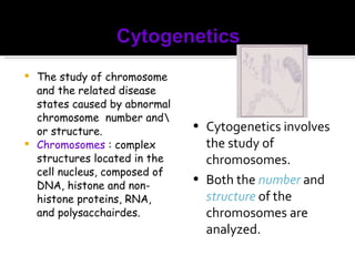
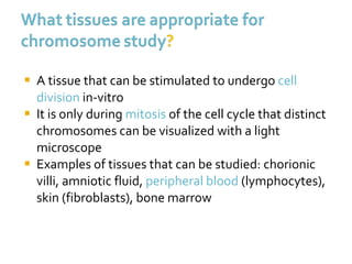
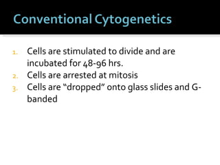
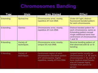
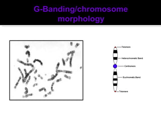
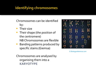
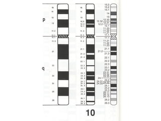
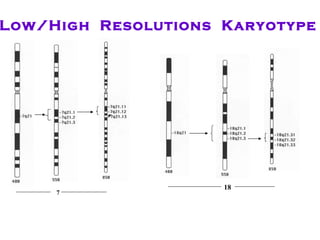
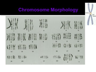
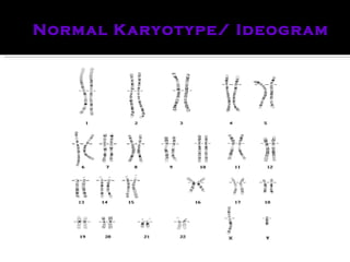
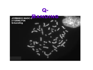
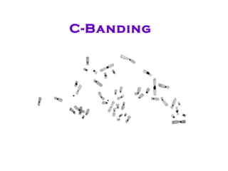
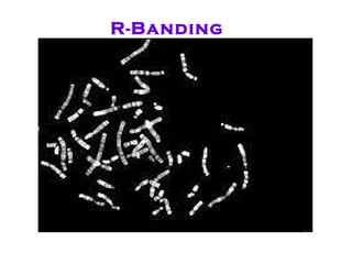
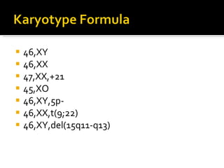
The study of chromosome and the related disease states caused by abnormal chromosome  number and\or structure. Chromosomes  : complex structures located in the cell nucleus, composed of DNA, histone and non-histone proteins, RNA, and polysacchairdes.  Cytogenetics involves the study of chromosomes. Both the  number  and  structure  of the chromosomes are analyzed.
A tissue that can be stimulated to undergo  cell   division  in-vitro It is only during  mitosis  of the cell cycle that distinct chromosomes can be visualized with a light microscope Examples of tissues that can be studied: chorionic villi, amniotic fluid,  peripheral blood  (lymphocytes), skin (fibroblasts), bone marrow
Cells are stimulated to divide and are incubated for 48-96 hrs. Cells are arrested at mitosis Cells are “dropped” onto glass slides and G-banded
 
Each chromatid represents one molecule of DNA
Effect Area Stained Stain Type Under UV light, distinct fluorescent banded pattern for each chromosome. Chromosome arms; mostly repetitive AT-rich DNA Quinacrine Q-banding Distinct banded pattern for each chromosome; same as Q-banding pattern except single additional band near centromere of chromosomes 1 and 16 Chromosome arms; mostly repetitive AT-rich DNA Giemsa G-banding Reverse banding pattern of that observed with Q- or G-banding Chromosome arms; mostly unique GC-rich DNA Variety of techniques R-banding Largest bands usually on chromosomes 1, 9, 16, and Y; chromosomes 7, 10, and 15 have medium-sized bands; size of C-bands highly variable from person to person Centromere region of each chromosome and distal portion of Y chromosome; highly repetitive, mostly AT-rich DNA Variety of techniques C-banding
Non-Banded  Karyotype
 
Chromosomes can be identified by: Their size Their shape (the position of the centromere)  NB Chromosomes are flexible Banding patterns produced by specific stains (Giemsa) Chromosomes are analysed by organising them into a  KARYOTYPE © Biologyreference.com
 
Short arm of the chromosome =  p Long arm of the chromosome =  q Bands are numbered independently on the short and long arms
 
7 18 Low/High  Resolutions  Karyotype
 
Normal Karyotype/ Ideogram
Q-Banding
C-Banding
R - Banding
Advantages 1-  Enable the entire genome to be viewed at one time. 2-  Suitable when a specific anomaly is suspected ( e.g.  Philadelphia in CML ) and as a general diagnostic tool to detect additional chr. Abnormalities commonly seen in disease progression of CML. Disadvantages 1-  Detect major structural abnormalities ( one band = 6mb of DNA ~ 150 genes ). 2-  Labor intensive and highly dependent upon operator experience and skills. Advantages  and Disadvantages of conventional technique
46,XY 46,XX 47,XX,+21 45,XO 46,XY,5p- 46,XX,t(9;22) 46,XY,del(15q11-q13)
Chromosomes can be identified by: Their size Their shape (the position of the centromere)  NB Chromosomes are flexible Banding patterns produced by specific stains (Giemsa) Chromosomes are analysed by organising them into a  KARYOTYPE © Biologyreference.com
A)   Polypoidy – change in complete sets of chromosomes (3n, 4n, etc) plants > animals. Autopolyploidy vs Allopolyploidy Plants    more yeild Animals    parthenogenesis B)  Aneuploidy – change in the no. of chromosomes nullisomy  2n-2 monosomy  2n-1 trisomy  2n+1 tetrasomy  2n+2 Gene dosage effect  1 - Sex-chromosomal aneuploids .  2 - Autosomal aneuploids .
 
 
Chromosome rearrangements: Effects Deletion.  pseudodominance  Duplication.  gene dosage Inversion – paracentric  positional pericentric  Translocation.  positional new linkage rearrangement
Extra single chromosomes or missing single chromosomes 2N + 1 2N - 1 Suffix:  “-somy” or “-somic” 2N + 1  =  trisomy 2N - 1 = monosomy 2N + 2 = tetrasomy
Generally arise through  non-disjunction at meiosis homologues or chromatids do not separate gametes contain 2 or no copies of one chromosome
 
Most aneuploidies in humans lead to such drastic effects, the fetus is spontaneously aborted early in development A few survive ‘til birth; some beyond
47, +21 1/700 live births over 60% of conceptions aborted spontaneously 20% stillborn incidence increases sharply w/ maternal age 1/300 for 35 year olds 1/22 for 45 year olds
characteristic facial appearance small nose, flat face, epicanthal fold single palmar crease mental retardation (avg. IQ < 50) multiple complications heart disease leukemia epilepsy some fertility
47; +18 1/8000 live births; maternal age affect low birth weight multiple dysmorphic features chin, ears, single palmar crease, clenched hands malformations of the brain, heart, kidneys, and other organs rarely survive beyond 1 year
47; + 13 1/20,000 live births; maternal age effect multiple dysmorphic features micropthalmia, cleft palate, clenched fists, polydactyly, ears and scalp abnormal… heart defects; systemic defects…. 50% die in first month; rarely survive beyond 1 year
1/500 live male births (?) often asymptomatic except for sterility, learning disabilities small testes; low testosterone levels poorly developed male 2 o  sexual charact. some female characteristics:  enlarged breasts, elongated limbs, increased incidence of “female” diseases: breast cancer, scoliosis, osteoporosis hormone therapy improves symptoms
1/1000 live male births? formerly called “criminal chromosome” 99% asymptomatic, though high incidence in penal institutions for the mentally subnormal  (20/1000) lower than average intelligence? (learning disabilities) above average height, tendency to severe acne
1/2500 live female births generally asymptomatic ‘til puberty lack of 2 o  sexual characteristics; amenorrhoea short stature; low hair line;  heart disease, renal malformations, ovaries generally underdeveloped, sterile hormone therapy helpful
1/700 live female births (?) generally asymptomatic 15 - 25% mildly mentally retarded some sterility
Changes in the numbers of genes deletions duplications Changes in the location of genes inversions translocations Robertsonian changes
Loss of a (generally small) segment of chromosome A  B  C  D  E  F  G A  B  D  E  F  G C
Arise through spontaneous breakage some chromosomes have fragile spots radiation, UV, chemicals, viruses may increase breakage
May arise through unequal crossing over A  B  C  D  E  F  G A  B  C  D  E  F  G x A  B  C  D  E  F  F  G A  B  C  D  E  G Deletion Duplication
Large deletions will most probably be lethal  Smaller deletions may allow survival E. coli :  deletions of up to 1% have been observed in living cells D. melanogaster  :  deletions of up to 0.1% observed
Cri-du-chat syndrome Micro deletion of chromosome 5 DiGeorge syndrome Micro deletion of chromosome 22 Angelman syndrome Micro deletion of chromosome 15 Prader-Willi syndrome Micro deletion of chromosome 15
lack of muscle tone in newborn poor swallowing reflex as adult - gross obesity mean I.Q. ~ 50 microdeletion of 15 developmentally delayed jerky movements stiff, fixed smile uncontrolled laughter abnormal E.E.G., epilepsy microdeletion of 15 Prader-Willi Angelman
Redundant segment of a chromosome A  B  C  D  E  F  G A  B  C  D  E  F  F G
 
180 o  reversal of chromosome segment A  B  C  D  E  F  G  H  I  J  K A  B  C  H  G  F  E  D  I  J  K 180 O
Produced through breakage and    reassociation of chromosome A B C D E F G
 
 
May change phenotype through “position effects” move active genes to sites generally inactive; lose gene function move inactive genes to sites generally active; gain gene function May act to preserve blocks of genes (specific alleles) which function well together
Exchange of segments between non-homologous chromosomes F A B C D E L M N O P Q
A  B  C D  E  F O  N  M  L Q  P Remember:  There are still two normal    chromosomes in the cell!   = translocation heterozygote
Translocation
Balanced Unbalanced Lead to impaired fertility complications to synapsis and segregation May lead to changes in phenotype position effects human cancers and translocations
Several human cancers are associated with reciprocal translocations Chronic myelocytic leukemia,  many acute leukemias Philadelphia chromosome Burkitt’s lymphoma  three translocations
All these involve oncogenes oncogenes:  genes involved in cell proliferation normally active only at specific times embryonic/fetal development, etc. shut off under normal conditions
Reciprocal translocation between chr.  22 and  chr. 9 9  9   22  22
Reciprocal translocation between chr.  22 and  chr. 9 9  9   22  22  Chr. 9 segment  has an oncogene; when moved, it becomes active
Translocations between chr. 8 and one of three others: 8 & 2  or  8 & 14  or  8 & 22 Chr.  8 has an oncogene Chr.  2, 14, 22 have genes coding for antibody production and their enhancer genes Oncogene becomes highly active under control of enhancers!
Fusions two chromosomes join to form one Fissions one chromosome splits to form two
Roberstsonian translocations
Familial Down Syndrome fusion of chromosomes 14 and 21 14  21  14+21 Carrier;  phenotypically normal produces normal, carrier, and  Down syndrome children
synapsis Three possible disjunctions
 
X X
X X X X
© 2007 Paul Billiet  ODWS Images believed to be in the Public Domain 23  unpaired  chromosomes 23  unpaired  chromosomes 23  unpaired  chromosomes 23  unpaired  chromosomes Fertilisation Child Father 23  pairs  of chromosomes Sex cells Meiosis Mother 23 pairs of chromosomes 23 pairs of chromosomes
A special type of cell division Used to make  sex cells Meiosis  halves  the numbers of chromosomes Meiosis picks one chromosome from each pair at random and places them in a sex cell. This results in enormous variation amongst the sex cells.   © 2007 Paul Billiet  ODWS
Y X X X Fertilisation Possible children Father Sex cells Meiosis Mother XX XY Chance of a girl 50%  Chance of a boy 50% © 2007 Paul Billiet  ODWS X Y X XX XY X XX XY
The sex of many animals is determined by genes but on chromosomes called  sex chromosomes The other chromosomes are called  autosomes One sex is homogametic The other sex is heterogametic © 2007 Paul Billiet  ODWS
© 2007 Paul Billiet  ODWS HOMOGAMETIC SEX HETEROGAMETIC SEX SEX DETERMINATION Female  XX Male  XY Presence of Y-chromosome = maleness (mammals and fish) Presence of second X-chromosome = femaleness (Drosophila, the fruit fly) Male  ZZ Female  ZW Birds, amphibians, reptiles, butterflies, moths. Female  XX Male  Xo Grasshoppers
Constant for each cell in the body (except sex cells which only have half sets). Constant throughout the life of an individual (you don’t lose or gain chromosomes) Constant for all members of a species © 2007 Paul Billiet  ODWS
Mouse Maize © A. Lane Rayburn Image believed to be in the Public Domain
© 2007 Paul Billiet  ODWS Organism Chromosome numbers Human 46 Chimpanzee 48 House Mouse 40 Maize 20
Image believed to be in the Public Domain

Cytogenetics

  • 1.
  • 2.
    The study ofchromosome and the related disease states caused by abnormal chromosome number and\or structure. Chromosomes : complex structures located in the cell nucleus, composed of DNA, histone and non-histone proteins, RNA, and polysacchairdes. Cytogenetics involves the study of chromosomes. Both the number and structure of the chromosomes are analyzed.
  • 3.
    A tissue thatcan be stimulated to undergo cell division in-vitro It is only during mitosis of the cell cycle that distinct chromosomes can be visualized with a light microscope Examples of tissues that can be studied: chorionic villi, amniotic fluid, peripheral blood (lymphocytes), skin (fibroblasts), bone marrow
  • 4.
    Cells are stimulatedto divide and are incubated for 48-96 hrs. Cells are arrested at mitosis Cells are “dropped” onto glass slides and G-banded
  • 5.
  • 6.
    Each chromatid representsone molecule of DNA
  • 7.
    Effect Area StainedStain Type Under UV light, distinct fluorescent banded pattern for each chromosome. Chromosome arms; mostly repetitive AT-rich DNA Quinacrine Q-banding Distinct banded pattern for each chromosome; same as Q-banding pattern except single additional band near centromere of chromosomes 1 and 16 Chromosome arms; mostly repetitive AT-rich DNA Giemsa G-banding Reverse banding pattern of that observed with Q- or G-banding Chromosome arms; mostly unique GC-rich DNA Variety of techniques R-banding Largest bands usually on chromosomes 1, 9, 16, and Y; chromosomes 7, 10, and 15 have medium-sized bands; size of C-bands highly variable from person to person Centromere region of each chromosome and distal portion of Y chromosome; highly repetitive, mostly AT-rich DNA Variety of techniques C-banding
  • 8.
  • 9.
  • 10.
    Chromosomes can beidentified by: Their size Their shape (the position of the centromere) NB Chromosomes are flexible Banding patterns produced by specific stains (Giemsa) Chromosomes are analysed by organising them into a KARYOTYPE © Biologyreference.com
  • 11.
  • 12.
    Short arm ofthe chromosome = p Long arm of the chromosome = q Bands are numbered independently on the short and long arms
  • 13.
  • 14.
    7 18 Low/High Resolutions Karyotype
  • 15.
  • 16.
  • 17.
  • 18.
  • 19.
  • 20.
    Advantages 1- Enable the entire genome to be viewed at one time. 2- Suitable when a specific anomaly is suspected ( e.g. Philadelphia in CML ) and as a general diagnostic tool to detect additional chr. Abnormalities commonly seen in disease progression of CML. Disadvantages 1- Detect major structural abnormalities ( one band = 6mb of DNA ~ 150 genes ). 2- Labor intensive and highly dependent upon operator experience and skills. Advantages and Disadvantages of conventional technique
  • 21.
    46,XY 46,XX 47,XX,+2145,XO 46,XY,5p- 46,XX,t(9;22) 46,XY,del(15q11-q13)
  • 22.
    Chromosomes can beidentified by: Their size Their shape (the position of the centromere) NB Chromosomes are flexible Banding patterns produced by specific stains (Giemsa) Chromosomes are analysed by organising them into a KARYOTYPE © Biologyreference.com
  • 23.
    A) Polypoidy – change in complete sets of chromosomes (3n, 4n, etc) plants > animals. Autopolyploidy vs Allopolyploidy Plants  more yeild Animals  parthenogenesis B) Aneuploidy – change in the no. of chromosomes nullisomy 2n-2 monosomy 2n-1 trisomy 2n+1 tetrasomy 2n+2 Gene dosage effect 1 - Sex-chromosomal aneuploids . 2 - Autosomal aneuploids .
  • 24.
  • 25.
  • 26.
    Chromosome rearrangements: EffectsDeletion. pseudodominance Duplication. gene dosage Inversion – paracentric positional pericentric Translocation. positional new linkage rearrangement
  • 27.
    Extra single chromosomesor missing single chromosomes 2N + 1 2N - 1 Suffix: “-somy” or “-somic” 2N + 1 = trisomy 2N - 1 = monosomy 2N + 2 = tetrasomy
  • 28.
    Generally arise through non-disjunction at meiosis homologues or chromatids do not separate gametes contain 2 or no copies of one chromosome
  • 29.
  • 30.
    Most aneuploidies inhumans lead to such drastic effects, the fetus is spontaneously aborted early in development A few survive ‘til birth; some beyond
  • 31.
    47, +21 1/700live births over 60% of conceptions aborted spontaneously 20% stillborn incidence increases sharply w/ maternal age 1/300 for 35 year olds 1/22 for 45 year olds
  • 32.
    characteristic facial appearancesmall nose, flat face, epicanthal fold single palmar crease mental retardation (avg. IQ < 50) multiple complications heart disease leukemia epilepsy some fertility
  • 33.
    47; +18 1/8000live births; maternal age affect low birth weight multiple dysmorphic features chin, ears, single palmar crease, clenched hands malformations of the brain, heart, kidneys, and other organs rarely survive beyond 1 year
  • 34.
    47; + 131/20,000 live births; maternal age effect multiple dysmorphic features micropthalmia, cleft palate, clenched fists, polydactyly, ears and scalp abnormal… heart defects; systemic defects…. 50% die in first month; rarely survive beyond 1 year
  • 35.
    1/500 live malebirths (?) often asymptomatic except for sterility, learning disabilities small testes; low testosterone levels poorly developed male 2 o sexual charact. some female characteristics: enlarged breasts, elongated limbs, increased incidence of “female” diseases: breast cancer, scoliosis, osteoporosis hormone therapy improves symptoms
  • 36.
    1/1000 live malebirths? formerly called “criminal chromosome” 99% asymptomatic, though high incidence in penal institutions for the mentally subnormal (20/1000) lower than average intelligence? (learning disabilities) above average height, tendency to severe acne
  • 37.
    1/2500 live femalebirths generally asymptomatic ‘til puberty lack of 2 o sexual characteristics; amenorrhoea short stature; low hair line; heart disease, renal malformations, ovaries generally underdeveloped, sterile hormone therapy helpful
  • 38.
    1/700 live femalebirths (?) generally asymptomatic 15 - 25% mildly mentally retarded some sterility
  • 39.
    Changes in thenumbers of genes deletions duplications Changes in the location of genes inversions translocations Robertsonian changes
  • 40.
    Loss of a(generally small) segment of chromosome A B C D E F G A B D E F G C
  • 41.
    Arise through spontaneousbreakage some chromosomes have fragile spots radiation, UV, chemicals, viruses may increase breakage
  • 42.
    May arise throughunequal crossing over A B C D E F G A B C D E F G x A B C D E F F G A B C D E G Deletion Duplication
  • 43.
    Large deletions willmost probably be lethal Smaller deletions may allow survival E. coli : deletions of up to 1% have been observed in living cells D. melanogaster : deletions of up to 0.1% observed
  • 44.
    Cri-du-chat syndrome Microdeletion of chromosome 5 DiGeorge syndrome Micro deletion of chromosome 22 Angelman syndrome Micro deletion of chromosome 15 Prader-Willi syndrome Micro deletion of chromosome 15
  • 45.
    lack of muscletone in newborn poor swallowing reflex as adult - gross obesity mean I.Q. ~ 50 microdeletion of 15 developmentally delayed jerky movements stiff, fixed smile uncontrolled laughter abnormal E.E.G., epilepsy microdeletion of 15 Prader-Willi Angelman
  • 46.
    Redundant segment ofa chromosome A B C D E F G A B C D E F F G
  • 47.
  • 48.
    180 o reversal of chromosome segment A B C D E F G H I J K A B C H G F E D I J K 180 O
  • 49.
    Produced through breakageand reassociation of chromosome A B C D E F G
  • 50.
    Produced through breakageand reassociation of chromosome A B C D E F G
  • 51.
  • 52.
  • 53.
    May change phenotypethrough “position effects” move active genes to sites generally inactive; lose gene function move inactive genes to sites generally active; gain gene function May act to preserve blocks of genes (specific alleles) which function well together
  • 54.
    Exchange of segmentsbetween non-homologous chromosomes F A B C D E L M N O P Q
  • 55.
    A B C D E F O N M L Q P Remember: There are still two normal chromosomes in the cell! = translocation heterozygote
  • 56.
  • 57.
    Balanced Unbalanced Leadto impaired fertility complications to synapsis and segregation May lead to changes in phenotype position effects human cancers and translocations
  • 58.
    Several human cancersare associated with reciprocal translocations Chronic myelocytic leukemia, many acute leukemias Philadelphia chromosome Burkitt’s lymphoma three translocations
  • 59.
    All these involveoncogenes oncogenes: genes involved in cell proliferation normally active only at specific times embryonic/fetal development, etc. shut off under normal conditions
  • 60.
    Reciprocal translocation betweenchr. 22 and chr. 9 9 9 22 22
  • 61.
    Reciprocal translocation betweenchr. 22 and chr. 9 9 9 22 22 Chr. 9 segment has an oncogene; when moved, it becomes active
  • 62.
    Translocations between chr.8 and one of three others: 8 & 2 or 8 & 14 or 8 & 22 Chr. 8 has an oncogene Chr. 2, 14, 22 have genes coding for antibody production and their enhancer genes Oncogene becomes highly active under control of enhancers!
  • 63.
    Fusions two chromosomesjoin to form one Fissions one chromosome splits to form two
  • 64.
  • 65.
    Familial Down Syndromefusion of chromosomes 14 and 21 14 21 14+21 Carrier; phenotypically normal produces normal, carrier, and Down syndrome children
  • 66.
  • 67.
  • 68.
  • 69.
  • 70.
    © 2007 PaulBilliet ODWS Images believed to be in the Public Domain 23 unpaired chromosomes 23 unpaired chromosomes 23 unpaired chromosomes 23 unpaired chromosomes Fertilisation Child Father 23 pairs of chromosomes Sex cells Meiosis Mother 23 pairs of chromosomes 23 pairs of chromosomes
  • 71.
    A special typeof cell division Used to make sex cells Meiosis halves the numbers of chromosomes Meiosis picks one chromosome from each pair at random and places them in a sex cell. This results in enormous variation amongst the sex cells. © 2007 Paul Billiet ODWS
  • 72.
    Y X XX Fertilisation Possible children Father Sex cells Meiosis Mother XX XY Chance of a girl 50% Chance of a boy 50% © 2007 Paul Billiet ODWS X Y X XX XY X XX XY
  • 73.
    The sex ofmany animals is determined by genes but on chromosomes called sex chromosomes The other chromosomes are called autosomes One sex is homogametic The other sex is heterogametic © 2007 Paul Billiet ODWS
  • 74.
    © 2007 PaulBilliet ODWS HOMOGAMETIC SEX HETEROGAMETIC SEX SEX DETERMINATION Female XX Male XY Presence of Y-chromosome = maleness (mammals and fish) Presence of second X-chromosome = femaleness (Drosophila, the fruit fly) Male ZZ Female ZW Birds, amphibians, reptiles, butterflies, moths. Female XX Male Xo Grasshoppers
  • 75.
    Constant for eachcell in the body (except sex cells which only have half sets). Constant throughout the life of an individual (you don’t lose or gain chromosomes) Constant for all members of a species © 2007 Paul Billiet ODWS
  • 76.
    Mouse Maize ©A. Lane Rayburn Image believed to be in the Public Domain
  • 77.
    © 2007 PaulBilliet ODWS Organism Chromosome numbers Human 46 Chimpanzee 48 House Mouse 40 Maize 20
  • 78.
    Image believed tobe in the Public Domain