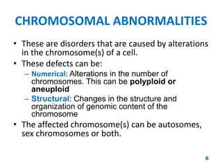
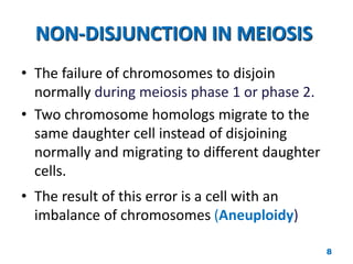
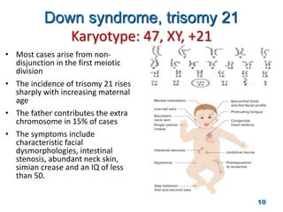
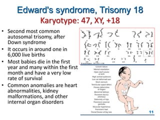
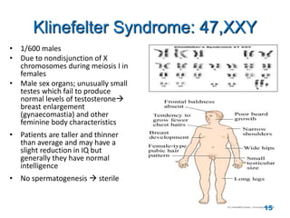
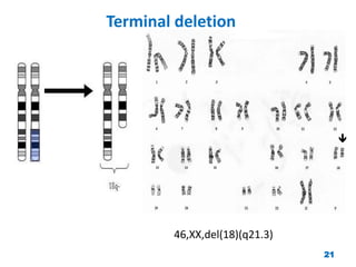
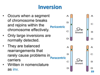
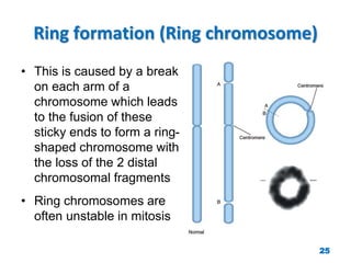
CHROMOSOMAL DISORDERS
1
CHROMOSOMES
• Rod-shaped filamentous bodies present in the
nucleus which are composed of DNA, RNA and
proteins (histones and non histones).
– These bodies are the units of heredity
• Human somatic cells contain 23 pairs of
chromosomes (diploid)
– 22 homologous autosomes and XX (females) and XY
(males)
2
EVENTS OF MITOSIS
INTERPHASE (G1, S, G2 phase)
• This phase prepares the cell for the mitotic
phase of cell division.
MITOTIC PHASE
Prophase.
• Formation of mitotic Spindle & pair of
centrosome
Prometaphase.
• Nuclear membrane dissolves
chromosomes to disperse & attach by
kinetochores to mitotic spindle
microtubules
Metaphase.
• Chromosomes condensed & line up at the
equatorial plane
Anaphase.
• Chromosomes separate at the centromere
and sister chromatids of each chromosome
become independent daughter
chromosomes
Telophase.
• Chromosomes de-condense from their
highly contracted state and the nuclear
membrane re-forms around each of the
two daughter nuclei with diploid
chromosomes. 3
EVENTS OF MEIOSIS I & II
4
KARYOTYPE
• It is a photographic
representation of a stained
metaphase spread in which
the chromosomes are
arranged in order of
decreasing length.
• N-diacetyl-N- methylcolchicine
– To arrest cell division at metaphase
• Giemsa stain
– To demonstrate the bands of
chromosomes.
5
CHROMOSOMAL ABNORMALITIES
• These are disorders that are caused by alterations
in the chromosome(s) of a cell.
• These defects can be:
– Numerical: Alterations in the number of
chromosomes. This can be polyploid or
aneuploid
– Structural: Changes in the structure and
organization of genomic content of the
chromosome
• The affected chromosome(s) can be autosomes,
sex chromosomes or both.
6
NUMERICAL CHROMOSOMAL
ANOMALIES
Numerical anomalies in autosomes
7
NON-DISJUNCTION IN MEIOSIS
• The failure of chromosomes to disjoin
normally during meiosis phase 1 or phase 2.
• Two chromosome homologs migrate to the
same daughter cell instead of disjoining
normally and migrating to different daughter
cells.
• The result of this error is a cell with an
imbalance of chromosomes (Aneuploidy)
8
9
Down syndrome, trisomy 21
Karyotype: 47, XY, +21
• Most cases arise from non-
disjunction in the first meiotic
division
• The incidence of trisomy 21 rises
sharply with increasing maternal
age
• The father contributes the extra
chromosome in 15% of cases
• The symptoms include
characteristic facial
dysmorphologies, intestinal
stenosis, abundant neck skin,
simian crease and an IQ of less
than 50.
10
Edward's syndrome, Trisomy 18
Karyotype: 47, XY, +18
• Second most common
autosomal trisomy, after
Down syndrome
• It occurs in around one in
6,000 live births
• Most babies die in the first
year and many within the first
month and have a very low
rate of survival
• Common anomalies are heart
abnormalities, kidney
malformations, and other
internal organ disorders
11
Patau Syndrome, Trisomy 13
Karyotype: 47, XY, +13
• 50% of these babies die
within the first month and
very few survive beyond the
first year.
• There are multiple
dysmorphic features.
• Most cases, as in Patau
syndrome, involve maternal
non-disjunction.
12
NUMERICAL CHROMOSOMAL
ANOMALIES
Numerical anomalies in Sex
chromosomes
13
Monosomy X (Turner’s syndrome,
45,XO)
• Occurs in 1 in 4000
phenotypic females
• As a result of paternal
meiotic nondisjunction
• The only viable monosomy
in humans
• Characteristics:
Webbed neck, Individuals are
genetically female, not mature
sexually, Sterile, Short stature,
Broad chest, Low hairline, Streak
ovaries, Normal intelligence,
Normal life span
14
Klinefelter Syndrome: 47,XXY
• 1/600 males
• Due to nondisjunction of X
chromosomes during meiosis I in
females
• Male sex organs; unusually small
testes which fail to produce
normal levels of testosterone
breast enlargement
(gynaecomastia) and other
feminine body characteristics
• Patients are taller and thinner
than average and may have a
slight reduction in IQ but
generally they have normal
intelligence
• No spermatogenesis  sterile
15
Mosaicism
• The presence of more than one
genetically distinct cell line in the
body
• A mosaic individual is made of 2
(or more) cell populations, coming
from only 1 zygote
• Denoted by a slash between the
various clones observed
– e.g. (46, XY / 47, XY, +21).
• Numerical mosaic anomaly is
usually due to a mitotic non-
disjunction
16
STRUCTURAL CHROMOSOMAL
ANOMALIES
17
Reciprocal Translocation
• Reciprocal translocation between chromosome 22 and the long arm
of chromosome 9 (the Philadelphia chromosome).
• The occurrence of this translocation in hematopoietic cells can
produce chronic myelogenous leukemia (CML) 18
Robertsonian translocation
• Short arms of two non-
homologous chromosomes are
lost and the long arms fuse at
the centromere to form a single
chromosome confined to the
acrocentric chromosomes (13,
14, 15, 21, and 22)
• Although carriers have only 45
chromosomes in each cell, they
are phenotypically unaffected
19
Deletion
• Loss of a segment from a chromosome, either
terminal or interstitial
• Invariably, but not always, results in the loss of
important genetic material
• Deletion is therefore an unbalanced
rearrangement.
• Indicated in nomenclature del
20
Terminal deletion
46,XX,del(18)(q21.3)

21
Interstitial deletion
karyotype description is as follows:
• 46: the total number of chromosomes.
• XY: the sex chromosomes (male).
• del(7): deletion in chromosome 7.
• (q11.23q21.2): breakpoints of the deleted
segment. 46,XY,del(7)(q11.23q21.2)
22
Inversion
Paracentric
Pericentric
• Occurs when a segment
of chromosome breaks
and rejoins within the
chromosome effectively.
• Only large inversions are
normally detected.
• They are balanced
rearrangements that
rarely cause problems in
carriers
• Written in nomenclature
as inv. 23
Isochromosome
Normal Isochromosome Normal Isochromosome
• The most probable explanation for
isochromosome is that the centromere has
divided transversely rather than
longitudinally
24
Ring formation (Ring chromosome)
• This is caused by a break
on each arm of a
chromosome which leads
to the fusion of these
sticky ends to form a ring-
shaped chromosome with
the loss of the 2 distal
chromosomal fragments
• Ring chromosomes are
often unstable in mitosis
25
Summary
• Chromosome disorders can be numerical or structural.
• Normal meiotic division results in four haploid gametes
• In trisomy, a single extra chromosome is present,
usually as a result of non-disjunction in the 1st or 2nd
meiotic division.
• Mosaicism arises from one zygote
• Structural abnormalities include translocations
(balanced or unbalanced), inversions, deletions,
isochromosomes & rings.
26
THANK YOU !
27

Chromosomal Disorders.pptx

  • 1.
  • 2.
    CHROMOSOMES • Rod-shaped filamentousbodies present in the nucleus which are composed of DNA, RNA and proteins (histones and non histones). – These bodies are the units of heredity • Human somatic cells contain 23 pairs of chromosomes (diploid) – 22 homologous autosomes and XX (females) and XY (males) 2
  • 3.
    EVENTS OF MITOSIS INTERPHASE(G1, S, G2 phase) • This phase prepares the cell for the mitotic phase of cell division. MITOTIC PHASE Prophase. • Formation of mitotic Spindle & pair of centrosome Prometaphase. • Nuclear membrane dissolves chromosomes to disperse & attach by kinetochores to mitotic spindle microtubules Metaphase. • Chromosomes condensed & line up at the equatorial plane Anaphase. • Chromosomes separate at the centromere and sister chromatids of each chromosome become independent daughter chromosomes Telophase. • Chromosomes de-condense from their highly contracted state and the nuclear membrane re-forms around each of the two daughter nuclei with diploid chromosomes. 3
  • 4.
  • 5.
    KARYOTYPE • It isa photographic representation of a stained metaphase spread in which the chromosomes are arranged in order of decreasing length. • N-diacetyl-N- methylcolchicine – To arrest cell division at metaphase • Giemsa stain – To demonstrate the bands of chromosomes. 5
  • 6.
    CHROMOSOMAL ABNORMALITIES • Theseare disorders that are caused by alterations in the chromosome(s) of a cell. • These defects can be: – Numerical: Alterations in the number of chromosomes. This can be polyploid or aneuploid – Structural: Changes in the structure and organization of genomic content of the chromosome • The affected chromosome(s) can be autosomes, sex chromosomes or both. 6
  • 7.
  • 8.
    NON-DISJUNCTION IN MEIOSIS •The failure of chromosomes to disjoin normally during meiosis phase 1 or phase 2. • Two chromosome homologs migrate to the same daughter cell instead of disjoining normally and migrating to different daughter cells. • The result of this error is a cell with an imbalance of chromosomes (Aneuploidy) 8
  • 9.
  • 10.
    Down syndrome, trisomy21 Karyotype: 47, XY, +21 • Most cases arise from non- disjunction in the first meiotic division • The incidence of trisomy 21 rises sharply with increasing maternal age • The father contributes the extra chromosome in 15% of cases • The symptoms include characteristic facial dysmorphologies, intestinal stenosis, abundant neck skin, simian crease and an IQ of less than 50. 10
  • 11.
    Edward's syndrome, Trisomy18 Karyotype: 47, XY, +18 • Second most common autosomal trisomy, after Down syndrome • It occurs in around one in 6,000 live births • Most babies die in the first year and many within the first month and have a very low rate of survival • Common anomalies are heart abnormalities, kidney malformations, and other internal organ disorders 11
  • 12.
    Patau Syndrome, Trisomy13 Karyotype: 47, XY, +13 • 50% of these babies die within the first month and very few survive beyond the first year. • There are multiple dysmorphic features. • Most cases, as in Patau syndrome, involve maternal non-disjunction. 12
  • 13.
  • 14.
    Monosomy X (Turner’ssyndrome, 45,XO) • Occurs in 1 in 4000 phenotypic females • As a result of paternal meiotic nondisjunction • The only viable monosomy in humans • Characteristics: Webbed neck, Individuals are genetically female, not mature sexually, Sterile, Short stature, Broad chest, Low hairline, Streak ovaries, Normal intelligence, Normal life span 14
  • 15.
    Klinefelter Syndrome: 47,XXY •1/600 males • Due to nondisjunction of X chromosomes during meiosis I in females • Male sex organs; unusually small testes which fail to produce normal levels of testosterone breast enlargement (gynaecomastia) and other feminine body characteristics • Patients are taller and thinner than average and may have a slight reduction in IQ but generally they have normal intelligence • No spermatogenesis  sterile 15
  • 16.
    Mosaicism • The presenceof more than one genetically distinct cell line in the body • A mosaic individual is made of 2 (or more) cell populations, coming from only 1 zygote • Denoted by a slash between the various clones observed – e.g. (46, XY / 47, XY, +21). • Numerical mosaic anomaly is usually due to a mitotic non- disjunction 16
  • 17.
  • 18.
    Reciprocal Translocation • Reciprocaltranslocation between chromosome 22 and the long arm of chromosome 9 (the Philadelphia chromosome). • The occurrence of this translocation in hematopoietic cells can produce chronic myelogenous leukemia (CML) 18
  • 19.
    Robertsonian translocation • Shortarms of two non- homologous chromosomes are lost and the long arms fuse at the centromere to form a single chromosome confined to the acrocentric chromosomes (13, 14, 15, 21, and 22) • Although carriers have only 45 chromosomes in each cell, they are phenotypically unaffected 19
  • 20.
    Deletion • Loss ofa segment from a chromosome, either terminal or interstitial • Invariably, but not always, results in the loss of important genetic material • Deletion is therefore an unbalanced rearrangement. • Indicated in nomenclature del 20
  • 21.
  • 22.
    Interstitial deletion karyotype descriptionis as follows: • 46: the total number of chromosomes. • XY: the sex chromosomes (male). • del(7): deletion in chromosome 7. • (q11.23q21.2): breakpoints of the deleted segment. 46,XY,del(7)(q11.23q21.2) 22
  • 23.
    Inversion Paracentric Pericentric • Occurs whena segment of chromosome breaks and rejoins within the chromosome effectively. • Only large inversions are normally detected. • They are balanced rearrangements that rarely cause problems in carriers • Written in nomenclature as inv. 23
  • 24.
    Isochromosome Normal Isochromosome NormalIsochromosome • The most probable explanation for isochromosome is that the centromere has divided transversely rather than longitudinally 24
  • 25.
    Ring formation (Ringchromosome) • This is caused by a break on each arm of a chromosome which leads to the fusion of these sticky ends to form a ring- shaped chromosome with the loss of the 2 distal chromosomal fragments • Ring chromosomes are often unstable in mitosis 25
  • 26.
    Summary • Chromosome disorderscan be numerical or structural. • Normal meiotic division results in four haploid gametes • In trisomy, a single extra chromosome is present, usually as a result of non-disjunction in the 1st or 2nd meiotic division. • Mosaicism arises from one zygote • Structural abnormalities include translocations (balanced or unbalanced), inversions, deletions, isochromosomes & rings. 26
  • 27.