SlideShare a Scribd company logo
1 of 72
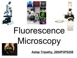
Fluorescence Microscopy Aalap Tripathy, 2004P3PS208
Then… Now…
What I will discuss ,[object Object],[object Object],[object Object]
 
A fluorescence microscope is basically a conventional light microscope with added features and components that extend its capabilities.  conventional microscope   fluorescence microscope   ,[object Object],[object Object],[object Object],[object Object]
[object Object],[object Object],TV Screen Sonoluminescence  is the emission of light by bubbles in a liquid excited by sound.
Fluorescence v/s Phosphorescence ,[object Object],[object Object],The term  phosphorescence  is often incorrectly considered synonymous with  luminescence
Basic Concepts ,[object Object],[object Object],[object Object],[object Object]
 
Zeiss   Axio  Imager Z1   Objective lenses:   Filtersets:   Camera:   Software:   ImageJ Imaris  4.2   Monchrome  Phase contrast  Oil         Imaging-Workstation:   Color
Why Prepare the Specimen ,[object Object],[object Object]
Preparation of Specimen Tagging of Proteins   Fluorescent Dyes   Immunofluorescence   Techniques Fluorescent Dyes Immunofluorescence   Tagging of Proteins   ,[object Object],[object Object],[object Object],[object Object],[object Object],[object Object],[object Object]
TYPES OF FLUOROPHORES USED ,[object Object],[object Object],[object Object],[object Object],[object Object]
[object Object],[object Object]
 
Cutaway diagram of a modern epi-fluorescence microscope
Working of the Fluorescence Microscope 1. Light source – epi-fluorescence lamphouse  2. Light of a specific wavelength (or defined band of wavelengths), is produced by passing multispectral light from an arc-discharge lamp through a wavelength selective  excitation  filter  3. Wavelengths passed by the excitation filter reflect from the surface of a  dichromatic  (also termed a  dichroic ) mirror or beamsplitter through the microscope objective to bathe the specimen with intense light
Working of the Fluorescence Microscope 4. If the specimen fluoresces, the emission light gathered by the objective passes back through the dichromatic mirror  5. It is Filtered by a  barrier  (or  emission ) filter, which blocks the unwanted excitation wavelengths
 
The “cube”
Working in greater detail 1. Excitation light travels along the illuminator  perpendicular to the optical axis of the microscope  2. The light then  impinges  upon the excitation filter where selection of the desired band and blockage of unwanted wavelength occurs.
3. Fluorescence emission produced by the illuminated specimen is gathered by the objective  4. Because the emitted light consists of longer wavelengths than the excitation illumination, it is able to pass through the dichromatic mirror and upward to the observation tubes or electronic detector.  Working in greater detail
The Dichroic Mirror ,[object Object],[object Object],dichroic, two color
 
The Dichroic Mirror dichroic, two color   ,[object Object],[object Object],[object Object],[object Object]
Total Internal Reflection in Prism Same Principle used in Dichromatic beam splitter
Modern fluorescence microscopes are capable of accommodating between four and six fluorescence cubes. This is where the “turret’s” come into picture.  The “cube” A specific combination of excitation filter, emission filter and dichroic mirror are needed
The Filters ,[object Object],[object Object],[object Object],[object Object],Fig: Light path through the filter cube in a fluorescence microscope.
Stoke’s Shift ,[object Object],[object Object],[object Object],[object Object],[object Object],[object Object]
Stoke’s Shift ,[object Object],Remember  Dichoric Mirror ???
Data for Alexa Fluor 555  ,[object Object],[object Object],[object Object],[object Object],[object Object]
The radiation collides with the atoms in the specimen and electrons are excited to a higher energy level. When they relax to a lower level, they emit light.  Principle of Fluorescence   1. Energy is absorbed by the atom which becomes excited. 2. The electron jumps to a higher energy level. 3. Soon, the electron drops back to the ground state, emitting a photon (or a packet of light) - the atom is fluorescing.
 
Visualizing The Cytoskeleton using Fluorescence Microscopy An Example of Fluorescent Dyes
Two cytoskeletal elements examined: Why use  Fluorescence Microscopy   ? ,[object Object],[object Object],Actin Microtubules
Let us test the effects of different drugs on the cytoskeleton and cell shape Nocodazole  prevents microtubule polymerization. Nocodazole Taxol  binds and stabilizes microtubules, Taxol Latrunculin  prevents actin polymerization.  Latrunculin TPA/PMA  causes a dramatic rearrangement of actin filaments TPA/PMA
Visualizing the cytoskeleton  using fluorescence microscopy 1)  Prepare samples: Fixation  - kills and immobilizes cells A. aldehydes - cross-link amino groups in proteins (formaldehyde, glutaraldehyde) B. alcohols - denature proteins, precipitate in place (methanol) Permeabilization  - detergents make proteins  accessible to staining reagents (Triton X100)
2) Staining Actin - phalloidin covalently linked to rhodamine (red) - binds to filamentous actin only Microtubules - immunofluorescence 1 o  ab: rabbit anti-tubulin; 2 o  ab: fluorescein anti-rabbit
3) Fluorescence microscopy excitation emission fluorescent molecule wavelength ex em intensity
Microtubules  = green DNA = blue interphase mitosis
mitosis
Green Fluorescent Protein (GFP) An Example of tagging proteins
Green Fluorescent Protein (GFP) ,[object Object],[object Object],[object Object],[object Object],[object Object],[object Object]
These transgenic mice express enhanced green fluorescent protein under the control of a chicken beta-actin promoter and cytomegalovirus enhancer
Why do this ?? developing transgenic mice to identify critical neuronal subpopulations and target them for electrophysiological recordings and biochemical analyses.
Some Pictures
 
Cotton A cross section of cotton stained with Rhodamine B.  Mammalian Cells Fluorescence double-labeling of mammalian cells. The DNA in the cell nuclei are shown in blue. Cytoplasmic fiber structures (microfilaments) are shown in green.  Photo: Petra Björk, Stockholm University
Researchers tag proteins with fluorophores to study the motion of these molecules. However, this creates an extremely complex motion picture (for example, in this image different colored particles move independently)
http://nobelprize.org/physics/educational/microscopes/fluorescence/fm.html Control of a fluorescence microscope
Figure 3: Problems with Fluorescence microscopy
Summary ,[object Object],[object Object],[object Object]
Future ,[object Object],[object Object],[object Object]
Some Pictures
Parainfluenza
 
 
Influenza


Fluorescent antibody detection
 
 
Fluorescent Antibody Staining
 
Fluoresence of chlorophyll-protein complex – Cytochrome b 6 f ,[object Object],[object Object]


Resources Kenneth R. Spring  - Scientific Consultant, Lusby, Maryland, 20657. Michael W. Davidson  - National High Magnetic Field Laboratory, 1800 East Paul Dirac Dr., The Florida State University, Tallahassee, Florida, 32310.

More Related Content

What's hot

Confocal microscopy Likhith K
Confocal microscopy Likhith KConfocal microscopy Likhith K
Confocal microscopy Likhith K
LIKHITHK1
 

What's hot (20)

PHASE MICROSCOPY
PHASE MICROSCOPYPHASE MICROSCOPY
PHASE MICROSCOPY
 
Fluorescence microscope by Subhankar Das
Fluorescence microscope by Subhankar DasFluorescence microscope by Subhankar Das
Fluorescence microscope by Subhankar Das
 
Phase contrast microscope
Phase contrast microscopePhase contrast microscope
Phase contrast microscope
 
Various types of microscopes and microscopy
Various types of microscopes and microscopy   Various types of microscopes and microscopy
Various types of microscopes and microscopy
 
Confocal microscope
Confocal microscopeConfocal microscope
Confocal microscope
 
Electron microscope
Electron microscopeElectron microscope
Electron microscope
 
Confocal microscopy
Confocal microscopyConfocal microscopy
Confocal microscopy
 
Phase contrast microscope
Phase contrast microscopePhase contrast microscope
Phase contrast microscope
 
Phase contrast microscope
Phase contrast microscopePhase contrast microscope
Phase contrast microscope
 
Light microscope
Light microscopeLight microscope
Light microscope
 
DARK FIELD MICROSCOPY
DARK FIELD MICROSCOPYDARK FIELD MICROSCOPY
DARK FIELD MICROSCOPY
 
Fluorescence microscopy
Fluorescence microscopy   Fluorescence microscopy
Fluorescence microscopy
 
Confocal microscopy by prabhat
Confocal microscopy by prabhatConfocal microscopy by prabhat
Confocal microscopy by prabhat
 
Principles and application of light, phase constrast and fluorescence microscope
Principles and application of light, phase constrast and fluorescence microscopePrinciples and application of light, phase constrast and fluorescence microscope
Principles and application of light, phase constrast and fluorescence microscope
 
Confocal microscopy Likhith K
Confocal microscopy Likhith KConfocal microscopy Likhith K
Confocal microscopy Likhith K
 
fluroscence microscopy final- ppt.pptx
fluroscence microscopy final- ppt.pptxfluroscence microscopy final- ppt.pptx
fluroscence microscopy final- ppt.pptx
 
Fluorescence microscopy presentation
Fluorescence microscopy presentationFluorescence microscopy presentation
Fluorescence microscopy presentation
 
Light microscope
Light microscopeLight microscope
Light microscope
 
Phase contrast microscope
Phase contrast microscopePhase contrast microscope
Phase contrast microscope
 
Stereo Microscope or Dissecting Miscrscope
Stereo Microscope or Dissecting MiscrscopeStereo Microscope or Dissecting Miscrscope
Stereo Microscope or Dissecting Miscrscope
 

Viewers also liked (6)

Scanning transmission electron microscope (2)
Scanning transmission electron microscope (2)Scanning transmission electron microscope (2)
Scanning transmission electron microscope (2)
 
Fluorescence spectroscopy
Fluorescence spectroscopyFluorescence spectroscopy
Fluorescence spectroscopy
 
Sem n tem
Sem n temSem n tem
Sem n tem
 
Phase contrast microscope
Phase contrast microscopePhase contrast microscope
Phase contrast microscope
 
Electron microscopy
Electron microscopyElectron microscopy
Electron microscopy
 
Scanning electron microscopy
Scanning electron microscopyScanning electron microscopy
Scanning electron microscopy
 

Similar to Fluorescence Microscopy

2 Microscopy and staining.pptx
2 Microscopy and staining.pptx2 Microscopy and staining.pptx
2 Microscopy and staining.pptx
NeelGamers
 

Similar to Fluorescence Microscopy (20)

FLUOROSCENCE & PHASE CONTRAST MICROSCOPY.pptx
FLUOROSCENCE & PHASE CONTRAST MICROSCOPY.pptxFLUOROSCENCE & PHASE CONTRAST MICROSCOPY.pptx
FLUOROSCENCE & PHASE CONTRAST MICROSCOPY.pptx
 
5. Fluorescence Microscope.pdf
5.            Fluorescence Microscope.pdf5.            Fluorescence Microscope.pdf
5. Fluorescence Microscope.pdf
 
Florescense microscopy
Florescense microscopyFlorescense microscopy
Florescense microscopy
 
Types of microscope
Types of microscopeTypes of microscope
Types of microscope
 
microscopy.pptx
microscopy.pptxmicroscopy.pptx
microscopy.pptx
 
Phase contrast & Flurosence microscope
Phase contrast & Flurosence microscopePhase contrast & Flurosence microscope
Phase contrast & Flurosence microscope
 
Microscope ppt, by jitendra kumar pandey,medical micro,2nd yr, mgm medical co...
Microscope ppt, by jitendra kumar pandey,medical micro,2nd yr, mgm medical co...Microscope ppt, by jitendra kumar pandey,medical micro,2nd yr, mgm medical co...
Microscope ppt, by jitendra kumar pandey,medical micro,2nd yr, mgm medical co...
 
Immunofluorescence
ImmunofluorescenceImmunofluorescence
Immunofluorescence
 
microscopy.ppt
microscopy.pptmicroscopy.ppt
microscopy.ppt
 
Types and applications of Microscopy.ppt
Types and applications of Microscopy.pptTypes and applications of Microscopy.ppt
Types and applications of Microscopy.ppt
 
Msc bioinstrumentation
Msc bioinstrumentation Msc bioinstrumentation
Msc bioinstrumentation
 
FLUORESCENCE MICROSCOPY .pptx
FLUORESCENCE MICROSCOPY .pptxFLUORESCENCE MICROSCOPY .pptx
FLUORESCENCE MICROSCOPY .pptx
 
FLUORESCENCE MICROSCOPY
FLUORESCENCE MICROSCOPYFLUORESCENCE MICROSCOPY
FLUORESCENCE MICROSCOPY
 
Fluorimetry
FluorimetryFluorimetry
Fluorimetry
 
2 Microscopy and staining.pptx
2 Microscopy and staining.pptx2 Microscopy and staining.pptx
2 Microscopy and staining.pptx
 
Phase contrast and Fluorescent microscope
Phase contrast and Fluorescent microscopePhase contrast and Fluorescent microscope
Phase contrast and Fluorescent microscope
 
Principles and application of fluorescence spectroscopy
Principles and application of fluorescence spectroscopyPrinciples and application of fluorescence spectroscopy
Principles and application of fluorescence spectroscopy
 
Fluorescence Microscope.pdf fluorescent microscopy
Fluorescence Microscope.pdf fluorescent microscopyFluorescence Microscope.pdf fluorescent microscopy
Fluorescence Microscope.pdf fluorescent microscopy
 
Fluorescence Microscopy
Fluorescence MicroscopyFluorescence Microscopy
Fluorescence Microscopy
 
Confocal microscope ppt and their working mechanism
Confocal microscope ppt and their working mechanismConfocal microscope ppt and their working mechanism
Confocal microscope ppt and their working mechanism
 

More from Aalap Tripathy

More from Aalap Tripathy (7)

Proud To Be BITSian
Proud To Be BITSianProud To Be BITSian
Proud To Be BITSian
 
Indian Independence
Indian IndependenceIndian Independence
Indian Independence
 
Intention based information retrieval
Intention based information retrievalIntention based information retrieval
Intention based information retrieval
 
Embryo Final
Embryo FinalEmbryo Final
Embryo Final
 
Abhigyaan
AbhigyaanAbhigyaan
Abhigyaan
 
Design of a campus network
Design of a campus networkDesign of a campus network
Design of a campus network
 
Taj Mahal or Tejo Mahalya
Taj Mahal or Tejo MahalyaTaj Mahal or Tejo Mahalya
Taj Mahal or Tejo Mahalya
 

Recently uploaded

IATP How-to Foreign Travel May 2024.pdff
IATP How-to Foreign Travel May 2024.pdffIATP How-to Foreign Travel May 2024.pdff
IATP How-to Foreign Travel May 2024.pdff
17thcssbs2
 
Liberal & Redical Feminism presentation.pptx
Liberal & Redical Feminism presentation.pptxLiberal & Redical Feminism presentation.pptx
Liberal & Redical Feminism presentation.pptx
Rizwan Abbas
 

Recently uploaded (20)

Incoming and Outgoing Shipments in 2 STEPS Using Odoo 17
Incoming and Outgoing Shipments in 2 STEPS Using Odoo 17Incoming and Outgoing Shipments in 2 STEPS Using Odoo 17
Incoming and Outgoing Shipments in 2 STEPS Using Odoo 17
 
Open Educational Resources Primer PowerPoint
Open Educational Resources Primer PowerPointOpen Educational Resources Primer PowerPoint
Open Educational Resources Primer PowerPoint
 
Application of Matrices in real life. Presentation on application of matrices
Application of Matrices in real life. Presentation on application of matricesApplication of Matrices in real life. Presentation on application of matrices
Application of Matrices in real life. Presentation on application of matrices
 
Mbaye_Astou.Education Civica_Human Rights.pptx
Mbaye_Astou.Education Civica_Human Rights.pptxMbaye_Astou.Education Civica_Human Rights.pptx
Mbaye_Astou.Education Civica_Human Rights.pptx
 
Operations Management - Book1.p - Dr. Abdulfatah A. Salem
Operations Management - Book1.p  - Dr. Abdulfatah A. SalemOperations Management - Book1.p  - Dr. Abdulfatah A. Salem
Operations Management - Book1.p - Dr. Abdulfatah A. Salem
 
Word Stress rules esl .pptx
Word Stress rules esl               .pptxWord Stress rules esl               .pptx
Word Stress rules esl .pptx
 
Capitol Tech Univ Doctoral Presentation -May 2024
Capitol Tech Univ Doctoral Presentation -May 2024Capitol Tech Univ Doctoral Presentation -May 2024
Capitol Tech Univ Doctoral Presentation -May 2024
 
Post Exam Fun(da) Intra UEM General Quiz 2024 - Prelims q&a.pdf
Post Exam Fun(da) Intra UEM General Quiz 2024 - Prelims q&a.pdfPost Exam Fun(da) Intra UEM General Quiz 2024 - Prelims q&a.pdf
Post Exam Fun(da) Intra UEM General Quiz 2024 - Prelims q&a.pdf
 
Telling Your Story_ Simple Steps to Build Your Nonprofit's Brand Webinar.pdf
Telling Your Story_ Simple Steps to Build Your Nonprofit's Brand Webinar.pdfTelling Your Story_ Simple Steps to Build Your Nonprofit's Brand Webinar.pdf
Telling Your Story_ Simple Steps to Build Your Nonprofit's Brand Webinar.pdf
 
The Last Leaf, a short story by O. Henry
The Last Leaf, a short story by O. HenryThe Last Leaf, a short story by O. Henry
The Last Leaf, a short story by O. Henry
 
“O BEIJO” EM ARTE .
“O BEIJO” EM ARTE                       .“O BEIJO” EM ARTE                       .
“O BEIJO” EM ARTE .
 
Removal Strategy _ FEFO _ Working with Perishable Products in Odoo 17
Removal Strategy _ FEFO _ Working with Perishable Products in Odoo 17Removal Strategy _ FEFO _ Working with Perishable Products in Odoo 17
Removal Strategy _ FEFO _ Working with Perishable Products in Odoo 17
 
Basic Civil Engg Notes_Chapter-6_Environment Pollution & Engineering
Basic Civil Engg Notes_Chapter-6_Environment Pollution & EngineeringBasic Civil Engg Notes_Chapter-6_Environment Pollution & Engineering
Basic Civil Engg Notes_Chapter-6_Environment Pollution & Engineering
 
Keeping Your Information Safe with Centralized Security Services
Keeping Your Information Safe with Centralized Security ServicesKeeping Your Information Safe with Centralized Security Services
Keeping Your Information Safe with Centralized Security Services
 
IATP How-to Foreign Travel May 2024.pdff
IATP How-to Foreign Travel May 2024.pdffIATP How-to Foreign Travel May 2024.pdff
IATP How-to Foreign Travel May 2024.pdff
 
Liberal & Redical Feminism presentation.pptx
Liberal & Redical Feminism presentation.pptxLiberal & Redical Feminism presentation.pptx
Liberal & Redical Feminism presentation.pptx
 
[GDSC YCCE] Build with AI Online Presentation
[GDSC YCCE] Build with AI Online Presentation[GDSC YCCE] Build with AI Online Presentation
[GDSC YCCE] Build with AI Online Presentation
 
Post Exam Fun(da) Intra UEM General Quiz - Finals.pdf
Post Exam Fun(da) Intra UEM General Quiz - Finals.pdfPost Exam Fun(da) Intra UEM General Quiz - Finals.pdf
Post Exam Fun(da) Intra UEM General Quiz - Finals.pdf
 
size separation d pharm 1st year pharmaceutics
size separation d pharm 1st year pharmaceuticssize separation d pharm 1st year pharmaceutics
size separation d pharm 1st year pharmaceutics
 
Salient features of Environment protection Act 1986.pptx
Salient features of Environment protection Act 1986.pptxSalient features of Environment protection Act 1986.pptx
Salient features of Environment protection Act 1986.pptx
 

Fluorescence Microscopy

  • 1. Fluorescence Microscopy Aalap Tripathy, 2004P3PS208
  • 3.
  • 4.  
  • 5.
  • 6.
  • 7.
  • 8.
  • 9.  
  • 10. Zeiss Axio Imager Z1 Objective lenses: Filtersets: Camera: Software: ImageJ Imaris 4.2 Monchrome Phase contrast Oil     Imaging-Workstation: Color
  • 11.
  • 12.
  • 13.
  • 14.
  • 15.  
  • 16. Cutaway diagram of a modern epi-fluorescence microscope
  • 17. Working of the Fluorescence Microscope 1. Light source – epi-fluorescence lamphouse 2. Light of a specific wavelength (or defined band of wavelengths), is produced by passing multispectral light from an arc-discharge lamp through a wavelength selective excitation filter 3. Wavelengths passed by the excitation filter reflect from the surface of a dichromatic (also termed a dichroic ) mirror or beamsplitter through the microscope objective to bathe the specimen with intense light
  • 18. Working of the Fluorescence Microscope 4. If the specimen fluoresces, the emission light gathered by the objective passes back through the dichromatic mirror 5. It is Filtered by a barrier (or emission ) filter, which blocks the unwanted excitation wavelengths
  • 19.  
  • 21. Working in greater detail 1. Excitation light travels along the illuminator perpendicular to the optical axis of the microscope 2. The light then impinges upon the excitation filter where selection of the desired band and blockage of unwanted wavelength occurs.
  • 22. 3. Fluorescence emission produced by the illuminated specimen is gathered by the objective 4. Because the emitted light consists of longer wavelengths than the excitation illumination, it is able to pass through the dichromatic mirror and upward to the observation tubes or electronic detector. Working in greater detail
  • 23.
  • 24.  
  • 25.
  • 26. Total Internal Reflection in Prism Same Principle used in Dichromatic beam splitter
  • 27. Modern fluorescence microscopes are capable of accommodating between four and six fluorescence cubes. This is where the “turret’s” come into picture. The “cube” A specific combination of excitation filter, emission filter and dichroic mirror are needed
  • 28.
  • 29.
  • 30.
  • 31.
  • 32. The radiation collides with the atoms in the specimen and electrons are excited to a higher energy level. When they relax to a lower level, they emit light. Principle of Fluorescence 1. Energy is absorbed by the atom which becomes excited. 2. The electron jumps to a higher energy level. 3. Soon, the electron drops back to the ground state, emitting a photon (or a packet of light) - the atom is fluorescing.
  • 33.  
  • 34. Visualizing The Cytoskeleton using Fluorescence Microscopy An Example of Fluorescent Dyes
  • 35.
  • 36. Let us test the effects of different drugs on the cytoskeleton and cell shape Nocodazole prevents microtubule polymerization. Nocodazole Taxol binds and stabilizes microtubules, Taxol Latrunculin prevents actin polymerization. Latrunculin TPA/PMA causes a dramatic rearrangement of actin filaments TPA/PMA
  • 37. Visualizing the cytoskeleton using fluorescence microscopy 1) Prepare samples: Fixation - kills and immobilizes cells A. aldehydes - cross-link amino groups in proteins (formaldehyde, glutaraldehyde) B. alcohols - denature proteins, precipitate in place (methanol) Permeabilization - detergents make proteins accessible to staining reagents (Triton X100)
  • 38. 2) Staining Actin - phalloidin covalently linked to rhodamine (red) - binds to filamentous actin only Microtubules - immunofluorescence 1 o ab: rabbit anti-tubulin; 2 o ab: fluorescein anti-rabbit
  • 39. 3) Fluorescence microscopy excitation emission fluorescent molecule wavelength ex em intensity
  • 40. Microtubules = green DNA = blue interphase mitosis
  • 42. Green Fluorescent Protein (GFP) An Example of tagging proteins
  • 43.
  • 44. These transgenic mice express enhanced green fluorescent protein under the control of a chicken beta-actin promoter and cytomegalovirus enhancer
  • 45. Why do this ?? developing transgenic mice to identify critical neuronal subpopulations and target them for electrophysiological recordings and biochemical analyses.
  • 47.  
  • 48. Cotton A cross section of cotton stained with Rhodamine B. Mammalian Cells Fluorescence double-labeling of mammalian cells. The DNA in the cell nuclei are shown in blue. Cytoplasmic fiber structures (microfilaments) are shown in green. Photo: Petra Björk, Stockholm University
  • 49. Researchers tag proteins with fluorophores to study the motion of these molecules. However, this creates an extremely complex motion picture (for example, in this image different colored particles move independently)
  • 51. Figure 3: Problems with Fluorescence microscopy
  • 52.
  • 53.
  • 56.  
  • 57.  
  • 59.  
  • 60.  
  • 61.  
  • 62.  
  • 64.  
  • 65.  
  • 67.  
  • 68.
  • 69.  
  • 70.  
  • 71.  
  • 72. Resources Kenneth R. Spring - Scientific Consultant, Lusby, Maryland, 20657. Michael W. Davidson - National High Magnetic Field Laboratory, 1800 East Paul Dirac Dr., The Florida State University, Tallahassee, Florida, 32310.