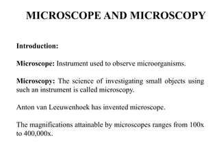
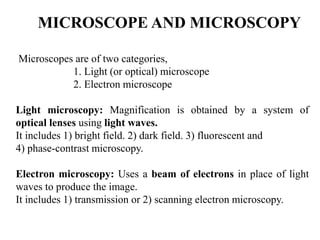
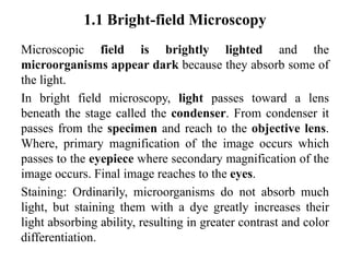
Microscopes and microscopy are introduced. There are two main types of microscopes - light microscopes, which use optical lenses and light, and electron microscopes, which use a beam of electrons. Light microscopes can use different techniques like brightfield, darkfield, fluorescence, and phase contrast. Electron microscopes have higher resolving power and include transmission electron microscopes and scanning electron microscopes. Sample preparation and staining are important for microscopy as they allow small and transparent specimens to be visualized.
2. MICROSCOPE AND MICROSCOPY
Introduction
Types of microscopy
Light Microscopy
1. Bright Field Microscopy
2. Dark Field Microscopy
3. Fluoroscent Microscopy
4. Phase Contrast Microscopy
Electron Microscopy
1. Transmission Electron Microscopy
2. Scanning Electron Microscopy
Limitation of Electron Microscopy
Difference Between Light and Electron Microscopy
Magnification and Numerical Aperture
3. MICROSCOPE AND MICROSCOPY
Introduction:
Microscope: Instrument used to observe microorganisms.
Microscopy: The science of investigating small objects using
such an instrument is called microscopy.
Anton van Leeuwenhoek has invented microscope.
The magnifications attainable by microscopes ranges from 100x
to 400,000x.
4. MICROSCOPE AND MICROSCOPY
Microscopes are of two categories,
1. Light (or optical) microscope
2. Electron microscope
Light microscopy: Magnification is obtained by a system of
optical lenses using light waves.
It includes 1) bright field. 2) dark field. 3) fluorescent and
4) phase-contrast microscopy.
Electron microscopy: Uses a beam of electrons in place of light
waves to produce the image.
It includes 1) transmission or 2) scanning electron microscopy.
17. objective
lenses
Total magnifications of 40X, 100X,
400X and 1000X can be obtained.
The shortest lens has the lowest power,
the longest one with the greatest power.
18. The high power objective lenses are
retractable. This means that if they hit a
slide, the end of the lens will push in (spring
loaded) thereby protecting the lens and the
slide.
objective
lenses
19. stage clips
Stage clips hold the slides in place. If
microscope has a mechanical stage, it
moves the slide around by turning two
knobs. One moves it left and right, the
other moves it up and down.
20. diaphragm
It controls the amount of light going through the specimen
Many microscopes have a rotating disk
under the stage. This diaphragm has
different sized holes and is used to vary
the intensity and size of the cone of light
22. 1.1 Bright-field Microscopy
Microscopic field is brightly lighted and the
microorganisms appear dark because they absorb some of
the light.
In bright field microscopy, light passes toward a lens
beneath the stage called the condenser. From condenser it
passes from the specimen and reach to the objective lens.
Where, primary magnification of the image occurs which
passes to the eyepiece where secondary magnification of the
image occurs. Final image reaches to the eyes.
Staining: Ordinarily, microorganisms do not absorb much
light, but staining them with a dye greatly increases their
light absorbing ability, resulting in greater contrast and color
differentiation.
23. 1.2 Dark-field Microscopy
To view a specimen in dark field, an
opaque disc is placed below the
condenser lens or special kind of
condenser, so that most of the light
directed through the condenser does
not enter the objective; the field is
essentially dark. However, some of
the light rays will be scattered
(diffracted) if the transparent medium
contains objects such as microbial
cells. This diffracted light will enter
the objective and reach the eye; thus
the object or microbial cell, in this
case, will appear bright under dark
microscopic field.
24. 1.3 Fluorescence Microscopy
• Many chemical substances are having
ability to absorb light of particular
wavelength and emit light of a longer
wavelength and lesser energy content.
Such substances are called fluorescent
and the phenomenon is termed
fluorescence.
• Excitation filter: Allows only blue
light to pass
• Dichromic mirror: reflect light of
certain colors but transmits light of
other colors
• Emission filter: Also called barrier
filter which blocks out blue light but
allow to pass green light
25. 1.4 Phase-Contrast Microscopy
• It is extremely valuable for studying living unstained
cells. It uses a conventional light microscope fitted with
a phase contrast objective and a phase contrast
condenser. This special optical system makes it possible
to distinguish unstained structures within a cell which
differ only slightly in their refractive index (thickness).
• Technique is based on the fact that light passing through
one material and into another material of a slightly
different refractive index or thickness will undergo a
change in phase. These differences in phase are translated
in brightness of the structures and hence are detected by
the eye.
27. Electron Microscopy
• An electron microscope (EM) is a type of
microscope that uses an electron beam to illuminate a
specimen and produce a magnified image.
• The resolving power of the electron microscope is
more than 100 times that of the light microscope and
it magnify image up to 400000 times, whereas,
magnification power of light microscope is up to 1000
times only.
• Electron microscopes are used to investigate the
ultrastructure of a wide range of biological and
inorganic specimens including microorganisms, cells,
large molecules, biopsy samples, metals, crystals etc.
28. Types of Electron microscope
1. Transmission Electron Microscope
2. Scanning Electron Microscope
29. 2.1 Transmission Electron Microscope
Component
Source: From the top down, the TEM consists of an emission source, which
may be a tungsten filament. By connecting this tungsten to a high voltage
source (typically ~100–300 kV) it begin to emit electrons into the vacuum.
Optics: Three stages of lenses are there. The stages are the condensor lenses,
the objective lenses, and the projector lenses.
Condensor lenses are responsible for primary beam formation
Objective lenses (When an electron beam passes through a thin-
section specimen of a material, electrons are scattered. A sophisticated system
of electromagnetic lenses focuses the scattered electrons into an image) focus
the beam that comes through the sample itself.
Projector lenses are used to expand the beam onto the phosphor
screen or other imaging device, such as film.
Display: The image can be photographically recorded by exposing a
photographic film to the electron beam, or a high-resolution phosphor may be
coupled to the sensor of a CCD (charge-coupled device) camera. The image
detected by the CCD may be displayed on a monitor or computer.
32. 2.2 Scanning Electron Microscope
A beam of electrons is produced at the top of the
microscope by an electron gun. The electron beam
follows a vertical path through the microscope, which is
held within a vacuum. The beam travels through
electromagnetic fields and lenses, which focus the beam
down toward the sample. Once the beam hits the
sample, backscattered electrons, secondary electron and
X-rays are ejected from the sample.
35. Limitation of Electron Microscopy
• The specimen being examined is in a chamber that is
under a very high vacuum. Thus cells cannot be
examined in a living state.
• Tedious sample preparation is required for electron
microscopy.
• Drying process (during sample preparation) may alter
some morphological characteristics.
• Due to the low penetration power of the electron
beam, necessitating the use of very thin sections to
reveal the internal structures of the cell.
36. Fixation
To fix specimen on slide.
Unfixed specimen may undergo through natural processes of
decomposition, and autolysis.
Autolysis is rapid in specimens that are rich in enzymes.
Fixation prevent sample degradation while at the same time
stabilizing and preserving the ultra-structure and morphology
characteristic of the native tissue.
Types
This procedure can either be accomplished through chemical or
physical means.
Physical methods for fixation include heat which is commonly
used for preparing bacterial smears
Staining
37. Basics of staining
Microorganisms cannot be easily visualized in living
state, not only because they are minute, but also because
they are transparent and particularly colorless when
suspended in aqueous medium.
To study their properties and to divide them into their
specific groups for diagnostic purposes, biological stains
and staining procedures in conjugation with light
microscopy have become an important tool in
microbiology.
38. Chemically, a stain or dye may be defined as an organic compound
containing a benzene ring plus a chromophore and auxochrome group.
1. Benzene: Colorless organic solvent.
2. Chromophore: Chemical group that imparts color to benzene
compound.
3. Auxochrome: Chemical group that impart the property of ionization
to the chromogen; enabling them to form salts and bind to the tissues
39. • The ability of a stain to bind with cell or cellular components such
as proteins or nucleic acids depends upon the electrical charge of the
chromogen portion, as well as on the cellular component to be
stained.
Types:
• Stains are broadly classified into two broad groups- acidic stains
(Negatively charged chromogen) and basic stains (Positively
charged chromogen).
• Acidic stains are anionic in nature i.e., upon ionization of the stain,
the chromogen portion exhibits a negative charge and therefore has
a strong affinity for the positive constituents of the cell. Positively
charged cellular components readily bind and accept the color of the
negatively charged anionic chromogen of the acidic stain.
• Basic stains are cationic in nature and upon ionization, its
chromogen portion exhibit a positive charge and therefore has a
strong affinity for the negative constituents of the cell.