Confocal microscopy
•Download as PPTX, PDF•
114 likes•58,436 views

Partial fulfilment of the Course.

Recommended
More Related Content
What's hot
What's hot (20)
Similar to Confocal microscopy
Similar to Confocal microscopy (20)
Recently uploaded
Recently uploaded (20)
Confocal microscopy
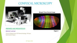
- 1. CONFOCAL MICROSCOPY PREPARED AND PRESENTED BY: Mahesh Lamsal Central Department Of Biotechnology, Kirtipur, Kathmandu Nepal. 1 25th March 2016
- 2. SLIDES INCLUDE Introduction History Instrumental design Principle Working mechanism Applications Advantages Limitations References 2
- 3. INTRODUCTION Confocal microscopy : (having the same focus ) An optical imaging technique for increasing optical resolution and contrast of a micrograph. Radiations emitted from laser cause sample to fluoresce. Uses pinhole screen to produce high resolution images. Eliminates out of focus. So images have better contrast and are less hazy. A series of thin slices of the specimen are assembled to generate a 3- dimensinal image. Is an updated version of fluorescence microscopy. 3
- 4. HISTORY Two investigators at Cambridge, Brad Amos and John White attempted to look at the mitotic divisions in the first few divisions in embryos of C. elegans. They were doing antitubulin immunofluorescence and were trying to determine the cleavage planes of the cells, but were frustrated in their attempt in that the majority of the fluorescence they observed was out of focus. They looked at the technique called confocal imaging which was first proposed by Nipkow and pioneered by a postdoc at Harvard named Minsky. He made the first stage scanning confocal microscope in 1957. By illuminating single point at a time, Minsky avoided most of the unwanted scattered light. For builiding the image, Minsky scanned the specimen by moving the stage rather than light rays. 4 Marvin Minsky
- 5. INSTRUMENTAL DESIGN 5 fig: Ray diagram for confocal microscope
- 6. 6 fig: schematic diagram confocal microscope
- 7. PRINCIPLE In confocal microscopy two pinholes are typically used: A pinhole is placed in front of the illumination source to allow transmission only through a small area This illumination pinhole is imaged onto the focal plane of the specimen, i.e. only a point of the specimen is illuminated at one time. Fluorescence excited in this manner at the focal plane is imaged onto a confocal pinhole placed right in front of the detector. Only fluorescence excited within the focal plane of the specimen will go through the detector pinhole. Scanning of small sections is done and joined them together for better view. 7
- 8. WORKING MECHANISM Confocal microscope incorporates 2 ideas: 1. Point-by-point illumination of the specimen. 2. Rejection of out of focus of light. Light source of very high intensity is used—Zirconium arc lamp in Minsky’s design & laser light source in modern design. a)Laser provides intense blue excitation light. b)The light reflects off a dichoric mirror, which directs it to an assembly of vertically and horizontally scanning mirrors. c)These motor driven mirrors scan the laser beam across the specimen. d) The specimen is scanned by moving the stage back & forth in the vertical & horizontal directions and optics are kept stationary. 8
- 9. Contd…. Dye in the specimen is excited by the laser light & fluoresces. The fluorescent (green) light is descanned by the same mirrors that are used to scan the excitation (blue) light from the laser beam then it passes through the dichoric mirror then it is focused on to pinhole. the light passing through the pinhole is measured by the detector such as photomultiplier tube. For visualization, detector is attached to the computer, which builds up the image at the rate of 0.1-1 second for single image 9
- 10. APPLICATIONS 1.Confocal microscopy allows analysis of fluorescent labelled thick specimens without physical sectioning. 10 Fig:Zebra fish embryo wholemount Neurons (green) Cell adhesion molecule (red) 2. Three-Dimensional Reconstruction of Specimen 3D shadow projection
- 11. 11 Because the images are detected by a computer rather than by eye, it is possible to detect more color differences. 3. More Colour Possibilities
- 12. APPLICATIONS 12 Fig:An Intestine Section Fig: Kidney cells (fluorescence vs Confocal microscope 4. Improved Resolution
- 13. 13 Fig: Colour coded image of actin filaments in a cancer cell
- 14. ADVANTAGES The specimen is everywhere illuminated axially, rather than at different angles, thereby avoiding optical aberrations. Entire field of view is illuminated uniformly. The field of view can be made larger than that of the static objective by controlling the amplitude of the stage movements. Image formed are of better resolution. Cells can be live or fixed. Serial optical sections can be collected. Taking a series of optical slices from different focus levels in the specimen generates a 3D data set. 14
- 15. DRAWBACKS Resolution : It has inherent resolution limitation due to diffraction. Maximum best resolution of confocal microscopy is typically about 200nm. Pin hole size : Strength of optical sectioning depends on the size of the pinhole. Intensity of the incident light Fluorophores : a)The fluorophore should tag the correct part of the specimen. b)Fluorophore should be sensitive enough for the given excitation wave length. c)It should not significantly alter the dynamics of the organism in the living specimen. Photobleaching: photochemical alteration of a dye or a fluorophore molecule such that it permanently is unable to fluoresce 15
- 16. REFERENCES Pawley JB (editor) (2006). Handbook of Biological Confocal Microscopy (3rd ed.). Berlin: Springer. ISBN 0-387-25921-X Confocal Microscopy - The Comprehensive Explanation www.microscopyu.com Marvin Minsky (1988). "Memoir on inventing the confocal scanning microscope". Scanning10 (4): 128–138. doi:10.1002/sca.4950100403. 16
- 17. 17