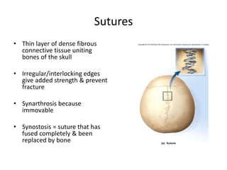
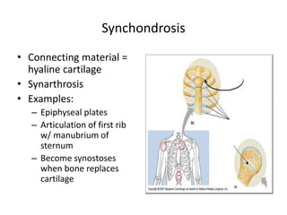
Downloaded 627 times
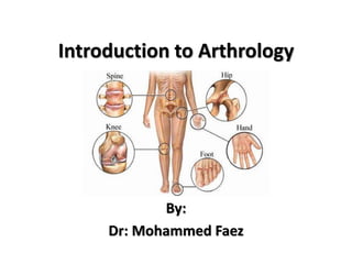


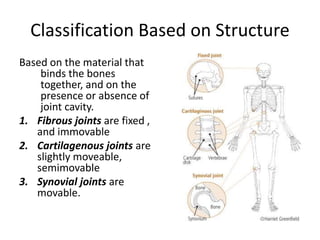


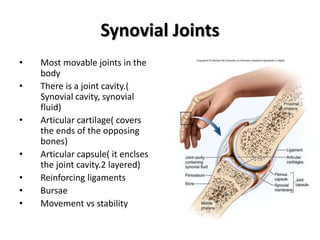


This document provides an introduction to arthrology, the study of joints. It defines arthrology and discusses the classification and basic structures of joints. Joints are classified based on both their function and structure. Based on structure, joints are categorized as fibrous, cartilaginous, or synovial. Synovial joints are the most common and complex type, allowing for free movement. The document outlines the key components of synovial joints and provides examples of different joint types, including ball and socket, hinge, pivot, saddle, condyloid, and gliding joints.


