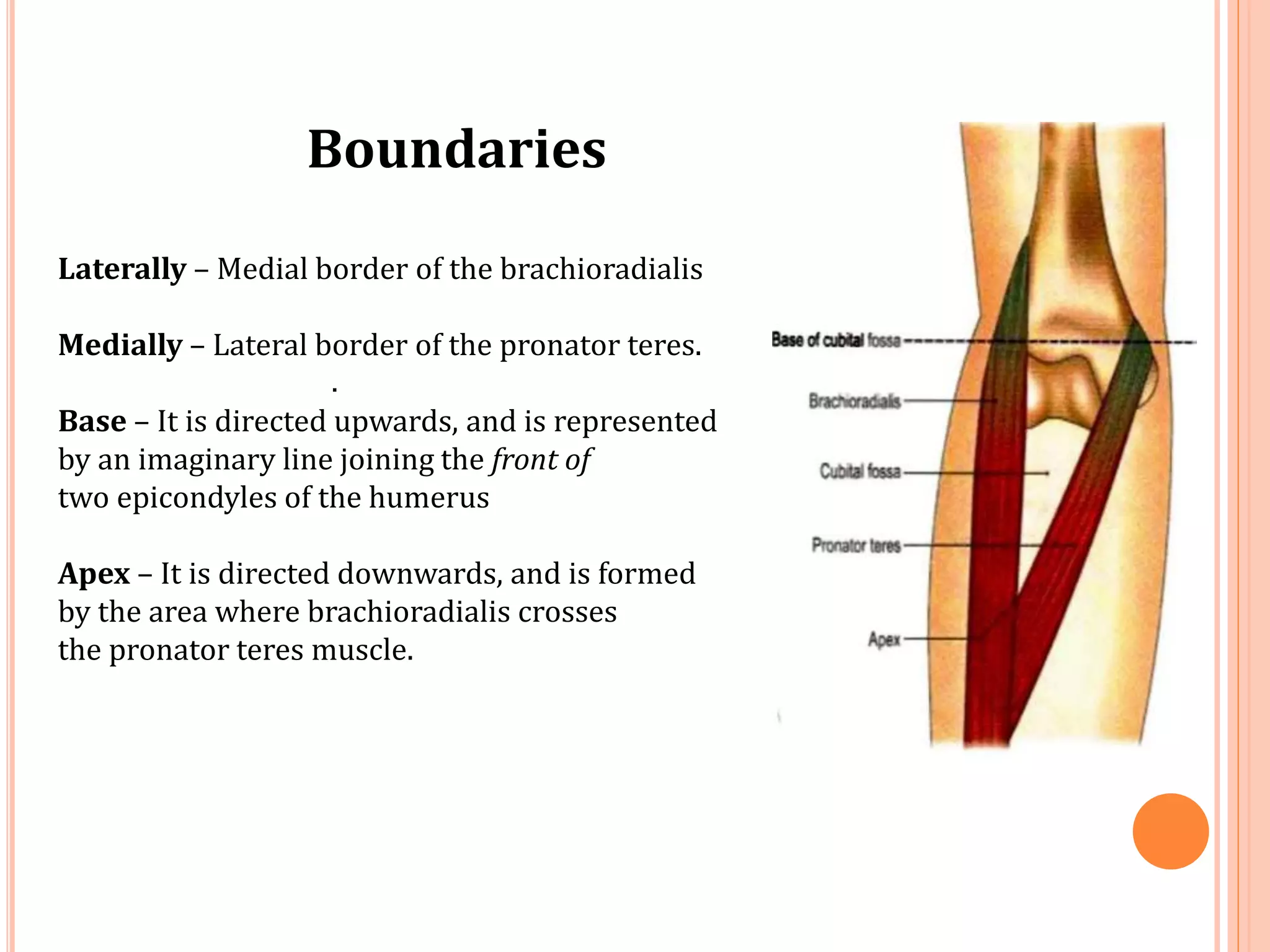
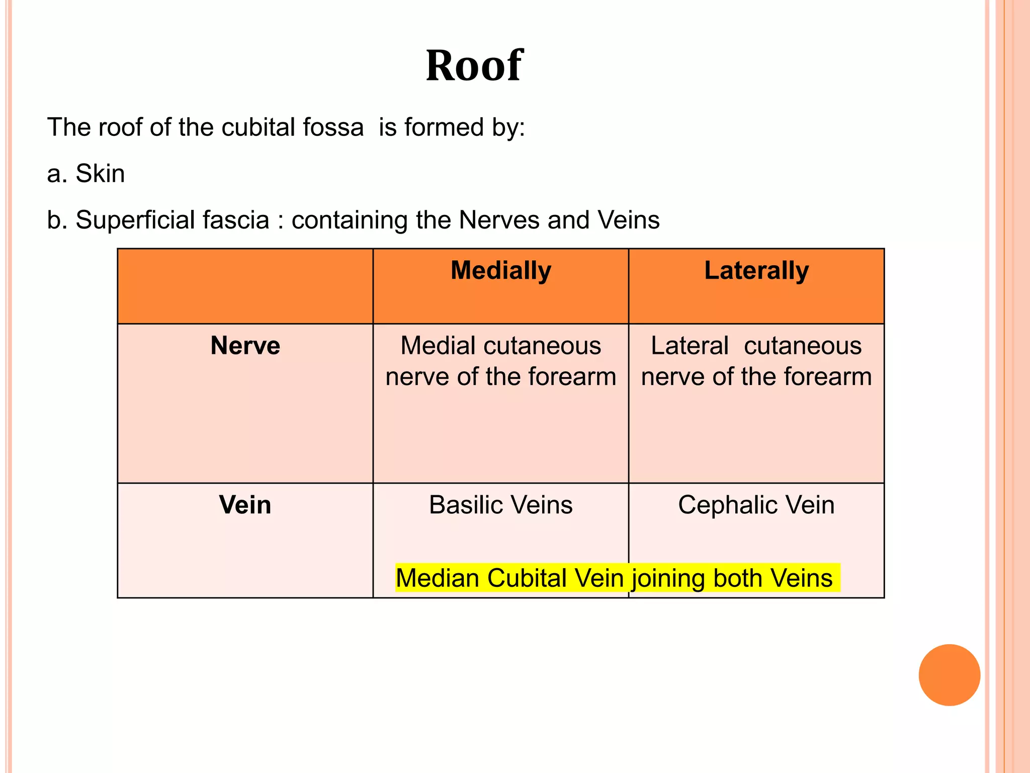
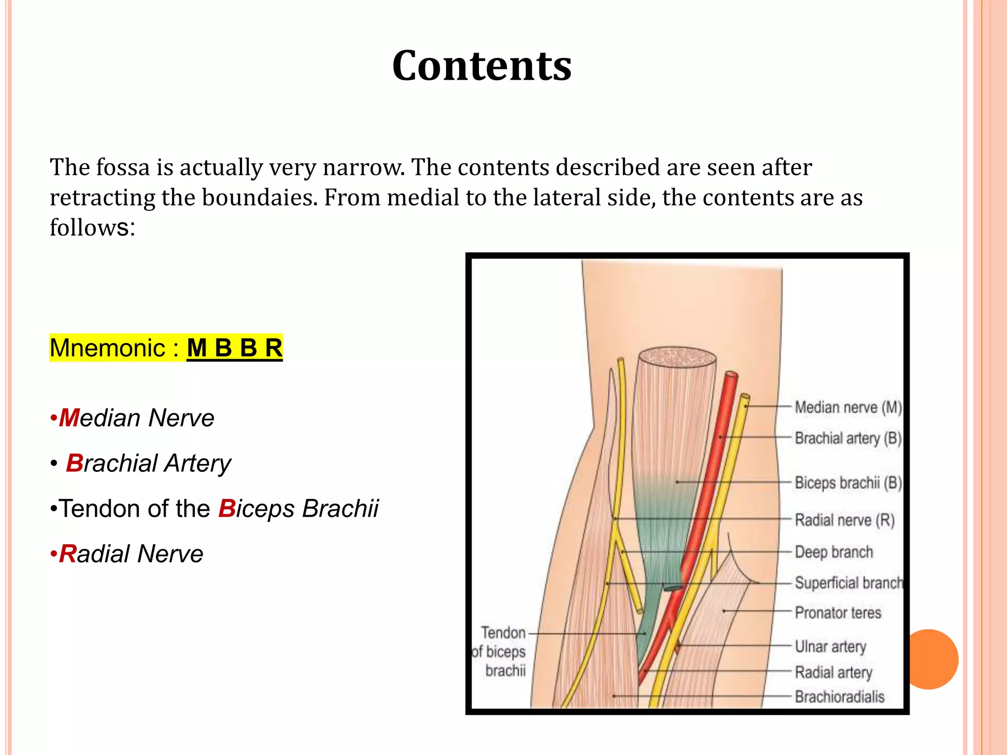
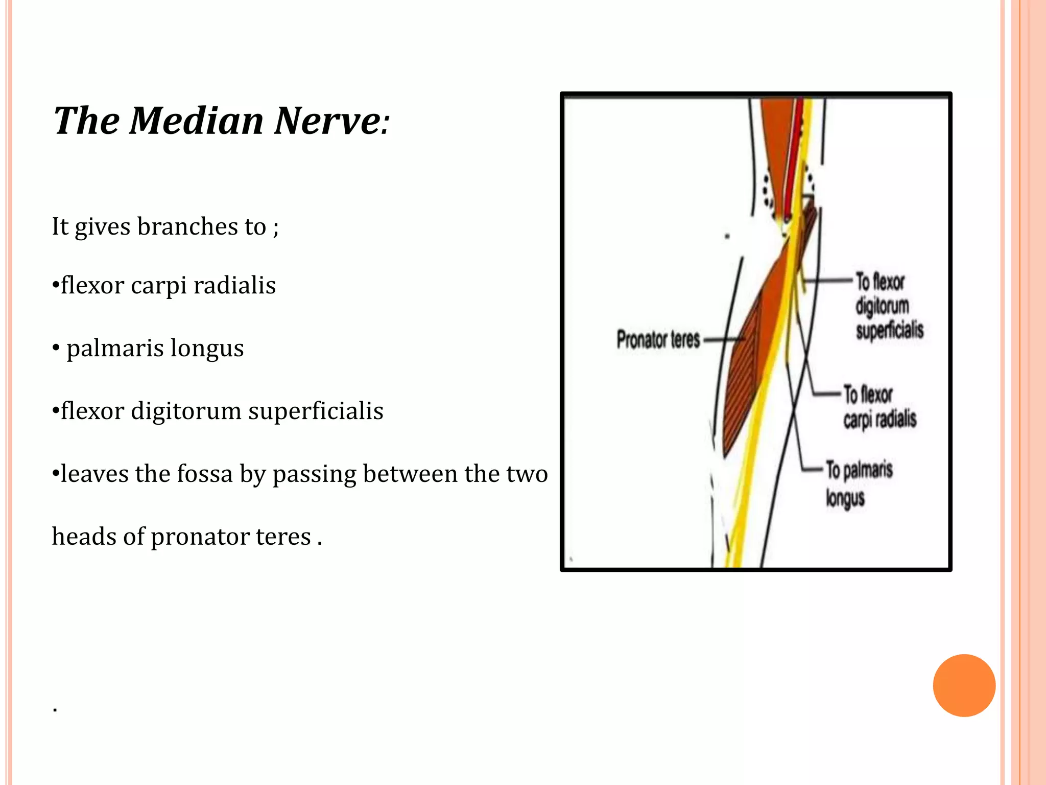
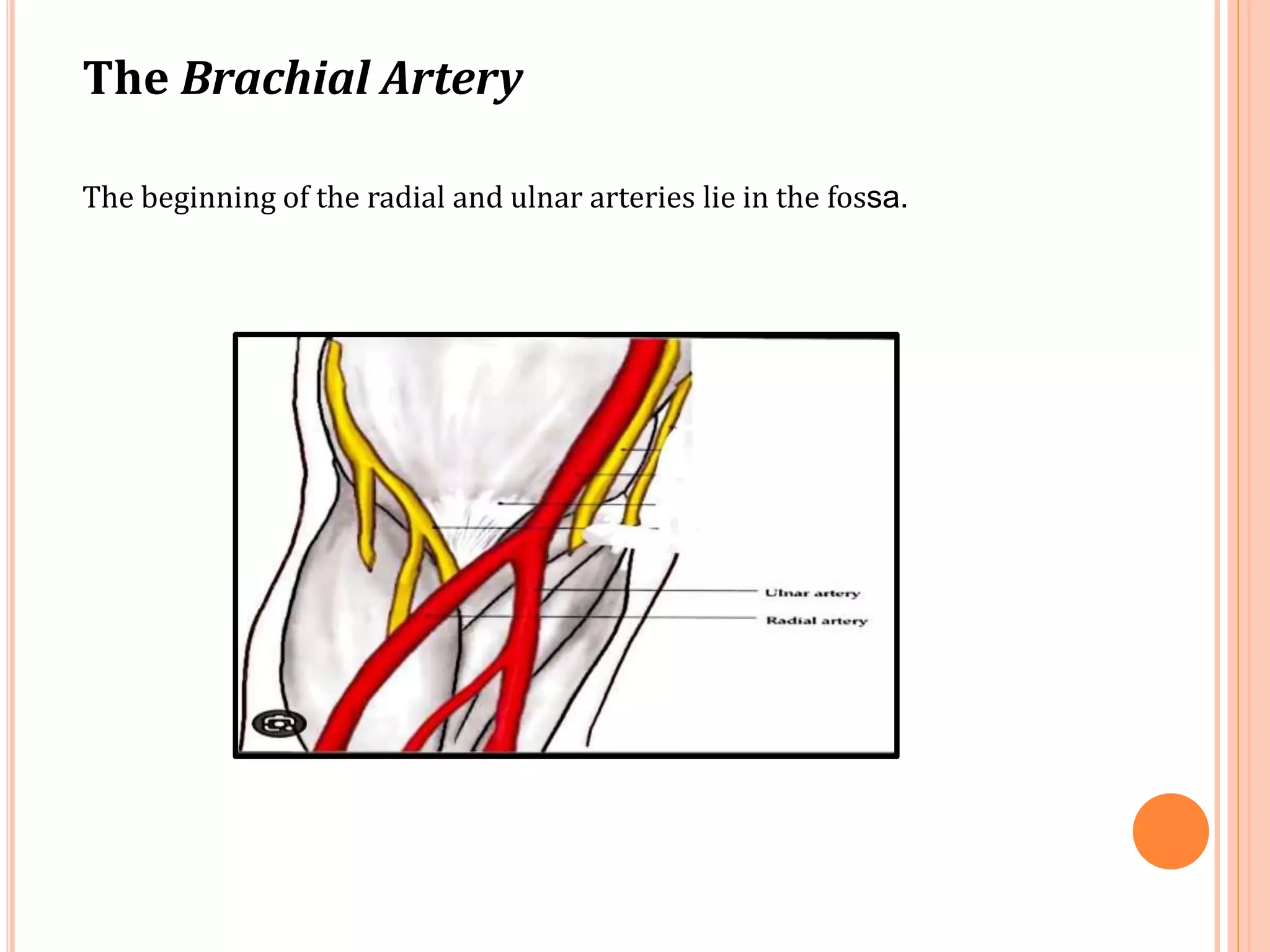
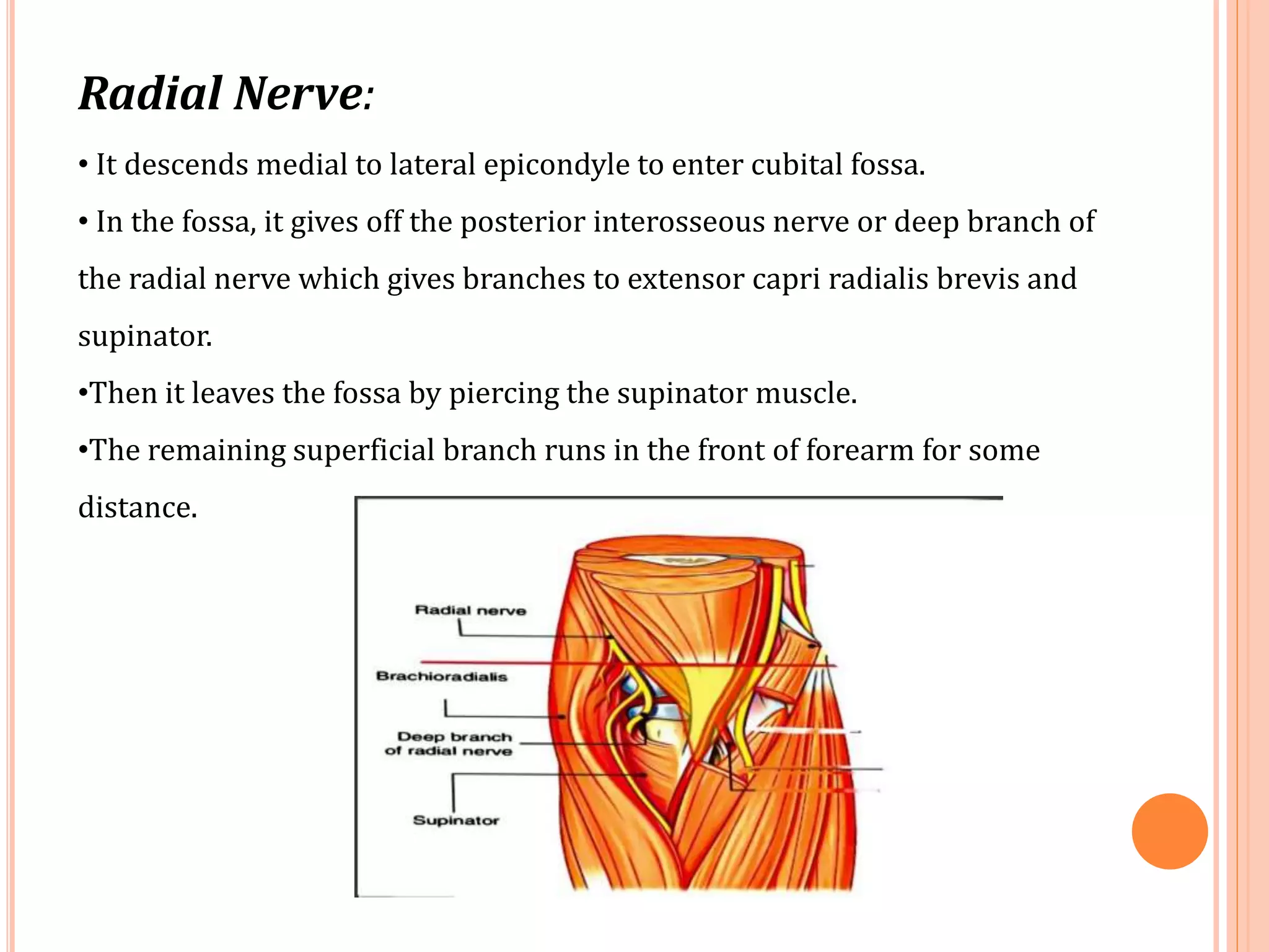
CUBITAL FOSSA
Presented by
Dr. Manisha Yadav (PT)
MPT ( Neuro )
INDEX
• Features
• Boundaries
• Roof
• Floor
• Content
• Clinal Anatomy
Features
Cubital (Latin cubitus, elbow) fossa is a triangular hollow .
(It is homologous with the popliteal fossa of the lower limb situated
on the back of the knee.)
Situation :-
Situated on the front of the elbow.
Boundaries
Laterally – Medial border of the brachioradialis
Medially – Lateral border of the pronator teres.
Base – It is directed upwards, and is represented
by an imaginary line joining the front of
two epicondyles of the humerus
Apex – It is directed downwards, and is formed
by the area where brachioradialis crosses
the pronator teres muscle.
.
Roof
The roof of the cubital fossa is formed by:
a. Skin
b. Superficial fascia : containing the Nerves and Veins
Medially Laterally
Nerve Medial cutaneous
nerve of the forearm
Lateral cutaneous
nerve of the forearm
Vein Basilic Veins Cephalic Vein
Median Cubital Vein joining both Veins
c. Deep fascia
d. Bicipital aponeurosis
FLOOR
IT IS FORMED BY:
I. BRACHIALIS
II. SUPINATOR SURROUNDING THE UPPER PART OF RADIUS
(a) Surface view
(b) Cross-sectional view
Contents
The fossa is actually very narrow. The contents described are seen after
retracting the boundaies. From medial to the lateral side, the contents are as
follows:
Mnemonic : M B B R
•Median Nerve
• Brachial Artery
•Tendon of the Biceps Brachii
•Radial Nerve
The Median Nerve:
It gives branches to ;
•flexor carpi radialis
• palmaris longus
•flexor digitorum superficialis
•leaves the fossa by passing between the two
heads of pronator teres .
.
The Brachial Artery
The beginning of the radial and ulnar arteries lie in the fossa.
Radial Artery Ulnar Artery
Its is smaller and more superficial
than the ulnar artery.
Its deep to both heads of pronator
teres and runs downwards and
medially
It gives off ;
•the radial recurrent branch
Ulnar artery gives off ;
• the anterior ulnar recurrent,
• the posterior ulnar recurrent
• the common interosseous
branches
The common interosseous branch divides into the:-
•Anterior Interosseous Arteries
•Posterior Interosseous Arteries
• Interosseous Recurrent Branch
The Tendon of the Biceps Brachii
Radial Nerve:
• It descends medial to lateral epicondyle to enter cubital fossa.
• In the fossa, it gives off the posterior interosseous nerve or deep branch of
the radial nerve which gives branches to extensor capri radialis brevis and
supinator.
•Then it leaves the fossa by piercing the supinator muscle.
•The remaining superficial branch runs in the front of forearm for some
distance.
CLINICAL ANATOMY
1. The Median cubital vein is often the vein of
choice for intravenous injections .
2. The Blood Pressure is universally recorded
by auscultating the brachial artery in front
of the elbow .
CUBITAL FOSSA.pptx

CUBITAL FOSSA.pptx

  • 1.
    CUBITAL FOSSA Presented by Dr.Manisha Yadav (PT) MPT ( Neuro )
  • 2.
    INDEX • Features • Boundaries •Roof • Floor • Content • Clinal Anatomy
  • 3.
    Features Cubital (Latin cubitus,elbow) fossa is a triangular hollow . (It is homologous with the popliteal fossa of the lower limb situated on the back of the knee.) Situation :- Situated on the front of the elbow.
  • 4.
    Boundaries Laterally – Medialborder of the brachioradialis Medially – Lateral border of the pronator teres. Base – It is directed upwards, and is represented by an imaginary line joining the front of two epicondyles of the humerus Apex – It is directed downwards, and is formed by the area where brachioradialis crosses the pronator teres muscle. .
  • 5.
    Roof The roof ofthe cubital fossa is formed by: a. Skin b. Superficial fascia : containing the Nerves and Veins Medially Laterally Nerve Medial cutaneous nerve of the forearm Lateral cutaneous nerve of the forearm Vein Basilic Veins Cephalic Vein Median Cubital Vein joining both Veins
  • 6.
    c. Deep fascia d.Bicipital aponeurosis
  • 7.
    FLOOR IT IS FORMEDBY: I. BRACHIALIS II. SUPINATOR SURROUNDING THE UPPER PART OF RADIUS (a) Surface view (b) Cross-sectional view
  • 8.
    Contents The fossa isactually very narrow. The contents described are seen after retracting the boundaies. From medial to the lateral side, the contents are as follows: Mnemonic : M B B R •Median Nerve • Brachial Artery •Tendon of the Biceps Brachii •Radial Nerve
  • 9.
    The Median Nerve: Itgives branches to ; •flexor carpi radialis • palmaris longus •flexor digitorum superficialis •leaves the fossa by passing between the two heads of pronator teres . .
  • 10.
    The Brachial Artery Thebeginning of the radial and ulnar arteries lie in the fossa.
  • 11.
    Radial Artery UlnarArtery Its is smaller and more superficial than the ulnar artery. Its deep to both heads of pronator teres and runs downwards and medially It gives off ; •the radial recurrent branch Ulnar artery gives off ; • the anterior ulnar recurrent, • the posterior ulnar recurrent • the common interosseous branches
  • 12.
    The common interosseousbranch divides into the:- •Anterior Interosseous Arteries •Posterior Interosseous Arteries • Interosseous Recurrent Branch
  • 13.
    The Tendon ofthe Biceps Brachii
  • 14.
    Radial Nerve: • Itdescends medial to lateral epicondyle to enter cubital fossa. • In the fossa, it gives off the posterior interosseous nerve or deep branch of the radial nerve which gives branches to extensor capri radialis brevis and supinator. •Then it leaves the fossa by piercing the supinator muscle. •The remaining superficial branch runs in the front of forearm for some distance.
  • 15.
    CLINICAL ANATOMY 1. TheMedian cubital vein is often the vein of choice for intravenous injections . 2. The Blood Pressure is universally recorded by auscultating the brachial artery in front of the elbow .