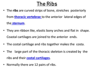
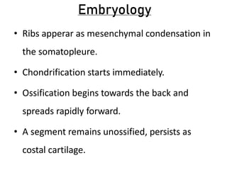
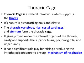
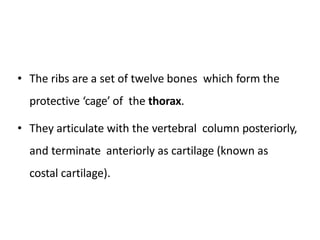
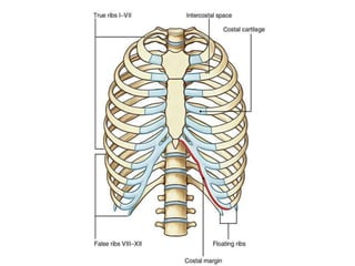
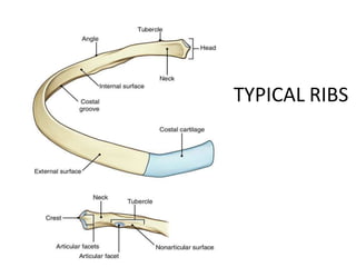
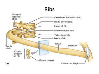
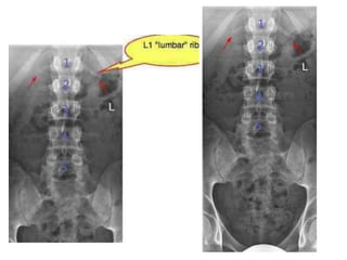
The ribs are curved strips of bone that form a protective cage around the thoracic cavity. There are typically 12 pairs of ribs that articulate posteriorly with thoracic vertebrae and terminate anteriorly as cartilage. The first 7 ribs connect directly to the sternum via costal cartilage, while the last 5 ribs connect to the cartilage of the rib above. The first, tenth, eleventh and twelfth ribs have distinguishing features compared to typical ribs. Ribs can fracture, and injury to multiple adjacent ribs can result in a flail chest.

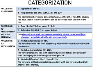


![Pump handle movement
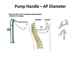
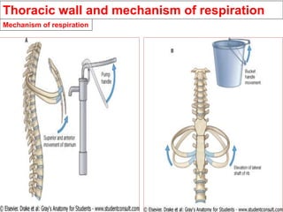
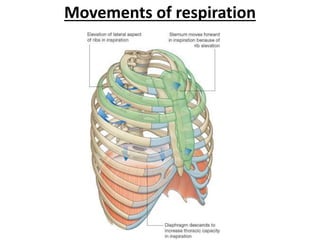
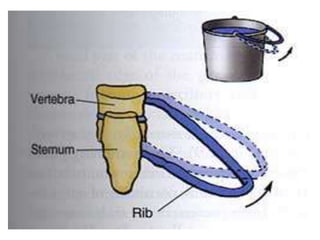
• On of the most important functions
of ribs and diaphragm, is the change in
volume of thorax that
helps inspiration and expiration.[2]
• In general, the ribs move around two axes.The
anterior end of the rib is lower than the
posterior end, therefore, during elevation of
the rib, the anterior end also moves forward](https://image.slidesharecdn.com/ribs-221112131905-1a22e7fd/85/RIBS-pptx-53-320.jpg)
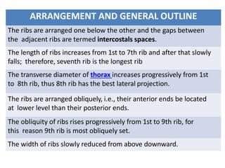
![• Movement at costovertebral joints 2 to 6
about a side-to-side axis results in raising and
lowering the sternal end of the rib, the
"pump-handle" movement.
• This occurs mostly in the vertebrosternal ribs.
In elevation, this increases the anteroposterior
diameter of the thorax.[4][](https://image.slidesharecdn.com/ribs-221112131905-1a22e7fd/85/RIBS-pptx-54-320.jpg)