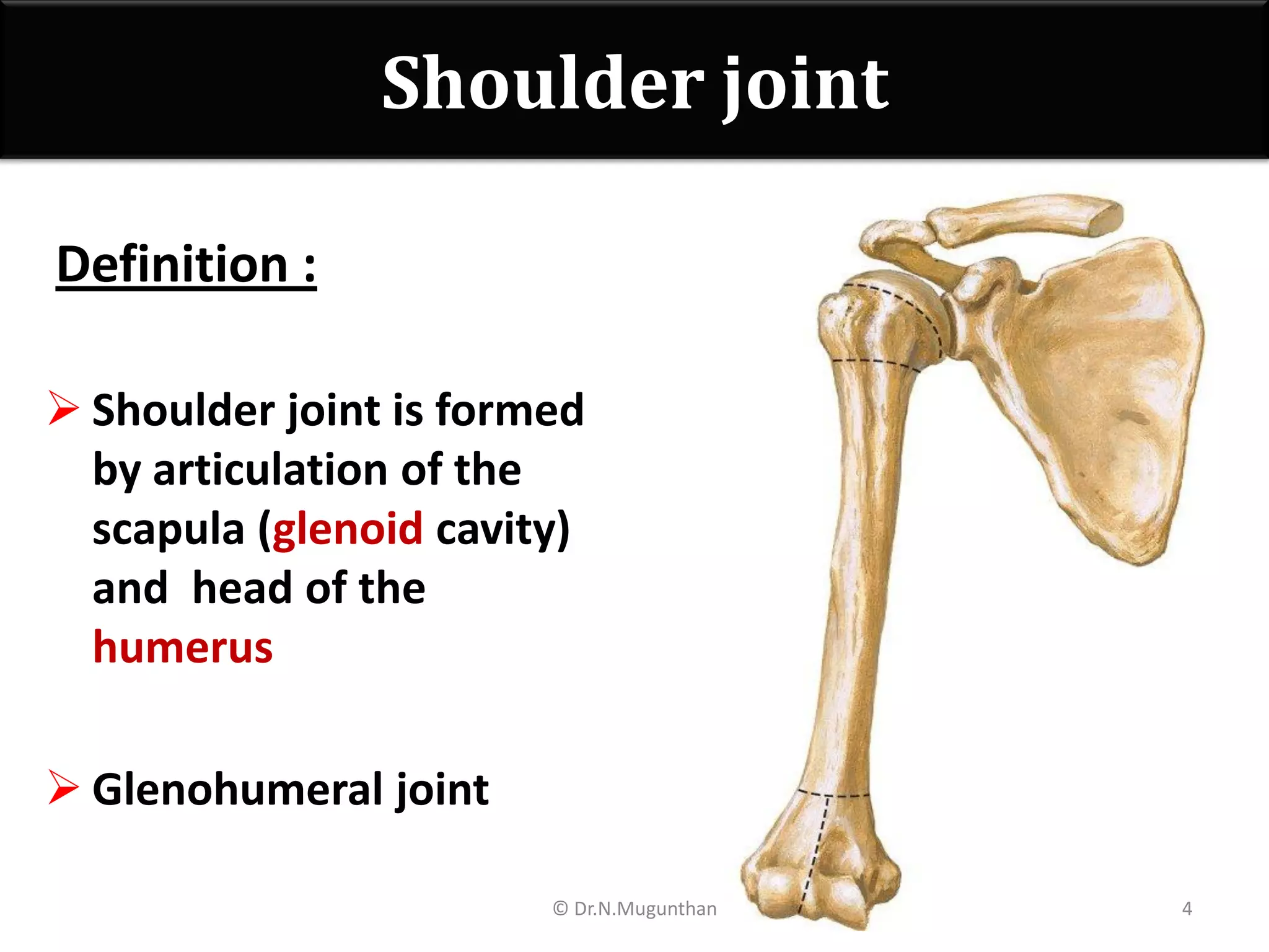
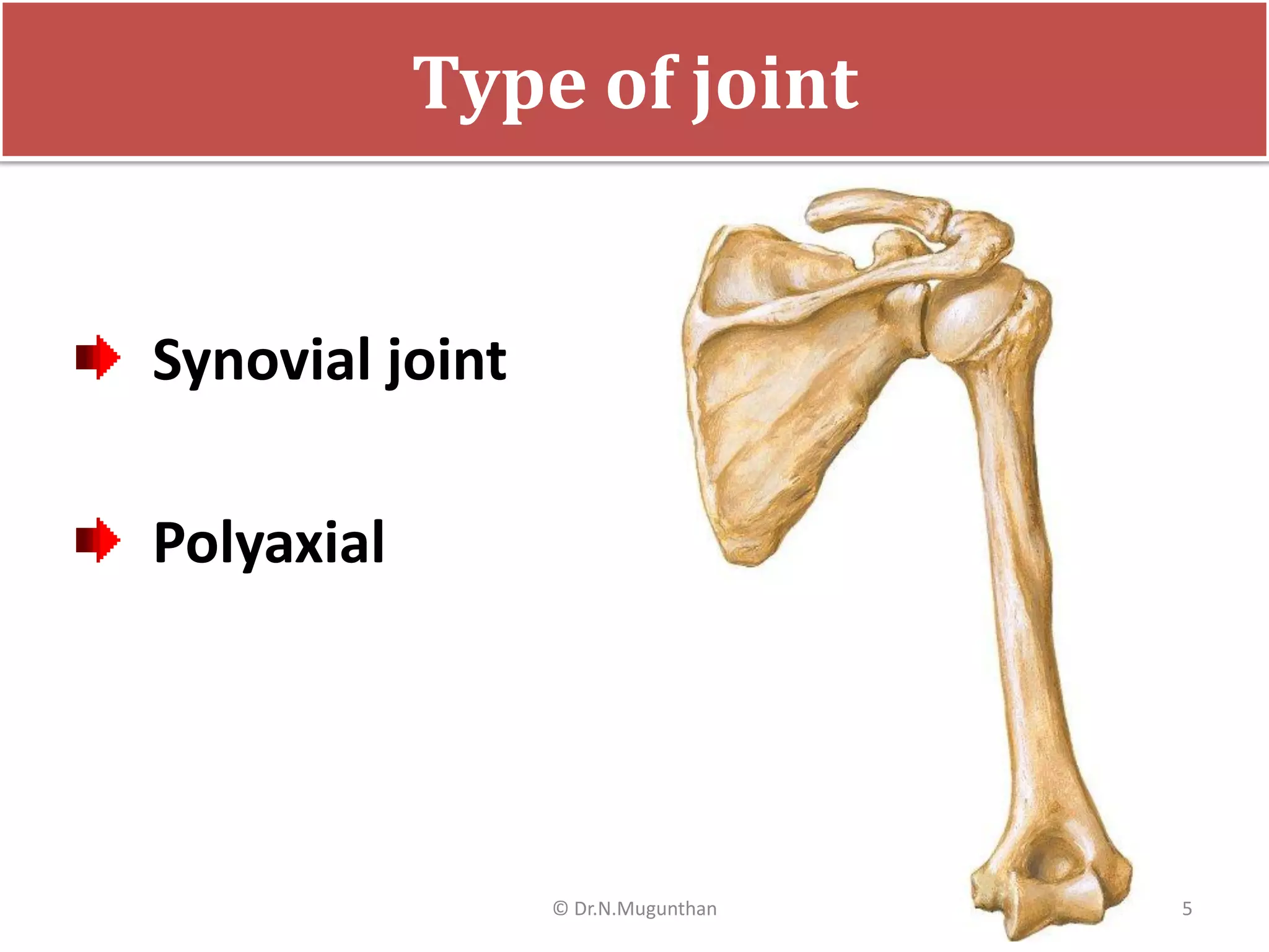
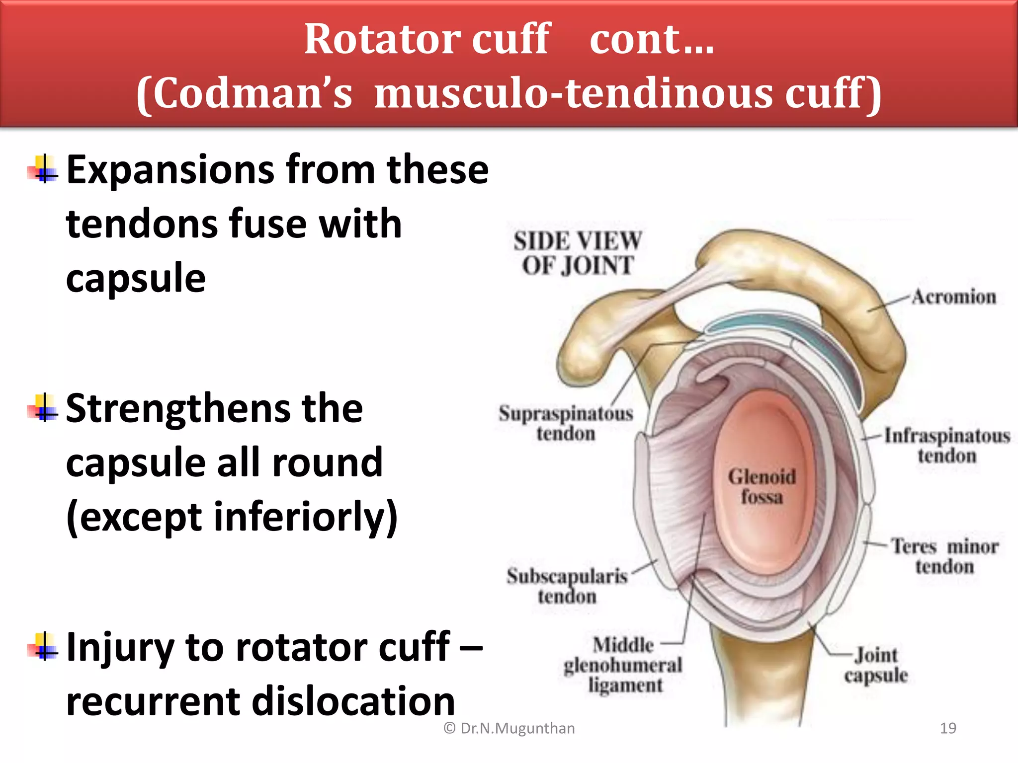
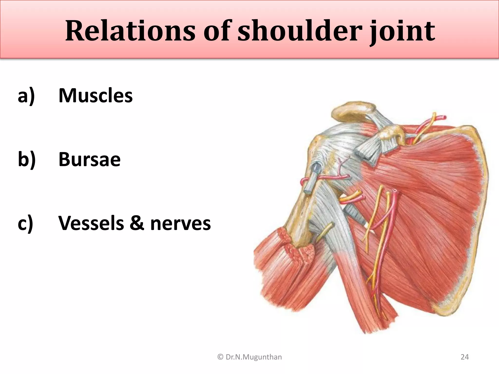
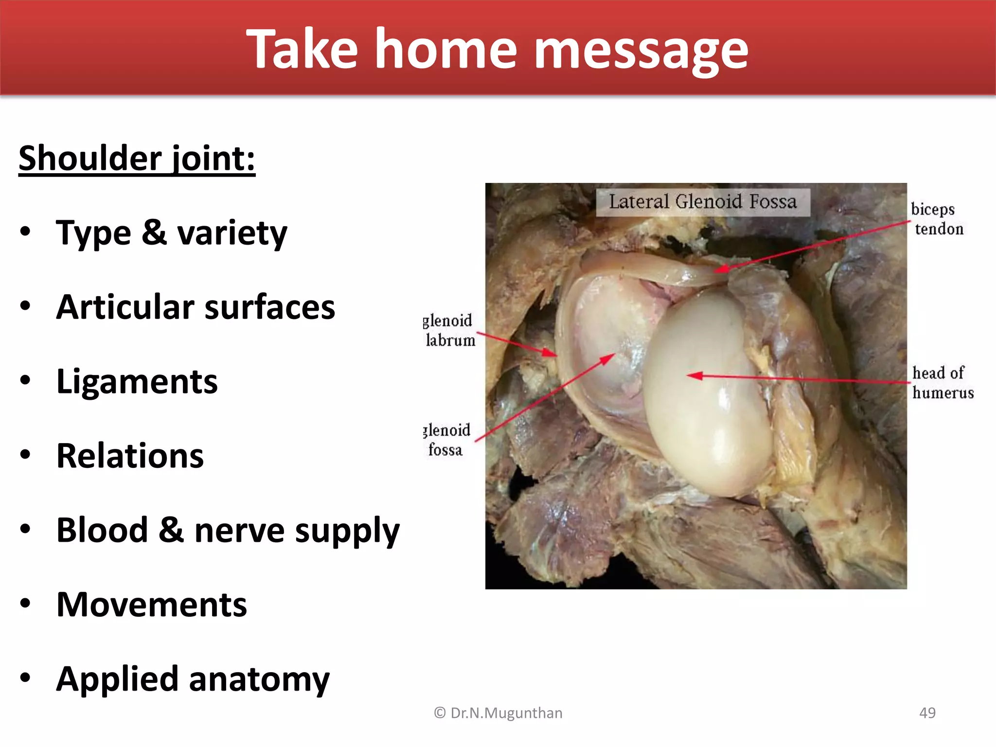
Shoulder Joint
Lecture by
Dr.N.Mugunthan.MBBS,MS,DNB,MNAMS,PhD
Associate Professor,
MGMC&RI.
1© Dr.N.Mugunthan
Learning objectives
Shoulder joint:
• Type & variety
• Articular surfaces
• Ligaments
• Relations
• Blood & nerve supply
• Movements
• Applied anatomy
LAQ: Describe the shoulder joint under following headings: a. articular
surfaces b. ligaments c. movements d. applied anatomy (2+3+3+2)2© Dr.N.Mugunthan
How to describe a joint
9. Movements
10. Muscles producing
movements
11. Unique features of
particular joint
12. Applied anatomy
1. Definition
2. Type of joint
3. Variety of joint
4. Articular surfaces
5. Ligaments
6. Relations
7. Blood supply
8. Nerve supply 3© Dr.N.Mugunthan
Shoulder joint
Definition :
 Shoulder joint is formed
by articulation of the
scapula (glenoid cavity)
and head of the
humerus
 Glenohumeral joint
4© Dr.N.Mugunthan
Type of joint
Synovial joint
Polyaxial
5© Dr.N.Mugunthan
Variety of joint
Ball and socket
variety
6© Dr.N.Mugunthan
Articular surfaces
Proximal articular surface :
Glenoid fossa of scapula
 Pyriform in shape
 Surface area, concavity
of glenoid fossa
increased by glenoidal
labrum
 Glenoidal labrum – fibro
cartilaginous ribbon like
structure
 Covered with hyaline
cartilage
7© Dr.N.Mugunthan
Articular surfaces cont…
Distal articular surface:
The head of humerus
Hemispherical in
shape
Covered with hyaline
cartilage
8© Dr.N.Mugunthan
Shoulder joint – weak joint
o Glenoid fossa is too small
& shallow
o to hold the head of the
humerus in place
o The head is four times
the size of glenoid cavity
o Structurally, it is a weak
joint
Stability of the joint is ?
9© Dr.N.Mugunthan
Ligaments
True ligament:
1. Capsule
Accessory ligaments:
2. Glenohumeral ligaments
3. Transverse humeral
ligament
4. Coracohumeral ligament
5. Secondary socket /
ligament
(Coracoacromial arch)
6. Glenoidal labrum
10© Dr.N.Mugunthan
Capsule
Loose fibrous covering
Inner surface lined with
synovial membrane
Attachments:
Proximal attachments:
Margins of glenoid fossa
(proximal to glenoid
labrum)
Distal attachments:
Anatomical neck of
humerus (except inferiorly,
extends to shaft for
1.25cm) 11© Dr.N.Mugunthan
Glenohumeral ligaments :
Three :
1. Superior band
2. Middle band
3. Inferior band
Seen on inner side of
ant. part of capsule
Accessory ligaments
13© Dr.N.Mugunthan
Attachments :
Proximally: (Glenoid)
 All 3 bands attached to
upper end of glenoid
fossa
Distal : (Humerus)
 Upper band – top of
lesser tuberosity
 Middle band – lower
part
 Lower band – shaft just
below lesser tuberosity
Glenohumeral ligaments cont…
14© Dr.N.Mugunthan
Transverse humeral ligament
Stretches between tops
of greater & lesser
tuberosities
Long head of biceps
tendon passes out deep
to this ligament
15© Dr.N.Mugunthan
Coracohumeral ligament
Attachment :
Lateral margin of root of
coracoid process to
Greater tuberosity of
humerus
All the accessory ligaments
attach either lesser/greater
tuberosity of humerus
16© Dr.N.Mugunthan
Coracoacromial ligament
(secondary socket)
Triangular band
Base –attached to lateral
margin of coracoid
process
Apex – attached to tip of
acromion
Coracoid process,ligament
& acromion together form
– coracoacromial arch
Which forms secondary
socket for the joint
17© Dr.N.Mugunthan
Rotator cuff
(Codman’s musculo-tendinous cuff)
Laxity & weakness of
joint is compensated
by this cuff
Derived from tendons
of – SITS
1. Supraspinatus
2. Infraspinatus
3. Teres minor
4. Subscapularis 18© Dr.N.Mugunthan
Expansions from these
tendons fuse with
capsule
Strengthens the
capsule all round
(except inferiorly)
Injury to rotator cuff –
recurrent dislocation
Rotator cuff cont…
(Codman’s musculo-tendinous cuff)
19© Dr.N.Mugunthan
Glenoidal labrum
Fibro cartilaginous ribbon
like structure
Attached to margins of
the glenoid cavity
Increase the depth of
glenoid cavity
Increase the articular
surface of glenoid
Lined with hyaline
cartilage
21© Dr.N.Mugunthan
Relations of shoulder joint
a) Muscles
b) Bursae
c) Vessels & nerves
24© Dr.N.Mugunthan
a) Muscles:
Anteriorly:
• Subscapularis,
coracobrachialis, short
head of biceps and
deltoid.
Posteriorly:
• Infraspinatus, teres
minor and deltoid.
Relations of shoulder joint
25© Dr.N.Mugunthan
a) Muscles: cont…
Superiorly:
• Long tendon of biceps
inside the capsule
• Supraspinatus outside
the capsule
Inferiorly:
• Long head of the triceps
 Deltoid – covers
superiorly, anteriorly,
posteriorly & laterally
Relations of shoulder joint
cont…
26© Dr.N.Mugunthan
Subacromial (Subdeltoid)
bursa:
 Lies between deltoid
muscle & capsule
 Does not communicate
with joint
 Extends between
supraspinatus and
acromion & coracoacromial
arch
Longest bursa in the body
b) Bursae related to shoulder joint cont…
30© Dr.N.Mugunthan
Blood supply
Arterial supply:
 Anterior circumflex
humeral artery
 Posterior circumflex
humeral artery
 Suprascapular artery
 Circumflex scapular
branch of Subscapular
artery.
Venous drainage :
 Corresponding veins
33© Dr.N.Mugunthan
Nerve supply
A twig from lateral
pectoral nerve
Suprascapular nerve
Axillary nerve
(posterior division)
34© Dr.N.Mugunthan
Movements @ shoulder joint
Flexion & extension
Adduction & abduction
Medial & lateral rotation
Circumduction
35© Dr.N.Mugunthan
Flexion & extension
Transverse axis :
Flexion:
• Pectoralis major
(clavicular part),
• Deltoid (anterior fibres)
coracobrachialis
• Assisted by biceps.
Extension:
• Deltoid (posterior fibres)
• Teres major,
• Latissimus dorsi and
pectoralis major
(sternocostal part)
36© Dr.N.Mugunthan
Abduction & adduction
Antero-posterior axis
Abduction:
• Supraspinatus : 0-30°
• Deltoid (middle fibers) 0-90°
• Serratus anterior &
trapezius: 90-180°
Adduction:
• Pectoralis major
• Latissimus dorsi
• Teres major
• Corachobrachialis
• Biceps (short head)
37© Dr.N.Mugunthan
Medial & lateral rotation
Longitudinal axis
Medial rotation:
• Pectoralis major
• Deltoid (anterior fibres)
• Latissimus dorsi
• Teres major and
subscapularis.
Lateral rotation:
• Infraspinatus
• Deltoid (posterior fibres)
and teres minor. 38© Dr.N.Mugunthan
Circumduction
• Combination of 3 axis
• Combination of all the
muscles around
shoulder
39© Dr.N.Mugunthan
Applied anatomy
• Referred pain from
diaphragm, pleura &
peritoneum (supplied by
phrenic nerve -C3,C4 &) to tip
of shoulder via supraclavicular
nerve (C3&C4)
• Dislocation
• Bursitis
• Supraspinatus Tendinitis
• Rupture of supraspinatus
• Rotator cuff injury
• Frozen shoulder
• Arthroscopy & arthroplasty
40© Dr.N.Mugunthan
Applied anatomy
• Referred pain from
diaphragm, pleura &
peritoneum (supplied by
phrenic nerve -C3,C4 &) to tip
of shoulder via supraclavicular
nerve (C3&C4)
• Dislocation
• Bursitis
• Supraspinatus Tendinitis
• Rupture of supraspinatus
• Rotator cuff injury
• Frozen shoulder
• Arthroscopy & arthroplasty
41© Dr.N.Mugunthan
Applied anatomy
• Referred pain from
diaphragm, pleura &
peritoneum (supplied by
phrenic nerve -C3,C4 &) to tip
of shoulder via supraclavicular
nerve (C3&C4)
• Dislocation
• Bursitis
• Supraspinatus Tendinitis
• Rupture of supraspinatus
• Rotator cuff injury
• Frozen shoulder
• Arthroscopy & arthroplasty
42© Dr.N.Mugunthan
Applied anatomy
• Referred pain from
diaphragm, pleura &
peritoneum (supplied by
phrenic nerve -C3,C4 &) to tip
of shoulder via supraclavicular
nerve (C3&C4)
• Dislocation
• Bursitis
• Supraspinatus tendinitis &
• Rupture of supraspinatus
• Rotator cuff injury
• Frozen shoulder
• Arthroscopy & arthroplasty
43© Dr.N.Mugunthan
Applied anatomy
• Referred pain from
diaphragm, pleura &
peritoneum (supplied by
phrenic nerve -C3,C4 &) to tip
of shoulder via supraclavicular
nerve (C3&C4)
• Dislocation
• Bursitis
• Supraspinatus Tendinitis
• Rupture of supraspinatus
• Rotator cuff injury
• Frozen shoulder
• Arthroscopy & arthroplasty
44© Dr.N.Mugunthan
Applied anatomy
• Referred pain from
diaphragm, pleura &
peritoneum (supplied by
phrenic nerve -C3,C4 &) to tip
of shoulder via supraclavicular
nerve (C3&C4)
• Dislocation
• Bursitis
• Tendinitis
• Rupture of supraspinatus
• Rotator cuff injury
• Frozen shoulder
• Arthroscopy & arthroplasty
45© Dr.N.Mugunthan
Applied anatomy
• Referred pain from
diaphragm, pleura &
peritoneum (supplied by
phrenic nerve -C3,C4 &) to tip
of shoulder via supraclavicular
nerve (C3&C4)
• Dislocation
• Bursitis
• Supraspinatus Tendinitis
• Rupture of supraspinatus
• Rotator cuff injury
• Frozen shoulder
• Arthroscopy & arthroplasty
46© Dr.N.Mugunthan
Take home message
Shoulder joint:
• Type & variety
• Articular surfaces
• Ligaments
• Relations
• Blood & nerve supply
• Movements
• Applied anatomy
49© Dr.N.Mugunthan
50© Dr.N.Mugunthan

Shoulder Joint -pdf lecture notes Dr.N.Mugunthan.M.S

  • 1.
  • 2.
    Learning objectives Shoulder joint: •Type & variety • Articular surfaces • Ligaments • Relations • Blood & nerve supply • Movements • Applied anatomy LAQ: Describe the shoulder joint under following headings: a. articular surfaces b. ligaments c. movements d. applied anatomy (2+3+3+2)2© Dr.N.Mugunthan
  • 3.
    How to describea joint 9. Movements 10. Muscles producing movements 11. Unique features of particular joint 12. Applied anatomy 1. Definition 2. Type of joint 3. Variety of joint 4. Articular surfaces 5. Ligaments 6. Relations 7. Blood supply 8. Nerve supply 3© Dr.N.Mugunthan
  • 4.
    Shoulder joint Definition : Shoulder joint is formed by articulation of the scapula (glenoid cavity) and head of the humerus  Glenohumeral joint 4© Dr.N.Mugunthan
  • 5.
    Type of joint Synovialjoint Polyaxial 5© Dr.N.Mugunthan
  • 6.
    Variety of joint Balland socket variety 6© Dr.N.Mugunthan
  • 7.
    Articular surfaces Proximal articularsurface : Glenoid fossa of scapula  Pyriform in shape  Surface area, concavity of glenoid fossa increased by glenoidal labrum  Glenoidal labrum – fibro cartilaginous ribbon like structure  Covered with hyaline cartilage 7© Dr.N.Mugunthan
  • 8.
    Articular surfaces cont… Distalarticular surface: The head of humerus Hemispherical in shape Covered with hyaline cartilage 8© Dr.N.Mugunthan
  • 9.
    Shoulder joint –weak joint o Glenoid fossa is too small & shallow o to hold the head of the humerus in place o The head is four times the size of glenoid cavity o Structurally, it is a weak joint Stability of the joint is ? 9© Dr.N.Mugunthan
  • 10.
    Ligaments True ligament: 1. Capsule Accessoryligaments: 2. Glenohumeral ligaments 3. Transverse humeral ligament 4. Coracohumeral ligament 5. Secondary socket / ligament (Coracoacromial arch) 6. Glenoidal labrum 10© Dr.N.Mugunthan
  • 11.
    Capsule Loose fibrous covering Innersurface lined with synovial membrane Attachments: Proximal attachments: Margins of glenoid fossa (proximal to glenoid labrum) Distal attachments: Anatomical neck of humerus (except inferiorly, extends to shaft for 1.25cm) 11© Dr.N.Mugunthan
  • 12.
    Glenohumeral ligaments : Three: 1. Superior band 2. Middle band 3. Inferior band Seen on inner side of ant. part of capsule Accessory ligaments 13© Dr.N.Mugunthan
  • 13.
    Attachments : Proximally: (Glenoid) All 3 bands attached to upper end of glenoid fossa Distal : (Humerus)  Upper band – top of lesser tuberosity  Middle band – lower part  Lower band – shaft just below lesser tuberosity Glenohumeral ligaments cont… 14© Dr.N.Mugunthan
  • 14.
    Transverse humeral ligament Stretchesbetween tops of greater & lesser tuberosities Long head of biceps tendon passes out deep to this ligament 15© Dr.N.Mugunthan
  • 15.
    Coracohumeral ligament Attachment : Lateralmargin of root of coracoid process to Greater tuberosity of humerus All the accessory ligaments attach either lesser/greater tuberosity of humerus 16© Dr.N.Mugunthan
  • 16.
    Coracoacromial ligament (secondary socket) Triangularband Base –attached to lateral margin of coracoid process Apex – attached to tip of acromion Coracoid process,ligament & acromion together form – coracoacromial arch Which forms secondary socket for the joint 17© Dr.N.Mugunthan
  • 17.
    Rotator cuff (Codman’s musculo-tendinouscuff) Laxity & weakness of joint is compensated by this cuff Derived from tendons of – SITS 1. Supraspinatus 2. Infraspinatus 3. Teres minor 4. Subscapularis 18© Dr.N.Mugunthan
  • 18.
    Expansions from these tendonsfuse with capsule Strengthens the capsule all round (except inferiorly) Injury to rotator cuff – recurrent dislocation Rotator cuff cont… (Codman’s musculo-tendinous cuff) 19© Dr.N.Mugunthan
  • 19.
    Glenoidal labrum Fibro cartilaginousribbon like structure Attached to margins of the glenoid cavity Increase the depth of glenoid cavity Increase the articular surface of glenoid Lined with hyaline cartilage 21© Dr.N.Mugunthan
  • 20.
    Relations of shoulderjoint a) Muscles b) Bursae c) Vessels & nerves 24© Dr.N.Mugunthan
  • 21.
    a) Muscles: Anteriorly: • Subscapularis, coracobrachialis,short head of biceps and deltoid. Posteriorly: • Infraspinatus, teres minor and deltoid. Relations of shoulder joint 25© Dr.N.Mugunthan
  • 22.
    a) Muscles: cont… Superiorly: •Long tendon of biceps inside the capsule • Supraspinatus outside the capsule Inferiorly: • Long head of the triceps  Deltoid – covers superiorly, anteriorly, posteriorly & laterally Relations of shoulder joint cont… 26© Dr.N.Mugunthan
  • 23.
    Subacromial (Subdeltoid) bursa:  Liesbetween deltoid muscle & capsule  Does not communicate with joint  Extends between supraspinatus and acromion & coracoacromial arch Longest bursa in the body b) Bursae related to shoulder joint cont… 30© Dr.N.Mugunthan
  • 24.
    Blood supply Arterial supply: Anterior circumflex humeral artery  Posterior circumflex humeral artery  Suprascapular artery  Circumflex scapular branch of Subscapular artery. Venous drainage :  Corresponding veins 33© Dr.N.Mugunthan
  • 25.
    Nerve supply A twigfrom lateral pectoral nerve Suprascapular nerve Axillary nerve (posterior division) 34© Dr.N.Mugunthan
  • 26.
    Movements @ shoulderjoint Flexion & extension Adduction & abduction Medial & lateral rotation Circumduction 35© Dr.N.Mugunthan
  • 27.
    Flexion & extension Transverseaxis : Flexion: • Pectoralis major (clavicular part), • Deltoid (anterior fibres) coracobrachialis • Assisted by biceps. Extension: • Deltoid (posterior fibres) • Teres major, • Latissimus dorsi and pectoralis major (sternocostal part) 36© Dr.N.Mugunthan
  • 28.
    Abduction & adduction Antero-posterioraxis Abduction: • Supraspinatus : 0-30° • Deltoid (middle fibers) 0-90° • Serratus anterior & trapezius: 90-180° Adduction: • Pectoralis major • Latissimus dorsi • Teres major • Corachobrachialis • Biceps (short head) 37© Dr.N.Mugunthan
  • 29.
    Medial & lateralrotation Longitudinal axis Medial rotation: • Pectoralis major • Deltoid (anterior fibres) • Latissimus dorsi • Teres major and subscapularis. Lateral rotation: • Infraspinatus • Deltoid (posterior fibres) and teres minor. 38© Dr.N.Mugunthan
  • 30.
    Circumduction • Combination of3 axis • Combination of all the muscles around shoulder 39© Dr.N.Mugunthan
  • 31.
    Applied anatomy • Referredpain from diaphragm, pleura & peritoneum (supplied by phrenic nerve -C3,C4 &) to tip of shoulder via supraclavicular nerve (C3&C4) • Dislocation • Bursitis • Supraspinatus Tendinitis • Rupture of supraspinatus • Rotator cuff injury • Frozen shoulder • Arthroscopy & arthroplasty 40© Dr.N.Mugunthan
  • 32.
    Applied anatomy • Referredpain from diaphragm, pleura & peritoneum (supplied by phrenic nerve -C3,C4 &) to tip of shoulder via supraclavicular nerve (C3&C4) • Dislocation • Bursitis • Supraspinatus Tendinitis • Rupture of supraspinatus • Rotator cuff injury • Frozen shoulder • Arthroscopy & arthroplasty 41© Dr.N.Mugunthan
  • 33.
    Applied anatomy • Referredpain from diaphragm, pleura & peritoneum (supplied by phrenic nerve -C3,C4 &) to tip of shoulder via supraclavicular nerve (C3&C4) • Dislocation • Bursitis • Supraspinatus Tendinitis • Rupture of supraspinatus • Rotator cuff injury • Frozen shoulder • Arthroscopy & arthroplasty 42© Dr.N.Mugunthan
  • 34.
    Applied anatomy • Referredpain from diaphragm, pleura & peritoneum (supplied by phrenic nerve -C3,C4 &) to tip of shoulder via supraclavicular nerve (C3&C4) • Dislocation • Bursitis • Supraspinatus tendinitis & • Rupture of supraspinatus • Rotator cuff injury • Frozen shoulder • Arthroscopy & arthroplasty 43© Dr.N.Mugunthan
  • 35.
    Applied anatomy • Referredpain from diaphragm, pleura & peritoneum (supplied by phrenic nerve -C3,C4 &) to tip of shoulder via supraclavicular nerve (C3&C4) • Dislocation • Bursitis • Supraspinatus Tendinitis • Rupture of supraspinatus • Rotator cuff injury • Frozen shoulder • Arthroscopy & arthroplasty 44© Dr.N.Mugunthan
  • 36.
    Applied anatomy • Referredpain from diaphragm, pleura & peritoneum (supplied by phrenic nerve -C3,C4 &) to tip of shoulder via supraclavicular nerve (C3&C4) • Dislocation • Bursitis • Tendinitis • Rupture of supraspinatus • Rotator cuff injury • Frozen shoulder • Arthroscopy & arthroplasty 45© Dr.N.Mugunthan
  • 37.
    Applied anatomy • Referredpain from diaphragm, pleura & peritoneum (supplied by phrenic nerve -C3,C4 &) to tip of shoulder via supraclavicular nerve (C3&C4) • Dislocation • Bursitis • Supraspinatus Tendinitis • Rupture of supraspinatus • Rotator cuff injury • Frozen shoulder • Arthroscopy & arthroplasty 46© Dr.N.Mugunthan
  • 38.
    Take home message Shoulderjoint: • Type & variety • Articular surfaces • Ligaments • Relations • Blood & nerve supply • Movements • Applied anatomy 49© Dr.N.Mugunthan
  • 39.