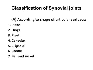
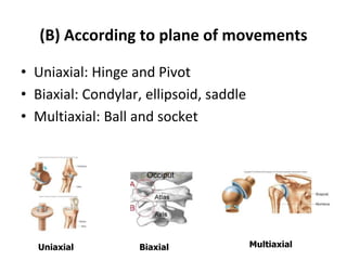
SlideShare a Scribd company logo
Arthrology
Dr. P. Vijayakumari M.P.T (O.G)
Consultant Physiotherapist
Arogya Welfare Trust (TVS)
Madurai
Introduction
• Arthrology: Study of joints
• Synonyms: Articulation (Latin)
Arthron (Greek)
• Joint: a junction between two or more bones
or cartilages.
• A device to permit movements
Classification of joints
Joints are classified according to structure and function-
(A) Functional (based on degree of mobility):
1.Synarthroses: immovable joints (cranial sutures in
adults, primary cartilaginous joints in
growing children).
2.Amphiarthroses: slightly movable joints (joints
between adjacent laminae of vertebrae).
3.Diarthroses: freely movable joints (synovial joints).
Functional classification (cont….)
Synarthroses Amphiarthroses Diarthroses
(B) Structural classification
Based on type of connecting tissue and presence or absence of
joint cavity:
1. Fibrous: composed of intervening fibrous tissue with no joint
cavity. Immovable or slightly movable.
2. Cartilaginous: articulating bones are united by cartilage
(hyaline or fibrocartilage) with no joint cavity.
3. Synovial: articulating bones are separated by a fluid-filled joint
cavity.
Structural classification (cont….)
Fibrous Cartilaginous Synovial
Fibrous joints
1. Sutures: articular surfaces are connected by a thin layer of
connective tissue. They are confined to the skull and
are immovable.
2. Syndesmoses: bones are connected by a considerably greater
amount of connective tissue than in sutures in
the form of interosseous ligaments and
membranes. Slight movement is permitted.
e.g. interosseous tibiofibular joints,
interroseous radioulnar joints.
3. Gomphoses (peg and socket joint): articulation of teeth in
alveolar sockets of mandible and maxilla by
periodontal ligament.
Fibrous joints (cont….)
Sutures Syndesmoses Gomphoses
Sutures
• Plane
• Serrate
• Denticulate
• Squamous
• Schindylesis
Cartilaginous joints
1.Primary (Synchondroses)
2.Secondary (Symphyses)
Synovial joints
• Most evolved and freely movable joints
• Have a joint cavity filled with synovial fluid
• Often termed as diarthrodial joints
• Components:
1. Fibrous capsule
2. Synovial membrane
3. Ligaments
4. Articular cartilage
5. Articular disc or meniscus
6. Bursae
7. Fat pads (Haversian glands)
Classification of Synovial joints
(A) According to shape of articular surfaces:
1. Plane
2. Hinge
3. Pivot
4. Condylar
5. Ellipsoid
6. Saddle
7. Ball and socket
Plane joints
• Articular surfaces are
nearly flat.
• Allow gliding
movements.
• e.g. intercarpal and
intertarsal joints.
Hinge joints (Ginglymus)
• Articular surfaces are
pulley shaped.
• Movements are
permitted only in one
plane (transverse
axis).
• Most common
joints.
• e.g. elbow, knee,
ankle and
interphalangeal joints.
Pivot joints (Trochoid joints)
• Rounded end of one bone
fits into
the concavity of another
bone.
• The rounded
part is surrounded by a
ligament.
• Limited rotation
around a central axis.
• e.g. superior radio-ulnar
and median atlanto-axial
joints.
Condylar joints
• Round articular surface
of one bone fits into a
socket-type articular
surface of another bone.
• Permit movements in
two directions.
• e.g. knee and temporo-
mandibular joints.
Ellipsoid joints
• Elliptical convex surface of
one bone articulates with
elliptical concave surface
of another.
• Permit movements in two
directions.
• Combination of
movements cause
circumduction.
• e.g. wrist and atlanto-
occipital joints.
Saddle (sellar) joints
• Articular surfaces are
reciprocally saddle
shaped i.e. concavo-
convex.
• Allows a
wide range of movement.
• e.g. first
carpo-metacarpal, sterno-
clavicular,
incudo-malleolar joints.
Ball and socket (spheroidal) joints
• Rounded convex surface
of one bone fits into the
cup-like socket of
another bone.
• Permits greatest range
of movements.
• e.g. shoulder, hip and
incudo-stapedial joints.
(B) According to plane of movements
• Uniaxial: Hinge and Pivot
• Biaxial: Condylar, ellipsoid, saddle
• Multiaxial: Ball and socket
Uniaxial Biaxial Multiaxial
(C) According to number of articulating bones
• Simple (2 bones)
• Compound (more than 2 bones)
Movements of synovial joints
1. Gliding (slipping)
2. Angular: flexion, extension, abduction, adduction
3. Rotary (circular): rotation (supination, pronation),
circumduction
Special movements:
1. Inversion: movement of the foot medially
2. Eversion: movement of the foot laterally
3. Protraction: movement of the mandible forward
4. Retraction: movement of the protracted part back
to its starting position
5. Elevation: lifting a body part superiorly
6. Depression: moving the elevated part inferiorly
7. Opposition: touching the thumb to the tips of
other fingers
REFERENCES
1. Cunningham’s Manual of Practical Anatomy, Vol.1.
2. Grant’s Method of Anatomy, 11th Edition.
3. Vishram Singh. General Anatomy, 1st Edition.

More Related Content

Similar to Joints.ppt

Classification and Applied Aspects of Joints
Classification and Applied Aspects of JointsClassification and Applied Aspects of Joints
Classification and Applied Aspects of Joints
Mathew Joseph
 
JOINTS.pptx
JOINTS.pptxJOINTS.pptx
Share_784_Joints.pdf
Share_784_Joints.pdfShare_784_Joints.pdf
Share_784_Joints.pdf
NITHYA718085
 
anatomy of joints
anatomy of jointsanatomy of joints
anatomy of joints
dr.supriti verma bhatnagar
 
1 точно ушул .pdf
1 точно ушул .pdf1 точно ушул .pdf
1 точно ушул .pdf
ssuserbf4af22
 
3JOINTS.pptx
3JOINTS.pptx3JOINTS.pptx
3JOINTS.pptx
Dr Danyal Khattak
 
Joints (arthrology)
Joints (arthrology)Joints (arthrology)
Joints (arthrology)
Dr Neeraj Tiwari
 
ABHI (TYPES OF JOINTS 2).docx
ABHI (TYPES OF  JOINTS 2).docxABHI (TYPES OF  JOINTS 2).docx
ABHI (TYPES OF JOINTS 2).docx
AshutoshPandey966553
 
Joints.ppt
Joints.pptJoints.ppt
Human anatomy (Classification Of Joints)
Human anatomy (Classification Of Joints)Human anatomy (Classification Of Joints)
Human anatomy (Classification Of Joints)
Mahmoud Bahnasi
 
Joint presentation description 4_5810042307021376986.pptx
Joint presentation description 4_5810042307021376986.pptxJoint presentation description 4_5810042307021376986.pptx
Joint presentation description 4_5810042307021376986.pptx
MontahaAlduais
 
Classification of joints
Classification of jointsClassification of joints
Classification of joints
Dr Lovely Jain
 
The Joints
The JointsThe Joints
The Joints
Abhay Rajpoot
 
Joint, types of joint By M Thiru murugan.pptx
Joint, types of joint By M Thiru murugan.pptxJoint, types of joint By M Thiru murugan.pptx
Joint, types of joint By M Thiru murugan.pptx
thiru murugan
 
Joints in human body
Joints in human bodyJoints in human body
Joints in human body
Dr Usha (Physio)
 
Articular system.pdf
Articular system.pdfArticular system.pdf
Articular system.pdf
myLord3
 
Arthrosyndesmology.ppt
Arthrosyndesmology.pptArthrosyndesmology.ppt
Arthrosyndesmology.ppt
yopixi9019
 

Similar to Joints.ppt (20)

Classification and Applied Aspects of Joints
Classification and Applied Aspects of JointsClassification and Applied Aspects of Joints
Classification and Applied Aspects of Joints
 
Arthrology
ArthrologyArthrology
Arthrology
 
JOINTS.pptx
JOINTS.pptxJOINTS.pptx
JOINTS.pptx
 
JOINt.pptx
JOINt.pptxJOINt.pptx
JOINt.pptx
 
Share_784_Joints.pdf
Share_784_Joints.pdfShare_784_Joints.pdf
Share_784_Joints.pdf
 
anatomy of joints
anatomy of jointsanatomy of joints
anatomy of joints
 
1 точно ушул .pdf
1 точно ушул .pdf1 точно ушул .pdf
1 точно ушул .pdf
 
3JOINTS.pptx
3JOINTS.pptx3JOINTS.pptx
3JOINTS.pptx
 
Joints (arthrology)
Joints (arthrology)Joints (arthrology)
Joints (arthrology)
 
ABHI (TYPES OF JOINTS 2).docx
ABHI (TYPES OF  JOINTS 2).docxABHI (TYPES OF  JOINTS 2).docx
ABHI (TYPES OF JOINTS 2).docx
 
Joints.ppt
Joints.pptJoints.ppt
Joints.ppt
 
Human anatomy (Classification Of Joints)
Human anatomy (Classification Of Joints)Human anatomy (Classification Of Joints)
Human anatomy (Classification Of Joints)
 
Joint presentation description 4_5810042307021376986.pptx
Joint presentation description 4_5810042307021376986.pptxJoint presentation description 4_5810042307021376986.pptx
Joint presentation description 4_5810042307021376986.pptx
 
Classification of joints
Classification of jointsClassification of joints
Classification of joints
 
The Joints
The JointsThe Joints
The Joints
 
Joints
JointsJoints
Joints
 
Joint, types of joint By M Thiru murugan.pptx
Joint, types of joint By M Thiru murugan.pptxJoint, types of joint By M Thiru murugan.pptx
Joint, types of joint By M Thiru murugan.pptx
 
Joints in human body
Joints in human bodyJoints in human body
Joints in human body
 
Articular system.pdf
Articular system.pdfArticular system.pdf
Articular system.pdf
 
Arthrosyndesmology.ppt
Arthrosyndesmology.pptArthrosyndesmology.ppt
Arthrosyndesmology.ppt
 

Recently uploaded

Flu Vaccine Alert in Bangalore Karnataka
Flu Vaccine Alert in Bangalore KarnatakaFlu Vaccine Alert in Bangalore Karnataka
Flu Vaccine Alert in Bangalore Karnataka
addon Scans
 
How STIs Influence the Development of Pelvic Inflammatory Disease.pptx
How STIs Influence the Development of Pelvic Inflammatory Disease.pptxHow STIs Influence the Development of Pelvic Inflammatory Disease.pptx
How STIs Influence the Development of Pelvic Inflammatory Disease.pptx
FFragrant
 
NVBDCP.pptx Nation vector borne disease control program
NVBDCP.pptx Nation vector borne disease control programNVBDCP.pptx Nation vector borne disease control program
NVBDCP.pptx Nation vector borne disease control program
Sapna Thakur
 
Knee anatomy and clinical tests 2024.pdf
Knee anatomy and clinical tests 2024.pdfKnee anatomy and clinical tests 2024.pdf
Knee anatomy and clinical tests 2024.pdf
vimalpl1234
 
Cervical & Brachial Plexus By Dr. RIG.pptx
Cervical & Brachial Plexus By Dr. RIG.pptxCervical & Brachial Plexus By Dr. RIG.pptx
Cervical & Brachial Plexus By Dr. RIG.pptx
Dr. Rabia Inam Gandapore
 
24 Upakrama.pptx class ppt useful in all
24 Upakrama.pptx class ppt useful in all24 Upakrama.pptx class ppt useful in all
24 Upakrama.pptx class ppt useful in all
DrSathishMS1
 
POST OPERATIVE OLIGURIA and its management
POST OPERATIVE OLIGURIA and its managementPOST OPERATIVE OLIGURIA and its management
POST OPERATIVE OLIGURIA and its management
touseefaziz1
 
263778731218 Abortion Clinic /Pills In Harare ,
263778731218 Abortion Clinic /Pills In Harare ,263778731218 Abortion Clinic /Pills In Harare ,
263778731218 Abortion Clinic /Pills In Harare ,
sisternakatoto
 
Maxilla, Mandible & Hyoid Bone & Clinical Correlations by Dr. RIG.pptx
Maxilla, Mandible & Hyoid Bone & Clinical Correlations by Dr. RIG.pptxMaxilla, Mandible & Hyoid Bone & Clinical Correlations by Dr. RIG.pptx
Maxilla, Mandible & Hyoid Bone & Clinical Correlations by Dr. RIG.pptx
Dr. Rabia Inam Gandapore
 
Non-respiratory Functions of the Lungs.pdf
Non-respiratory Functions of the Lungs.pdfNon-respiratory Functions of the Lungs.pdf
Non-respiratory Functions of the Lungs.pdf
MedicoseAcademics
 
ANATOMY AND PHYSIOLOGY OF URINARY SYSTEM.pptx
ANATOMY AND PHYSIOLOGY OF URINARY SYSTEM.pptxANATOMY AND PHYSIOLOGY OF URINARY SYSTEM.pptx
ANATOMY AND PHYSIOLOGY OF URINARY SYSTEM.pptx
Swetaba Besh
 
HOT NEW PRODUCT! BIG SALES FAST SHIPPING NOW FROM CHINA!! EU KU DB BK substit...
HOT NEW PRODUCT! BIG SALES FAST SHIPPING NOW FROM CHINA!! EU KU DB BK substit...HOT NEW PRODUCT! BIG SALES FAST SHIPPING NOW FROM CHINA!! EU KU DB BK substit...
HOT NEW PRODUCT! BIG SALES FAST SHIPPING NOW FROM CHINA!! EU KU DB BK substit...
GL Anaacs
 
Ocular injury ppt Upendra pal optometrist upums saifai etawah
Ocular injury  ppt  Upendra pal  optometrist upums saifai etawahOcular injury  ppt  Upendra pal  optometrist upums saifai etawah
Ocular injury ppt Upendra pal optometrist upums saifai etawah
pal078100
 
Ozempic: Preoperative Management of Patients on GLP-1 Receptor Agonists
Ozempic: Preoperative Management of Patients on GLP-1 Receptor Agonists  Ozempic: Preoperative Management of Patients on GLP-1 Receptor Agonists
Ozempic: Preoperative Management of Patients on GLP-1 Receptor Agonists
Saeid Safari
 
New Drug Discovery and Development .....
New Drug Discovery and Development .....New Drug Discovery and Development .....
New Drug Discovery and Development .....
NEHA GUPTA
 
Physiology of Chemical Sensation of smell.pdf
Physiology of Chemical Sensation of smell.pdfPhysiology of Chemical Sensation of smell.pdf
Physiology of Chemical Sensation of smell.pdf
MedicoseAcademics
 
Physiology of Special Chemical Sensation of Taste
Physiology of Special Chemical Sensation of TastePhysiology of Special Chemical Sensation of Taste
Physiology of Special Chemical Sensation of Taste
MedicoseAcademics
 
Alcohol_Dr. Jeenal Mistry MD Pharmacology.pdf
Alcohol_Dr. Jeenal Mistry MD Pharmacology.pdfAlcohol_Dr. Jeenal Mistry MD Pharmacology.pdf
Alcohol_Dr. Jeenal Mistry MD Pharmacology.pdf
Dr Jeenal Mistry
 
Hemodialysis: Chapter 3, Dialysis Water Unit - Dr.Gawad
Hemodialysis: Chapter 3, Dialysis Water Unit - Dr.GawadHemodialysis: Chapter 3, Dialysis Water Unit - Dr.Gawad
Hemodialysis: Chapter 3, Dialysis Water Unit - Dr.Gawad
NephroTube - Dr.Gawad
 
BENIGN PROSTATIC HYPERPLASIA.BPH. BPHpdf
BENIGN PROSTATIC HYPERPLASIA.BPH. BPHpdfBENIGN PROSTATIC HYPERPLASIA.BPH. BPHpdf
BENIGN PROSTATIC HYPERPLASIA.BPH. BPHpdf
DR SETH JOTHAM
 

Recently uploaded (20)

Flu Vaccine Alert in Bangalore Karnataka
Flu Vaccine Alert in Bangalore KarnatakaFlu Vaccine Alert in Bangalore Karnataka
Flu Vaccine Alert in Bangalore Karnataka
 
How STIs Influence the Development of Pelvic Inflammatory Disease.pptx
How STIs Influence the Development of Pelvic Inflammatory Disease.pptxHow STIs Influence the Development of Pelvic Inflammatory Disease.pptx
How STIs Influence the Development of Pelvic Inflammatory Disease.pptx
 
NVBDCP.pptx Nation vector borne disease control program
NVBDCP.pptx Nation vector borne disease control programNVBDCP.pptx Nation vector borne disease control program
NVBDCP.pptx Nation vector borne disease control program
 
Knee anatomy and clinical tests 2024.pdf
Knee anatomy and clinical tests 2024.pdfKnee anatomy and clinical tests 2024.pdf
Knee anatomy and clinical tests 2024.pdf
 
Cervical & Brachial Plexus By Dr. RIG.pptx
Cervical & Brachial Plexus By Dr. RIG.pptxCervical & Brachial Plexus By Dr. RIG.pptx
Cervical & Brachial Plexus By Dr. RIG.pptx
 
24 Upakrama.pptx class ppt useful in all
24 Upakrama.pptx class ppt useful in all24 Upakrama.pptx class ppt useful in all
24 Upakrama.pptx class ppt useful in all
 
POST OPERATIVE OLIGURIA and its management
POST OPERATIVE OLIGURIA and its managementPOST OPERATIVE OLIGURIA and its management
POST OPERATIVE OLIGURIA and its management
 
263778731218 Abortion Clinic /Pills In Harare ,
263778731218 Abortion Clinic /Pills In Harare ,263778731218 Abortion Clinic /Pills In Harare ,
263778731218 Abortion Clinic /Pills In Harare ,
 
Maxilla, Mandible & Hyoid Bone & Clinical Correlations by Dr. RIG.pptx
Maxilla, Mandible & Hyoid Bone & Clinical Correlations by Dr. RIG.pptxMaxilla, Mandible & Hyoid Bone & Clinical Correlations by Dr. RIG.pptx
Maxilla, Mandible & Hyoid Bone & Clinical Correlations by Dr. RIG.pptx
 
Non-respiratory Functions of the Lungs.pdf
Non-respiratory Functions of the Lungs.pdfNon-respiratory Functions of the Lungs.pdf
Non-respiratory Functions of the Lungs.pdf
 
ANATOMY AND PHYSIOLOGY OF URINARY SYSTEM.pptx
ANATOMY AND PHYSIOLOGY OF URINARY SYSTEM.pptxANATOMY AND PHYSIOLOGY OF URINARY SYSTEM.pptx
ANATOMY AND PHYSIOLOGY OF URINARY SYSTEM.pptx
 
HOT NEW PRODUCT! BIG SALES FAST SHIPPING NOW FROM CHINA!! EU KU DB BK substit...
HOT NEW PRODUCT! BIG SALES FAST SHIPPING NOW FROM CHINA!! EU KU DB BK substit...HOT NEW PRODUCT! BIG SALES FAST SHIPPING NOW FROM CHINA!! EU KU DB BK substit...
HOT NEW PRODUCT! BIG SALES FAST SHIPPING NOW FROM CHINA!! EU KU DB BK substit...
 
Ocular injury ppt Upendra pal optometrist upums saifai etawah
Ocular injury  ppt  Upendra pal  optometrist upums saifai etawahOcular injury  ppt  Upendra pal  optometrist upums saifai etawah
Ocular injury ppt Upendra pal optometrist upums saifai etawah
 
Ozempic: Preoperative Management of Patients on GLP-1 Receptor Agonists
Ozempic: Preoperative Management of Patients on GLP-1 Receptor Agonists  Ozempic: Preoperative Management of Patients on GLP-1 Receptor Agonists
Ozempic: Preoperative Management of Patients on GLP-1 Receptor Agonists
 
New Drug Discovery and Development .....
New Drug Discovery and Development .....New Drug Discovery and Development .....
New Drug Discovery and Development .....
 
Physiology of Chemical Sensation of smell.pdf
Physiology of Chemical Sensation of smell.pdfPhysiology of Chemical Sensation of smell.pdf
Physiology of Chemical Sensation of smell.pdf
 
Physiology of Special Chemical Sensation of Taste
Physiology of Special Chemical Sensation of TastePhysiology of Special Chemical Sensation of Taste
Physiology of Special Chemical Sensation of Taste
 
Alcohol_Dr. Jeenal Mistry MD Pharmacology.pdf
Alcohol_Dr. Jeenal Mistry MD Pharmacology.pdfAlcohol_Dr. Jeenal Mistry MD Pharmacology.pdf
Alcohol_Dr. Jeenal Mistry MD Pharmacology.pdf
 
Hemodialysis: Chapter 3, Dialysis Water Unit - Dr.Gawad
Hemodialysis: Chapter 3, Dialysis Water Unit - Dr.GawadHemodialysis: Chapter 3, Dialysis Water Unit - Dr.Gawad
Hemodialysis: Chapter 3, Dialysis Water Unit - Dr.Gawad
 
BENIGN PROSTATIC HYPERPLASIA.BPH. BPHpdf
BENIGN PROSTATIC HYPERPLASIA.BPH. BPHpdfBENIGN PROSTATIC HYPERPLASIA.BPH. BPHpdf
BENIGN PROSTATIC HYPERPLASIA.BPH. BPHpdf
 

Joints.ppt

  • 1. Arthrology Dr. P. Vijayakumari M.P.T (O.G) Consultant Physiotherapist Arogya Welfare Trust (TVS) Madurai
  • 2. Introduction • Arthrology: Study of joints • Synonyms: Articulation (Latin) Arthron (Greek) • Joint: a junction between two or more bones or cartilages. • A device to permit movements
  • 3. Classification of joints Joints are classified according to structure and function- (A) Functional (based on degree of mobility): 1.Synarthroses: immovable joints (cranial sutures in adults, primary cartilaginous joints in growing children). 2.Amphiarthroses: slightly movable joints (joints between adjacent laminae of vertebrae). 3.Diarthroses: freely movable joints (synovial joints).
  • 5. (B) Structural classification Based on type of connecting tissue and presence or absence of joint cavity: 1. Fibrous: composed of intervening fibrous tissue with no joint cavity. Immovable or slightly movable. 2. Cartilaginous: articulating bones are united by cartilage (hyaline or fibrocartilage) with no joint cavity. 3. Synovial: articulating bones are separated by a fluid-filled joint cavity.
  • 7. Fibrous joints 1. Sutures: articular surfaces are connected by a thin layer of connective tissue. They are confined to the skull and are immovable. 2. Syndesmoses: bones are connected by a considerably greater amount of connective tissue than in sutures in the form of interosseous ligaments and membranes. Slight movement is permitted. e.g. interosseous tibiofibular joints, interroseous radioulnar joints. 3. Gomphoses (peg and socket joint): articulation of teeth in alveolar sockets of mandible and maxilla by periodontal ligament.
  • 8. Fibrous joints (cont….) Sutures Syndesmoses Gomphoses
  • 9. Sutures • Plane • Serrate • Denticulate • Squamous • Schindylesis
  • 11. Synovial joints • Most evolved and freely movable joints • Have a joint cavity filled with synovial fluid • Often termed as diarthrodial joints • Components: 1. Fibrous capsule 2. Synovial membrane 3. Ligaments 4. Articular cartilage 5. Articular disc or meniscus 6. Bursae 7. Fat pads (Haversian glands)
  • 12. Classification of Synovial joints (A) According to shape of articular surfaces: 1. Plane 2. Hinge 3. Pivot 4. Condylar 5. Ellipsoid 6. Saddle 7. Ball and socket
  • 13. Plane joints • Articular surfaces are nearly flat. • Allow gliding movements. • e.g. intercarpal and intertarsal joints.
  • 14. Hinge joints (Ginglymus) • Articular surfaces are pulley shaped. • Movements are permitted only in one plane (transverse axis). • Most common joints. • e.g. elbow, knee, ankle and interphalangeal joints.
  • 15. Pivot joints (Trochoid joints) • Rounded end of one bone fits into the concavity of another bone. • The rounded part is surrounded by a ligament. • Limited rotation around a central axis. • e.g. superior radio-ulnar and median atlanto-axial joints.
  • 16. Condylar joints • Round articular surface of one bone fits into a socket-type articular surface of another bone. • Permit movements in two directions. • e.g. knee and temporo- mandibular joints.
  • 17. Ellipsoid joints • Elliptical convex surface of one bone articulates with elliptical concave surface of another. • Permit movements in two directions. • Combination of movements cause circumduction. • e.g. wrist and atlanto- occipital joints.
  • 18. Saddle (sellar) joints • Articular surfaces are reciprocally saddle shaped i.e. concavo- convex. • Allows a wide range of movement. • e.g. first carpo-metacarpal, sterno- clavicular, incudo-malleolar joints.
  • 19. Ball and socket (spheroidal) joints • Rounded convex surface of one bone fits into the cup-like socket of another bone. • Permits greatest range of movements. • e.g. shoulder, hip and incudo-stapedial joints.
  • 20. (B) According to plane of movements • Uniaxial: Hinge and Pivot • Biaxial: Condylar, ellipsoid, saddle • Multiaxial: Ball and socket Uniaxial Biaxial Multiaxial
  • 21. (C) According to number of articulating bones • Simple (2 bones) • Compound (more than 2 bones)
  • 22. Movements of synovial joints 1. Gliding (slipping) 2. Angular: flexion, extension, abduction, adduction 3. Rotary (circular): rotation (supination, pronation), circumduction
  • 23. Special movements: 1. Inversion: movement of the foot medially 2. Eversion: movement of the foot laterally 3. Protraction: movement of the mandible forward 4. Retraction: movement of the protracted part back to its starting position 5. Elevation: lifting a body part superiorly 6. Depression: moving the elevated part inferiorly 7. Opposition: touching the thumb to the tips of other fingers
  • 24. REFERENCES 1. Cunningham’s Manual of Practical Anatomy, Vol.1. 2. Grant’s Method of Anatomy, 11th Edition. 3. Vishram Singh. General Anatomy, 1st Edition.