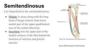
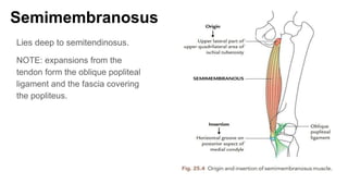
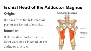
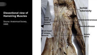
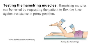
2. Contents
1. Introduction to Hamstring Muscles
2. Features
3. Actions
4. Various Hamstring Muscles
a. Semitendinosus
b. Biceps Femoris
c. Semimembranosus
d. Ischial head of the Adductor Magnus
5. Clinical Significance of Hamstring
Muscles
3. What are the
Hamstring
Muscles?
The muscles on the back of the thigh
are called the Hamstring Muscles.
The hamstring muscles are:
1. Semitendinosus
2. Semimembranosus
3. Biceps femoris (long head)
4. Ischial head of adductor magnus
4. Features of Hamstring Muscles:
The characteristic features of hamstring muscles are:
1. All arise from the ischial tuberosity.
2. All are inserted into one of the bones of the leg.
3. All are supplied by tibial part of the sciatic nerve.
4. All are flexors of the knee and extensors of the hip joint.
5. ● Nerve supply: Tibial part of sciatic nerve
(L5,S1,S2)
● Blood supply: from the perforating branches of the
deep femoral artery, also known as the profunda
femoris artery. The profunda femoris is a branch of
the femoral artery.
6. ● Actions:
○ Chief flexors of the knee
○ Medial rotator of the leg in semi flexed
knee
○ Weak extensors of the hip.
9. Semitendinosus
Lies Superficial to the semimembranosus.
● Origin: It arises along with the long
head of biceps femoris from lower
medial part of the upper quadrilateral
area of the ischial tuberosity.
● Insertion: into the upper part of the
medial surfaces of the tibia behind the
insertion of sartorius and gracilis
muscles
Source: BD Chaurasia’s Human Anatomy
10. Biceps Femoris
It has two heads of origin-
1. Long head, arises from lower medial part of the upper
quadrilateral area of the ischial tuberosity in common with the
semitendinosus and also from the lower part of the sacrotuberous
ligament.
2. Short head, arises from the lower part of the lateral lip of the
linea aspera and upper two-third of the lateral supracondylar line.
11. Insertion of Biceps Femoris
The two heads unite in the lower third of the thigh to
form a conjoint tendon, which slopes downward and
laterally to be inserted on to the head of fibula in front of
the styloid process.
12. ● Nerve supply of Biceps Femoris:
1. Long head, by the tibial part of the sciatic nerve.
2. Short head, by the common peroneal part of the sciatic nerve.
● Actions:
○ Chief flexors of the knee
○ Medial rotator of the leg in semi flexed knee
○ Weak extensors of the hip.
13. Semimembranosus
Lies deep to semitendinosus.
NOTE: expansions from the
tendon form the oblique popliteal
ligament and the fascia covering
the popliteus.
14. Ischial Head of the Adductor Magnus
Origin:
It arises from the inferolateral
part of the ischial tuberosity.
Insertion:
It descends almost vertically
downward to be inserted on the
adductor tubercle.
16. Clinical Significance of Hamstring Muscles
• If hamstring muscles are paralyzed, the patient tends to fall
forward because the gluteus maximus muscle cannot maintain
the necessary tone to stand upright.
• In ancient warfare, the hamstring tendons of soldiers were cut
so that they could not run. This was termed “hamstringing” the
enemy.
17. Source: BD Chaurasia’s Human Anatomy
Testing the hamstring muscles: Hamstring muscles
can be tested by requesting the patient to flex the knee
against resistance in prone position.
18. Source: BD Chaurasia’s Human Anatomy
The inflammation of semimembranosus bursa is called
semimembranosus bursitis. The bursa becomes more prominent
during extension of knee, and disappears during flexion of knee.
20. Take Away
Points
Conventionally,
● the short head of Biceps Femoris is not
included in the hamstring group (as it originates
from lateral lip of the linea aspera and not from
the ischial tuberosity).
● Adductor Magnus is included amongst the
Hamstrings groups of muscles despite
reaching only up to the Adductor Tubercle of
the femur and not inserting into leg bone
(Tibia).
21. Why is the Adductor Magnus included in
Hamstrings group?
The Adductor Magnus is included in Hamstrings group of muscles
because the tibial collateral ligament of the knee joint morphologically
represents the degenerated tendon of this muscle, which is attached
below on the tibia.