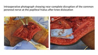
Traumatic Knee
Dislocations
Evaluation, Management, and Surgical
Treatment
James R. Lachman, Saqib Rehman,Paul S. Pipitone
Orthop Clin N Am - (2015)
PRESENTED BY:
DHRUV GOEL
INTRODUCTION
• Incidences :< 0.02% of all musculoskeletal injuries.
• This number is most likely an underestimate caused by spontaneous
reductions and missed diagnosis.
• Most knee dislocations are the result of high-energy mechanisms .
• Careful history and physical examination in a systematic approach will
aid in identifying patients at risk for this injury.
MECHANISM
• M:F=2.5 :1
• High-energy mechanism, the most common:
• motor vehicle collision (up to 50%).
• sports injuries (up to 33%)
• simple falls(up to 12%)
• fourth subset, designated ultralow energy, has been recently described.
Classification:
The first description of a knee dislocation : closed versus open
Incidence : open knee dislocations varies between sources from 15% to
>35%
LATERAL DISLOCATION
ANTEROLATERAL DISLOCATION
V
C
N
III L ACL / PCL / LCL+PLC MCL intact
IV ACL / PCL / MCL / LCL+PLC
III M ACL / PCL / MCL LCL+PLC intact
Schenck 1992
II
arterial injury
nerve injury
fracture dislocation
Anatomic Classification of Knee Dislocations
I single cruciate + collateral
ACL + collateral
PCL + collateral
ACL / PCL collaterals intact
EVALUATION
Acute Assessment
Initial evaluation
PRIMARY SURVEY
ATLS support protocol
SECONDARY SURVEY
careful evaluation of the
neurologic and vascular status of the
affected limb
• In patients with a spontaneously reduced knee dislocation, identifying
those at risk for vascular or soft tissue compromise is much more
difficult.
• Subtle signs of bruising or swelling surrounding the knee may suggest
capsular disruption.
Vascular Examination
• The incidence ranges in the literature from less than 5% up to 65%.
• Historically,high-energy mechanisms resulting in a hyperextension
moment were thought to be more likely to cause vascular compromise.
• Recent review of available literature did not demonstrate an association
between direction of dislocation and vascular insult.
• A standard examination includes palpating dorsalis pedis and posterior
tibial pulses bilaterally and assessing for any asymmetry.
• In the absence of any asymmetry, further assessment is not necessary.
• Bilateral ankle-brachial-indices (ABI) evaluations in the initial
assessment is critical.
• cutoff <0.9, the sensitivity of ABI in detecting vascular injury requiring
surgical intervention approaches 100%.
Routine Angiogram
• concept of a routine angiogram for all suspected knee dislocations has been
the matter of debate.
Currently,
Angiography
(routine computed tomographic angiography [CTA] or magnetic resonance
angiography [MRA] )
is recommended for patients demonstrating insufficient perfusion or any
asymmetry in physical examination
• Green and Allen described the importance of timely identification of vascular
injury.
• Of the patients who were identified with vascular compromise,
those treated surgically within 8 hours had a significantly
lower amputation rate (11%) than those treated after 8 hours (86%)
Vascular Anatomy
• Popliteal artery at risk for being
tethered
• Adductor hiatus
• Soleus arch
• If blood flow through popliteal artery
disrupted :blood supply is inadequate
distally
Neurologic Examination
• The physical examination should include a detailed neurologic examination
including
Sensory examination:
Sensation in the
Tibial
Deep peroneal
Superficial peroneal
distributions to light touch, pinprick,
and temperature if available
Motor examination
including
flexor and extensor hallucis longus,
tibialis anterior
gastrocnemius
is important to establish the baseline
function
• The incidence of nerve injury : 4.5% to 40.0%.
• Most commonly, the common peroneal is the injured nerve, though
isolated tibial nerve palsy has been reported.
• The fibular neck tethers the CPN proximally, and the fibrous arches of the
intermuscular septum tethers distally.
• Contrary to intuition, the reported incidence of nerve injury in ultralow-
energy knee dislocations is higher (44.4%) than the incidence in the
higher-energy trauma patients.
Intraoperative photograph showing near-complete disruption of the common
peroneal nerve at the popliteal hiatus after knee dislocation
RADIOGRAPHIC EVALUATION
Immediate
• After confirmation of limb perfusion and before physical examination
of ligamentous integrity, standard views of the knee are obtained
immediately after reduction.
• Associated fracture has a reported incidence ranging from 10% to
20%.
• Fibular head (arcuate fracture), tibial spine, and lateral tibial condyle
(Segond fracture)avulsions are common.
Medial joint space widening Classic arcuate fragment Segond fracture
Secondary
• After the limb is reduced, vascular injury is ruled out and grossly
unstable knees are stabilized advanced imaging is appropriate.
• Computed tomography (CT), MRI, or both may be appropriate
EXAMINATION OF KNEE STABILITY
Clues to ligament injury in a spontaneously reduced knee dislocation are
• any asymmetry in the joint space
• minor subluxations in any direction
• Segond fractures.
• Examination of ligamentous integrity is often limited secondary to patient
discomfort.
• Intra-articular injection of lidocaine after aspiration of any hemarthrosis
can aid in patient comfort.
Anatomy – 4 groups of ligaments
• ACL
• PCL
• MCL, posteromedial capsule
• LCL & PLC (popliteofibular
ligament, popliteus, capsule, ITB,
biceps femoris)
Knee Examination
• Special Tests (ligaments)
• Valgus and Varus Stress Tests
(MCL/LCL)
• Lachman’s & Anterior Drawer
(ACL)
• Posterior Drawer & Posterior Sag
Test (PCL)
• Postero-lateral corner
MCL Stability
Apply Valgus
or Medial
Stress
AT 30°
FLEXION
LCL Stability
Apply Varus
or Lateral
Stress
Test of ACL
At 90° Flexion
At 30° Flexion
(more sensitive)
Posterior Sag
Posterior Drawer
• The Lachman test and anterior drawer :ACL rupture.
• Varus/Valgus stressing : MCL/LCL compromise.
• Posterior sag :PCL disruption
• are the most reliable maneuvers in the acute setting.
• The most common ligament injury pattern is both cruciates and the medial
ligament complex .
• The posterolateral corner (PLC) is second most common.
• In addition, reports of tendon injury (patella, popliteus, and biceps femoris)
are present.
• Intraoperative photographs demonstrating varus and valgus stress
examination and fluoroscopic images:
Reduction Technique
After palpation of the surface anatomy, gentle in-line traction
attempting to bring the knee into extension is enough to reduce a
dislocated knee.
• No manual pressure be used to aid in any direction,especially in the
popliteal fossa, to avoid iatrogenic neurovascular injury.
• DIMPLE SIGN: closed reduction will be unsuccessful.
SKIN DIMPLING
DIMPLE SIGN
Buttonhole of medial femoral
condyle through soft tissues
(capsule, MCL, retinaculum,
vastus medialis)
Closed Reduction Maneuver
POSITION of DISLOCATION
(Tibia relative to Femur)
•Anterior
• Traction & elevation of distal femur
•Posterior
• Traction & extension of proximal tibia
•Lateral / Medial
• Traction & correctional translation
•Rotational
• Traction & correctional derotation
• Once the knee is reduced, repeat neurolovascular examination.
• With any vascular compromise or asymmetry in ABI(< 0.9), surgical
exploration is warranted.
TREATMENT
Nonoperative Versus Operative Treatment
• Management of knee dislocations is a topic of hot debate.
• Direct ligament repair versus ligament reconstruction
• use of autograft versus allograft tissue for reconstruction
• arthroscopic versus open treatment
• and timing of treatment are all areas of controversy
Early Treatment
Arterial injuries
• Arterial injuries require immediate exploration and vascular surgery
consultation.
• Reexamination is an important mechanism to prevent a missed
arterial injury.
• An evolving ischemia, changing pulse examination, or change in ABI
measurements can all be detected if routine reexamination is part of
standard treatment
Ligament injuries
• Determination of ligamentous injury in the emergency room will
govern the next step.
Postreduction knee
UNSTABLE
Temporizing knee-spanning
external fixator may be placed to
provide stability
and allow soft tissues to calm
down
STABLE
Placement into a knee
immobilizer is recommended
instead of circumferential
splinting or casting
SURGICAL TECHNIQUE
Acute Surgical Intervention
• Acute ligament reconstruction (earlier than 3 weeks), should be done
as an open procedure because of the capsular disruption precluding
arthroscopic assistance.
• If the PLC is involved, a lateral curvilinear incision is used.
• Access to the cruciates and MCL : a midline skin incision with medial
para-patellar arthrotomy.
• Posteromedial corner :Posteromedial approach
Sequence of Ligament Reconstruction
• Cruciate ligament reconstruction typically precedes PLC reconstruction
• PCL ACL COLLATERAL LIGAMENTS
PLC RECONSTRUCTION
Delayed Surgical Intervention
Arthroscopy is a preferred method
• Direct repair of the collaterals is no longer possible and
reconstruction with autograft or allograft is necessary.
Postoperative Complications
• Neurovascular injury
• tourniquet problems
• wound problems
• compartment syndrome
• complex regional pain syndrome
• knee stiffness
• persistent laxity
• osteonecrosis
• posttraumatic osteoarthritis
• Deep vein thrombosis
Outcomes
• Levy and colleagues conducted a systematic review that supports early
operative intervention defined as within 3 weeks compared with delayed
intervention
• Higher-energy injuries were shown to have inferior outcomes to lower-
energy mechanisms
• Lower-energy injuries were correlated with an increased incidence of
neurological injuries
• The incidence of posttraumatic osteoarthritis has been reported as high as
87%
Key points
• Knee dislocation is a relatively uncommon but often missed diagnosis
leading to significant morbidity.
• Serial examination of a suspected knee dislocation is essential in the
prevention of missed arterial injury.
• Currently, angiography is recommended for patients demonstrating
insufficient perfusion or any asymmetry in physical examination.
• Clinicians must be aware of the existence of an irreducible knee
dislocation.
• Use caution during reduction and cognizant of signs (dimple sign,
excessive force required for reduction, joint asymmetry after
reduction attempt).
THANK YOU

Knee

  • 1.
    Traumatic Knee Dislocations Evaluation, Management,and Surgical Treatment James R. Lachman, Saqib Rehman,Paul S. Pipitone Orthop Clin N Am - (2015) PRESENTED BY: DHRUV GOEL
  • 2.
    INTRODUCTION • Incidences :<0.02% of all musculoskeletal injuries. • This number is most likely an underestimate caused by spontaneous reductions and missed diagnosis. • Most knee dislocations are the result of high-energy mechanisms . • Careful history and physical examination in a systematic approach will aid in identifying patients at risk for this injury.
  • 3.
    MECHANISM • M:F=2.5 :1 •High-energy mechanism, the most common: • motor vehicle collision (up to 50%). • sports injuries (up to 33%) • simple falls(up to 12%) • fourth subset, designated ultralow energy, has been recently described.
  • 4.
    Classification: The first descriptionof a knee dislocation : closed versus open Incidence : open knee dislocations varies between sources from 15% to >35%
  • 6.
  • 8.
    V C N III L ACL/ PCL / LCL+PLC MCL intact IV ACL / PCL / MCL / LCL+PLC III M ACL / PCL / MCL LCL+PLC intact Schenck 1992 II arterial injury nerve injury fracture dislocation Anatomic Classification of Knee Dislocations I single cruciate + collateral ACL + collateral PCL + collateral ACL / PCL collaterals intact
  • 9.
    EVALUATION Acute Assessment Initial evaluation PRIMARYSURVEY ATLS support protocol SECONDARY SURVEY careful evaluation of the neurologic and vascular status of the affected limb
  • 10.
    • In patientswith a spontaneously reduced knee dislocation, identifying those at risk for vascular or soft tissue compromise is much more difficult. • Subtle signs of bruising or swelling surrounding the knee may suggest capsular disruption.
  • 11.
    Vascular Examination • Theincidence ranges in the literature from less than 5% up to 65%. • Historically,high-energy mechanisms resulting in a hyperextension moment were thought to be more likely to cause vascular compromise. • Recent review of available literature did not demonstrate an association between direction of dislocation and vascular insult.
  • 12.
    • A standardexamination includes palpating dorsalis pedis and posterior tibial pulses bilaterally and assessing for any asymmetry. • In the absence of any asymmetry, further assessment is not necessary. • Bilateral ankle-brachial-indices (ABI) evaluations in the initial assessment is critical. • cutoff <0.9, the sensitivity of ABI in detecting vascular injury requiring surgical intervention approaches 100%.
  • 13.
    Routine Angiogram • conceptof a routine angiogram for all suspected knee dislocations has been the matter of debate. Currently, Angiography (routine computed tomographic angiography [CTA] or magnetic resonance angiography [MRA] ) is recommended for patients demonstrating insufficient perfusion or any asymmetry in physical examination
  • 14.
    • Green andAllen described the importance of timely identification of vascular injury. • Of the patients who were identified with vascular compromise, those treated surgically within 8 hours had a significantly lower amputation rate (11%) than those treated after 8 hours (86%)
  • 15.
    Vascular Anatomy • Poplitealartery at risk for being tethered • Adductor hiatus • Soleus arch • If blood flow through popliteal artery disrupted :blood supply is inadequate distally
  • 18.
    Neurologic Examination • Thephysical examination should include a detailed neurologic examination including Sensory examination: Sensation in the Tibial Deep peroneal Superficial peroneal distributions to light touch, pinprick, and temperature if available Motor examination including flexor and extensor hallucis longus, tibialis anterior gastrocnemius is important to establish the baseline function
  • 19.
    • The incidenceof nerve injury : 4.5% to 40.0%. • Most commonly, the common peroneal is the injured nerve, though isolated tibial nerve palsy has been reported. • The fibular neck tethers the CPN proximally, and the fibrous arches of the intermuscular septum tethers distally. • Contrary to intuition, the reported incidence of nerve injury in ultralow- energy knee dislocations is higher (44.4%) than the incidence in the higher-energy trauma patients.
  • 20.
    Intraoperative photograph showingnear-complete disruption of the common peroneal nerve at the popliteal hiatus after knee dislocation
  • 21.
    RADIOGRAPHIC EVALUATION Immediate • Afterconfirmation of limb perfusion and before physical examination of ligamentous integrity, standard views of the knee are obtained immediately after reduction. • Associated fracture has a reported incidence ranging from 10% to 20%. • Fibular head (arcuate fracture), tibial spine, and lateral tibial condyle (Segond fracture)avulsions are common.
  • 22.
    Medial joint spacewidening Classic arcuate fragment Segond fracture
  • 23.
    Secondary • After thelimb is reduced, vascular injury is ruled out and grossly unstable knees are stabilized advanced imaging is appropriate. • Computed tomography (CT), MRI, or both may be appropriate
  • 24.
    EXAMINATION OF KNEESTABILITY Clues to ligament injury in a spontaneously reduced knee dislocation are • any asymmetry in the joint space • minor subluxations in any direction • Segond fractures. • Examination of ligamentous integrity is often limited secondary to patient discomfort. • Intra-articular injection of lidocaine after aspiration of any hemarthrosis can aid in patient comfort.
  • 25.
    Anatomy – 4groups of ligaments • ACL • PCL • MCL, posteromedial capsule • LCL & PLC (popliteofibular ligament, popliteus, capsule, ITB, biceps femoris)
  • 27.
    Knee Examination • SpecialTests (ligaments) • Valgus and Varus Stress Tests (MCL/LCL) • Lachman’s & Anterior Drawer (ACL) • Posterior Drawer & Posterior Sag Test (PCL) • Postero-lateral corner
  • 28.
    MCL Stability Apply Valgus orMedial Stress AT 30° FLEXION LCL Stability Apply Varus or Lateral Stress
  • 29.
    Test of ACL At90° Flexion At 30° Flexion (more sensitive)
  • 30.
  • 31.
    • The Lachmantest and anterior drawer :ACL rupture. • Varus/Valgus stressing : MCL/LCL compromise. • Posterior sag :PCL disruption • are the most reliable maneuvers in the acute setting. • The most common ligament injury pattern is both cruciates and the medial ligament complex . • The posterolateral corner (PLC) is second most common. • In addition, reports of tendon injury (patella, popliteus, and biceps femoris) are present.
  • 32.
    • Intraoperative photographsdemonstrating varus and valgus stress examination and fluoroscopic images:
  • 33.
    Reduction Technique After palpationof the surface anatomy, gentle in-line traction attempting to bring the knee into extension is enough to reduce a dislocated knee. • No manual pressure be used to aid in any direction,especially in the popliteal fossa, to avoid iatrogenic neurovascular injury. • DIMPLE SIGN: closed reduction will be unsuccessful.
  • 34.
    SKIN DIMPLING DIMPLE SIGN Buttonholeof medial femoral condyle through soft tissues (capsule, MCL, retinaculum, vastus medialis)
  • 35.
    Closed Reduction Maneuver POSITIONof DISLOCATION (Tibia relative to Femur) •Anterior • Traction & elevation of distal femur •Posterior • Traction & extension of proximal tibia •Lateral / Medial • Traction & correctional translation •Rotational • Traction & correctional derotation
  • 36.
    • Once theknee is reduced, repeat neurolovascular examination. • With any vascular compromise or asymmetry in ABI(< 0.9), surgical exploration is warranted.
  • 37.
    TREATMENT Nonoperative Versus OperativeTreatment • Management of knee dislocations is a topic of hot debate. • Direct ligament repair versus ligament reconstruction • use of autograft versus allograft tissue for reconstruction • arthroscopic versus open treatment • and timing of treatment are all areas of controversy
  • 38.
    Early Treatment Arterial injuries •Arterial injuries require immediate exploration and vascular surgery consultation. • Reexamination is an important mechanism to prevent a missed arterial injury. • An evolving ischemia, changing pulse examination, or change in ABI measurements can all be detected if routine reexamination is part of standard treatment
  • 39.
    Ligament injuries • Determinationof ligamentous injury in the emergency room will govern the next step. Postreduction knee UNSTABLE Temporizing knee-spanning external fixator may be placed to provide stability and allow soft tissues to calm down STABLE Placement into a knee immobilizer is recommended instead of circumferential splinting or casting
  • 40.
    SURGICAL TECHNIQUE Acute SurgicalIntervention • Acute ligament reconstruction (earlier than 3 weeks), should be done as an open procedure because of the capsular disruption precluding arthroscopic assistance. • If the PLC is involved, a lateral curvilinear incision is used. • Access to the cruciates and MCL : a midline skin incision with medial para-patellar arthrotomy. • Posteromedial corner :Posteromedial approach
  • 41.
    Sequence of LigamentReconstruction • Cruciate ligament reconstruction typically precedes PLC reconstruction • PCL ACL COLLATERAL LIGAMENTS PLC RECONSTRUCTION
  • 42.
    Delayed Surgical Intervention Arthroscopyis a preferred method • Direct repair of the collaterals is no longer possible and reconstruction with autograft or allograft is necessary.
  • 43.
    Postoperative Complications • Neurovascularinjury • tourniquet problems • wound problems • compartment syndrome • complex regional pain syndrome • knee stiffness • persistent laxity • osteonecrosis • posttraumatic osteoarthritis • Deep vein thrombosis
  • 44.
    Outcomes • Levy andcolleagues conducted a systematic review that supports early operative intervention defined as within 3 weeks compared with delayed intervention • Higher-energy injuries were shown to have inferior outcomes to lower- energy mechanisms • Lower-energy injuries were correlated with an increased incidence of neurological injuries • The incidence of posttraumatic osteoarthritis has been reported as high as 87%
  • 45.
    Key points • Kneedislocation is a relatively uncommon but often missed diagnosis leading to significant morbidity. • Serial examination of a suspected knee dislocation is essential in the prevention of missed arterial injury. • Currently, angiography is recommended for patients demonstrating insufficient perfusion or any asymmetry in physical examination.
  • 46.
    • Clinicians mustbe aware of the existence of an irreducible knee dislocation. • Use caution during reduction and cognizant of signs (dimple sign, excessive force required for reduction, joint asymmetry after reduction attempt).
  • 47.