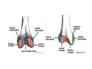
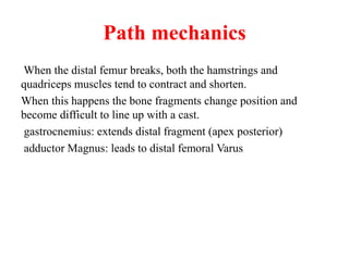
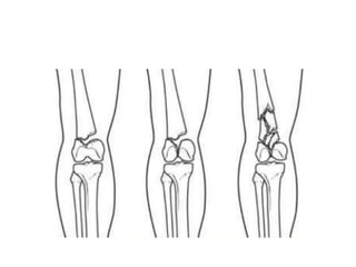
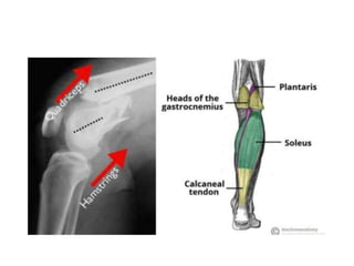
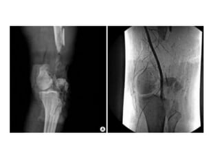
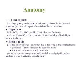
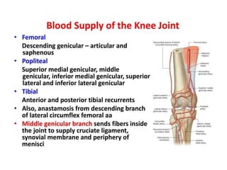
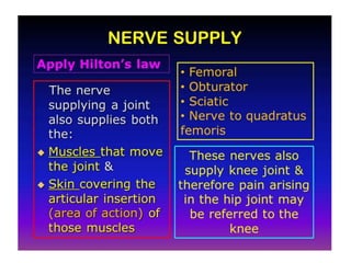
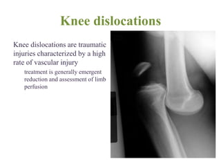
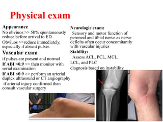
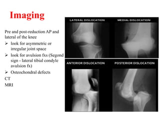
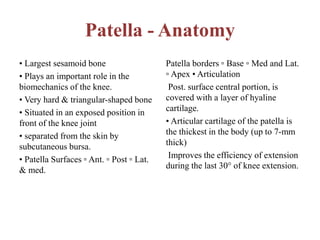
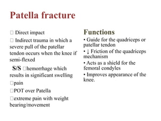
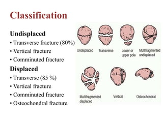
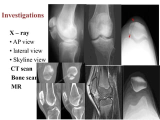
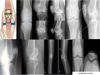
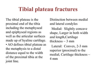
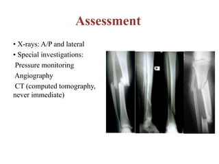
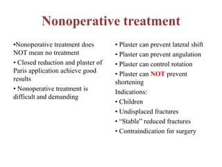
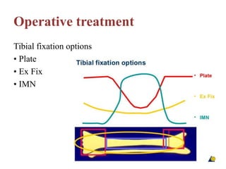
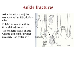
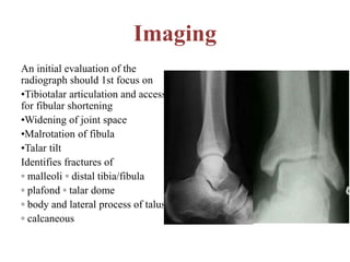
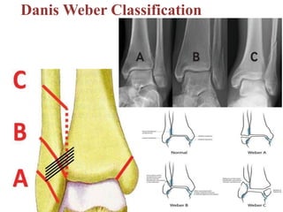
Lower limb trauma...
Presentedby
Dr. KarrarKareem
Orthopedic surgeon
Distal femur fracture
Definition Fractures of the thighbone that occur
just above the knee joint are called distal femur
fractures.
Anatomy
Basic anatomy of femur - only bone in the thigh.
- It is classed as a long bone, and is the longest bone in the body.
- The main function of the femur is to transmit forces from the
tibia to the hip joint.
Articulate proximally with hip bone forming hip joint and distally
with tibia & patella forming knee joint
Divided into three areas: proximal, shaft and distal
The distal end is characterised by the presence of the medial and
lateral condyles, which articulate with the tibia and patella,
forming the knee joint.
Medial and lateral condyles – Rounded areas at the end of the
femur. The posterior and inferior surfaces articulate with the tibia
and menisci of the knee, while the anterior surface articulates
with the patella.
Medial and lateral epicondyles – Bony elevations on the non-
articular areas of the condyles. They are the area of attachment of
some muscles and the collateral ligaments of the knee joint.
Path mechanics
When the distal femur breaks, both the hamstrings and
quadriceps muscles tend to contract and shorten.
When this happens the bone fragments change position and
become difficult to line up with a cast.
gastrocnemius: extends distal fragment (apex posterior)
adductor Magnus: leads to distal femoral Varus
Investigations
X-ray .. obtain standard AP and Lateral
CT .. obtain with frontal and sagittal reconstructions useful for
establishing intra-articular involvement
Angiography : indicated when diminished distal pulses after
gross alignment restored, consider if associated with knee
dislocation
Hoffa fracture
Is a type of supracondylar distal femoral fracture and is
characterized by an associated fracture component in the coronal
plane.
• Hoffa fractures are intra-articular and are characterised by a
fracture in the coronal plane.
• Hoffa fragments are more commonly unicondylar and usually
originate from the lateral femoral condyle.
They can be occasionally bicondylar.
Treatment
Non - Operative
Skeletal traction
Casting and bracing for 6 weeks : indications (rare) >> - non
displaced fractures - non ambulatory patient - patient with
significant comorbidities
Operative
External fixation temporizing measure until soft
tissues permit internal fixation, or until patient is stable
avoid pin placement in area of planned plate placement if
possible
ORIF indications : 1- displaced fracture 2- intra-articular
fracture 3- nonunion - goals : 1- need anatomic reduction of joint
2- stable fixation of articular component to shaft to permit early
motion 3- preserve vascularity
Retrograde IM Nail
Postoperative
Early ROM of knee important
non-weight bearing or toe touch weight-bearing for 6-8 weeks,
up to 10-12 weeks if comminuted * quadriceps and hamstring
strength exercises
Anatomy
 The knee joint:
Is a hinge type synovial joint, which mainly allows for flexion and
extension (and a small degree of medial and lateral rotation)
 Ligaments
PCL, ACL, LCL, MCL, and PLC are all at risk for injury
main stabilizers of the knee given the limited stability afforded by the
bony articulations
 Blood supply
popliteal artery injuries occur often due to tethering at the popliteal fossa
 proximal - fibrous tunnel at the adductor hiatus
 distal - fibrous tunnel at soleus muscle
geniculate arteries may provide collateral flow and palpable pulses
masking a limb-threatening vascular injury
Knee dislocations
Knee dislocations are traumatic
injuries characterized by a high
rate of vascular injury
treatment is generally emergent
reduction and assessment of limb
perfusion
Epidemiology
 incidence
0.02% of orthopedic
injuries
likely underreported as
approximately 50% self-
reduce and are
misdiagnosed
 demographics
4:1 male to female
 location
tibiofemoral articulation
(knee joint)
 risk factors
morbid obesity is a risk
factor for "ultra-low
energy" knee
dislocations with
activities of daily living
Mechanism of injury
 high-energy vs low
energy
 hyperextension injury
leads to anterior
dislocations
 posteriorly directed
force across the
proximal tibia
(dashboard injuries)
leads to posterior
dislocations
Associated injuries:
 Vascular injury
 Nerve injury >> peroneal
25%, tibial n. less common
 Fractures in about 60%
 Soft tissue injury
Classification
• Kennedy
classification
Direction of
displacement of the tibia
• anterior 30-50%
• posterior 30-40%
• lateral 13%
• medial 3%
• rotational 4%
Schenck Classification
 KD I injury to the ACL or
PC
 KD II Injury to ACL and
PCL
 KD III injury to ACL, PCL,
and PMC or PLC
 KD IV Injury to ACL, PCL,
PMC, and PLC
 KD V Multiligamentous
injury with periarticular
fracture
Physical exam
Appearance
No obviuos >> 50% spontaneously
reduce before arrival to ED
Obviuos >>reduce immediately,
especially if absent pulses
Vascular exam
if pulses are present and normal
If ABI >0.9 >> then monitor with
serial examination
If ABI <0.9 >> perform an arterial
duplex ultrasound or CT angiography
if arterial injury confirmed then
consult vascular surgery
Neurologic exam:
Sensory and motor function of
peroneal and tibial nerve as nerve
deficits often occur concomitantly
with vascular injuries
Stability:
Assess ACL, PCL, MCL,
LCL, and PLC
diagnosis based on instability
Imaging
Pre and post-reduction AP and
lateral of the knee
 look for asymmetric or
irregular joint space
 look for avulsion fxs (Segond
sign - lateral tibial condyle
avulsion fx)
 Osteochondral defects
CT
MRI
Treatment
Nonoperative
>> Emergent closed reduction
followed by vascular
assessment/consult
>> Immobilization as
definitive management
Operative
Emergent open reduction
> irreducible knee
> posterolateral dislocation
> open fracture-dislocation
> obesity
> compartment syndrome
> vascular injury
external fixation
delayed ligamentous
reconstruction/repair
Complications of knee dislocation
Vascular compromise
>> 40-50% in anterior or posterior
dislocations
>> KD IV injuries have the highest
Rx >> emergent vascular repair and
prophylactic fasciotomies
Stiffness (arthrofibrosis)
most common complication (38%)
more common in delayed
mobilization
Mx by early reconstruction and
motion, arthroscopic lysis of
adhesion, and manipulation under
anesthesia
Laxity and instability:
37% of some instability, however,
redislocation is uncommon
Peroneal nerve injury
25% occurrence of a peroneal nerve
injury
50% recover partially
posterolateral dislocations
Rx >> AFO to prevent equinus,
neurolysis or exploration later on,
tendon transfers if chronic nerve
palsy persists
Patella - Anatomy
• Largest sesamoid bone
• Plays an important role in the
biomechanics of the knee.
• Very hard & triangular-shaped bone
• Situated in an exposed position in
front of the knee joint
• separated from the skin by
subcutaneous bursa.
• Patella Surfaces ▫ Ant. ▫ Post ▫ Lat.
& med.
Patella borders ▫ Base ▫ Med and Lat.
▫ Apex • Articulation
Post. surface central portion, is
covered with a layer of hyaline
cartilage.
• Articular cartilage of the patella is
the thickest in the body (up to 7-mm
thick)
Improves the efficiency of extension
during the last 30° of knee extension.
Patella fracture
Direct impact
Indirect trauma in which a
severe pull of the patellar
tendon occurs when the knee if
semi-flexed
S/S hemorrhage which
results in significant swelling
pain
POT over Patella
extreme pain with weight
bearing/movement
Functions
• Guide for the quadriceps or
patellar tendon
• ↓ Friction of the quadriceps
mechanism
• Acts as a shield for the
femoral condyles
• Improves appearance of the
knee.
Classification
Undisplaced
• Transverse fracture (80%)
• Vertical fracture
• Comminuted fracture
Displaced
• Transverse (85 %)
• Vertical fracture
• Comminuted fracture
• Osteochondral fracture
Investigations
X – ray
• AP view
• lateral view
• Skyline view
CT scan
Bone scan
MR
Treatment
Non operative
–For non displaced fracture cylinder
cast: extending from the groin to just
above the malleoli for 4 to 6 weeks.
–Followed by physiotherapy-
quadriceps strengthening exercise
Operative
• Tension band wiring.
• Patellectomy
1. Partial:for proximal pole fracture;
major fragment is preserved
2. Complete: for comminuted
fractures. >> Knee should be
immobilized for 3 to 6 weeks in a
long leg cast at 10degrees flexion for
both partial and complete
patellectomy.
• Open reduction and internal
fixation for transverse fracture
Complications
• Refracture • Non union • AVN •
OA • Stiffness • Patellar
instability Incomplete extension
Tibial plateau fractures
The tibial plateau is the
proximal end of the tibia
including the metaphyseal
and epiphyseal regions as
well as the articular surfaces
made up of hyaline cartilage.
• AO defines tibial plateau as
the metaphysis to a distal
distance equal to the width
of the proximal tibia at the
joint line.
Distinction between medial
and lateral condyles
Medial: Slightly concave
shape, Larger in both width
and length,Cartilage
thickness ~ 3 mm
Lateral: Convex, 2-3 mm
superior (proximal) to the
medial, Cartilage thickness ~
4 mm
Mechanism of injury and Classification
Force directed medially
(valgus deformity) or
laterally (varus deformity)
or both.
Axial compressive force.
Both axial force and
force from the side.
• Shatzker classification – Six
types
Type I: Split-wedge
Type II: Split +depression
Type III: pure depression
Type IV: Split fracture of
medial plateau
Type V: bicondylar fracture
Type VI: total disconnection
from the diaphysis
Hohl moore and Shatzker
Evaluation
- X ray AP view Lateral
view
- CT with3D reconstruction -
Better visualisation -
Preoperative planning
- MRI Useful for evaluating
injuries of menisci, cruciate
& collateral ligaments and
soft tissue envelope
- Arteriography for any
vascular injury in question
Management
• Non-operative management: –
Indicated for non-displaced or
minimally displaced fractures
• Method:
– Protected weight bearing and
early range-of-knee motion in a
hinged fracture brace.
– Isometric quadriceps exercises
and progressive passive, active-
assisted, and active range-of-knee
motion exercises.
– Partial-weight bearing (30-40
Ib) for 8 to 12 weeks with
progression to full weight
bearing.
Operative treatment
Accepted range of articular
depression varies from < 2 mm
to 1 cm – Instability > 10
degrees of nearly extended
knee compared to the
contralateral side – Open
fractures – Associated
compartment syndrome –
Associated vascular injury
Goals of treatment: –
reconstruction of the articular
surface – re-establishment of
tibial alignment • Treatment
involves reducing and
buttressing of elevated articular
segments with bone graft • Soft
tissue reconstruction including
menisci and ligaments •
Spanning external fixator as a
temporizing measure in
patients with high-energy
injuries or significant soft
tissue injury. • Arthroscopy
Tibial Shaft Fracture
Most common long bone
fracture - 492,000
fractures/year
• Most common open fracture
• Significant cost - 569,000
hospital days
- Major cause of disability
• Significant complications
- 50,000 nonunions/year
Assessment
• History: velocity of injury
• Clinical examination
• General ATLS (advanced
trauma life support)
• Local soft tissues
• Neurovascular
Assessment
• X-rays: A/P and lateral
• Special investigations:
Pressure monitoring
Angiography
CT (computed tomography,
never immediate)
Nonoperative treatment
•Nonoperative treatment does
NOT mean no treatment
• Closed reduction and plaster of
Paris application achieve good
results
• Nonoperative treatment is
difficult and demanding
• Plaster can prevent lateral shift
• Plaster can prevent angulation
• Plaster can control rotation
• Plaster can NOT prevent
shortening
Indications:
• Children
• Undisplaced fractures
• “Stable” reduced fractures
• Contraindication for surgery
Operative treatment
Tibial fixation options
• Plate
• Ex Fix
• IMN
Ankle fractures
Ankle is a three bone joint
composed of the tibia, fibula an
talus
Talus articulates with the
tibial plafond superiorly
Itsconsidered saddle-shaped
with the dome itself is wider
anteriorly than posteriorly
Imaging
An initial evaluation of the
radiograph should 1st focus on
•Tibiotalar articulation and access
for fibular shortening
•Widening of joint space
•Malrotation of fibula
•Talar tilt
Identifies fractures of
◦ malleoli ◦ distal tibia/fibula
◦ plafond ◦ talar dome
◦ body and lateral process of talus
◦ calcaneous
Danis Weber Classification
Thank you

lower limb trauma conhhhhhhhhhht....pptx

  • 1.
    Lower limb trauma... Presentedby Dr.KarrarKareem Orthopedic surgeon
  • 2.
    Distal femur fracture DefinitionFractures of the thighbone that occur just above the knee joint are called distal femur fractures.
  • 3.
    Anatomy Basic anatomy offemur - only bone in the thigh. - It is classed as a long bone, and is the longest bone in the body. - The main function of the femur is to transmit forces from the tibia to the hip joint. Articulate proximally with hip bone forming hip joint and distally with tibia & patella forming knee joint Divided into three areas: proximal, shaft and distal
  • 4.
    The distal endis characterised by the presence of the medial and lateral condyles, which articulate with the tibia and patella, forming the knee joint. Medial and lateral condyles – Rounded areas at the end of the femur. The posterior and inferior surfaces articulate with the tibia and menisci of the knee, while the anterior surface articulates with the patella. Medial and lateral epicondyles – Bony elevations on the non- articular areas of the condyles. They are the area of attachment of some muscles and the collateral ligaments of the knee joint.
  • 6.
    Path mechanics When thedistal femur breaks, both the hamstrings and quadriceps muscles tend to contract and shorten. When this happens the bone fragments change position and become difficult to line up with a cast. gastrocnemius: extends distal fragment (apex posterior) adductor Magnus: leads to distal femoral Varus
  • 9.
    Investigations X-ray .. obtainstandard AP and Lateral CT .. obtain with frontal and sagittal reconstructions useful for establishing intra-articular involvement Angiography : indicated when diminished distal pulses after gross alignment restored, consider if associated with knee dislocation
  • 10.
    Hoffa fracture Is atype of supracondylar distal femoral fracture and is characterized by an associated fracture component in the coronal plane. • Hoffa fractures are intra-articular and are characterised by a fracture in the coronal plane. • Hoffa fragments are more commonly unicondylar and usually originate from the lateral femoral condyle. They can be occasionally bicondylar.
  • 12.
    Treatment Non - Operative Skeletaltraction Casting and bracing for 6 weeks : indications (rare) >> - non displaced fractures - non ambulatory patient - patient with significant comorbidities
  • 13.
    Operative External fixation temporizingmeasure until soft tissues permit internal fixation, or until patient is stable avoid pin placement in area of planned plate placement if possible ORIF indications : 1- displaced fracture 2- intra-articular fracture 3- nonunion - goals : 1- need anatomic reduction of joint 2- stable fixation of articular component to shaft to permit early motion 3- preserve vascularity Retrograde IM Nail
  • 16.
    Postoperative Early ROM ofknee important non-weight bearing or toe touch weight-bearing for 6-8 weeks, up to 10-12 weeks if comminuted * quadriceps and hamstring strength exercises
  • 18.
    Anatomy  The kneejoint: Is a hinge type synovial joint, which mainly allows for flexion and extension (and a small degree of medial and lateral rotation)  Ligaments PCL, ACL, LCL, MCL, and PLC are all at risk for injury main stabilizers of the knee given the limited stability afforded by the bony articulations  Blood supply popliteal artery injuries occur often due to tethering at the popliteal fossa  proximal - fibrous tunnel at the adductor hiatus  distal - fibrous tunnel at soleus muscle geniculate arteries may provide collateral flow and palpable pulses masking a limb-threatening vascular injury
  • 21.
    Knee dislocations Knee dislocationsare traumatic injuries characterized by a high rate of vascular injury treatment is generally emergent reduction and assessment of limb perfusion
  • 22.
    Epidemiology  incidence 0.02% oforthopedic injuries likely underreported as approximately 50% self- reduce and are misdiagnosed  demographics 4:1 male to female  location tibiofemoral articulation (knee joint)  risk factors morbid obesity is a risk factor for "ultra-low energy" knee dislocations with activities of daily living
  • 23.
    Mechanism of injury high-energy vs low energy  hyperextension injury leads to anterior dislocations  posteriorly directed force across the proximal tibia (dashboard injuries) leads to posterior dislocations Associated injuries:  Vascular injury  Nerve injury >> peroneal 25%, tibial n. less common  Fractures in about 60%  Soft tissue injury
  • 24.
    Classification • Kennedy classification Direction of displacementof the tibia • anterior 30-50% • posterior 30-40% • lateral 13% • medial 3% • rotational 4% Schenck Classification  KD I injury to the ACL or PC  KD II Injury to ACL and PCL  KD III injury to ACL, PCL, and PMC or PLC  KD IV Injury to ACL, PCL, PMC, and PLC  KD V Multiligamentous injury with periarticular fracture
  • 25.
    Physical exam Appearance No obviuos>> 50% spontaneously reduce before arrival to ED Obviuos >>reduce immediately, especially if absent pulses Vascular exam if pulses are present and normal If ABI >0.9 >> then monitor with serial examination If ABI <0.9 >> perform an arterial duplex ultrasound or CT angiography if arterial injury confirmed then consult vascular surgery Neurologic exam: Sensory and motor function of peroneal and tibial nerve as nerve deficits often occur concomitantly with vascular injuries Stability: Assess ACL, PCL, MCL, LCL, and PLC diagnosis based on instability
  • 26.
    Imaging Pre and post-reductionAP and lateral of the knee  look for asymmetric or irregular joint space  look for avulsion fxs (Segond sign - lateral tibial condyle avulsion fx)  Osteochondral defects CT MRI
  • 27.
    Treatment Nonoperative >> Emergent closedreduction followed by vascular assessment/consult >> Immobilization as definitive management Operative Emergent open reduction > irreducible knee > posterolateral dislocation > open fracture-dislocation > obesity > compartment syndrome > vascular injury external fixation delayed ligamentous reconstruction/repair
  • 28.
    Complications of kneedislocation Vascular compromise >> 40-50% in anterior or posterior dislocations >> KD IV injuries have the highest Rx >> emergent vascular repair and prophylactic fasciotomies Stiffness (arthrofibrosis) most common complication (38%) more common in delayed mobilization Mx by early reconstruction and motion, arthroscopic lysis of adhesion, and manipulation under anesthesia Laxity and instability: 37% of some instability, however, redislocation is uncommon Peroneal nerve injury 25% occurrence of a peroneal nerve injury 50% recover partially posterolateral dislocations Rx >> AFO to prevent equinus, neurolysis or exploration later on, tendon transfers if chronic nerve palsy persists
  • 29.
    Patella - Anatomy •Largest sesamoid bone • Plays an important role in the biomechanics of the knee. • Very hard & triangular-shaped bone • Situated in an exposed position in front of the knee joint • separated from the skin by subcutaneous bursa. • Patella Surfaces ▫ Ant. ▫ Post ▫ Lat. & med. Patella borders ▫ Base ▫ Med and Lat. ▫ Apex • Articulation Post. surface central portion, is covered with a layer of hyaline cartilage. • Articular cartilage of the patella is the thickest in the body (up to 7-mm thick) Improves the efficiency of extension during the last 30° of knee extension.
  • 30.
    Patella fracture Direct impact Indirecttrauma in which a severe pull of the patellar tendon occurs when the knee if semi-flexed S/S hemorrhage which results in significant swelling pain POT over Patella extreme pain with weight bearing/movement Functions • Guide for the quadriceps or patellar tendon • ↓ Friction of the quadriceps mechanism • Acts as a shield for the femoral condyles • Improves appearance of the knee.
  • 31.
    Classification Undisplaced • Transverse fracture(80%) • Vertical fracture • Comminuted fracture Displaced • Transverse (85 %) • Vertical fracture • Comminuted fracture • Osteochondral fracture
  • 32.
    Investigations X – ray •AP view • lateral view • Skyline view CT scan Bone scan MR
  • 33.
    Treatment Non operative –For nondisplaced fracture cylinder cast: extending from the groin to just above the malleoli for 4 to 6 weeks. –Followed by physiotherapy- quadriceps strengthening exercise Operative • Tension band wiring. • Patellectomy 1. Partial:for proximal pole fracture; major fragment is preserved 2. Complete: for comminuted fractures. >> Knee should be immobilized for 3 to 6 weeks in a long leg cast at 10degrees flexion for both partial and complete patellectomy. • Open reduction and internal fixation for transverse fracture Complications • Refracture • Non union • AVN • OA • Stiffness • Patellar instability Incomplete extension
  • 35.
    Tibial plateau fractures Thetibial plateau is the proximal end of the tibia including the metaphyseal and epiphyseal regions as well as the articular surfaces made up of hyaline cartilage. • AO defines tibial plateau as the metaphysis to a distal distance equal to the width of the proximal tibia at the joint line. Distinction between medial and lateral condyles Medial: Slightly concave shape, Larger in both width and length,Cartilage thickness ~ 3 mm Lateral: Convex, 2-3 mm superior (proximal) to the medial, Cartilage thickness ~ 4 mm
  • 37.
    Mechanism of injuryand Classification Force directed medially (valgus deformity) or laterally (varus deformity) or both. Axial compressive force. Both axial force and force from the side. • Shatzker classification – Six types Type I: Split-wedge Type II: Split +depression Type III: pure depression Type IV: Split fracture of medial plateau Type V: bicondylar fracture Type VI: total disconnection from the diaphysis
  • 38.
  • 40.
    Evaluation - X rayAP view Lateral view - CT with3D reconstruction - Better visualisation - Preoperative planning - MRI Useful for evaluating injuries of menisci, cruciate & collateral ligaments and soft tissue envelope - Arteriography for any vascular injury in question
  • 41.
    Management • Non-operative management:– Indicated for non-displaced or minimally displaced fractures • Method: – Protected weight bearing and early range-of-knee motion in a hinged fracture brace. – Isometric quadriceps exercises and progressive passive, active- assisted, and active range-of-knee motion exercises. – Partial-weight bearing (30-40 Ib) for 8 to 12 weeks with progression to full weight bearing.
  • 42.
    Operative treatment Accepted rangeof articular depression varies from < 2 mm to 1 cm – Instability > 10 degrees of nearly extended knee compared to the contralateral side – Open fractures – Associated compartment syndrome – Associated vascular injury Goals of treatment: – reconstruction of the articular surface – re-establishment of tibial alignment • Treatment involves reducing and buttressing of elevated articular segments with bone graft • Soft tissue reconstruction including menisci and ligaments • Spanning external fixator as a temporizing measure in patients with high-energy injuries or significant soft tissue injury. • Arthroscopy
  • 44.
    Tibial Shaft Fracture Mostcommon long bone fracture - 492,000 fractures/year • Most common open fracture • Significant cost - 569,000 hospital days - Major cause of disability • Significant complications - 50,000 nonunions/year Assessment • History: velocity of injury • Clinical examination • General ATLS (advanced trauma life support) • Local soft tissues • Neurovascular
  • 45.
    Assessment • X-rays: A/Pand lateral • Special investigations: Pressure monitoring Angiography CT (computed tomography, never immediate)
  • 46.
    Nonoperative treatment •Nonoperative treatmentdoes NOT mean no treatment • Closed reduction and plaster of Paris application achieve good results • Nonoperative treatment is difficult and demanding • Plaster can prevent lateral shift • Plaster can prevent angulation • Plaster can control rotation • Plaster can NOT prevent shortening Indications: • Children • Undisplaced fractures • “Stable” reduced fractures • Contraindication for surgery
  • 47.
    Operative treatment Tibial fixationoptions • Plate • Ex Fix • IMN
  • 48.
    Ankle fractures Ankle isa three bone joint composed of the tibia, fibula an talus Talus articulates with the tibial plafond superiorly Itsconsidered saddle-shaped with the dome itself is wider anteriorly than posteriorly
  • 49.
    Imaging An initial evaluationof the radiograph should 1st focus on •Tibiotalar articulation and access for fibular shortening •Widening of joint space •Malrotation of fibula •Talar tilt Identifies fractures of ◦ malleoli ◦ distal tibia/fibula ◦ plafond ◦ talar dome ◦ body and lateral process of talus ◦ calcaneous
  • 50.
  • 51.