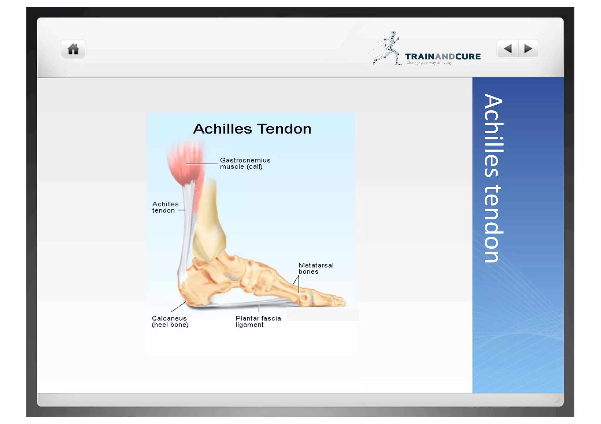
The Achilles tendon connects the calf muscles to the heel bone and allows walking, running, and jumping by tightening as the calf muscles contract. Achilles tendon ruptures are most common in recreational athletes aged 30-50 and can result from a sudden forced movement of the foot or overuse. While nonsurgical treatment is a option, surgery is often recommended to repair the torn tendon and speed recovery through physical therapy.
Tendoachilles rupture and its managementRohan Vakta
Achilles tendon is the strongest tendon of body. There are many causes of its rupture. It can be acute or chronic rupture. Management of chronic rupture by semitendinosus tendon is mentioned here.
Dr. Ankur Mittal presented on diagnostic tests and imaging for Achilles tendon injuries. [1] Ultrasound is often used to determine tendon thickness and gap size for complete ruptures and is inexpensive and fast. [2] MRI is better for detecting incomplete tears and planning surgery for chronic tears but is more expensive. [3] Imaging is rarely needed for acute cases but can help with chronic cases for diagnosis and surgical planning.
SPORTS INJURY I Dr.RAJAT JANGIR JAIPUR
#aclsurgeryjaipur #aclsurgeryhindia #aclsurgerytaekwondo
Acl reconstruction in jaipur | Acl reconstruction in taekwondo | Acl injury in football player surgery | Acl reconstruction surgery in football | acl surgery | Acl surgery ke baad physiotherapy | Acl surgery in jaipur | acl surgery recovery | Best acl surgeon in jaipur | Best ligament doctor in hindi | Best acl surgeon in india | Meniscus repair surgery in jaipur | Sports injury doctor | Acl injury in football players | Acl injury in taekwondo | acl tear | Best knee surgeon in jaipur
#allinsideacl #internalbrace #drrajatjangir #bestaclsurgeon #aclexpert #bestkneesurgeon
To Know more about ACL Injury, Click the links below:
1. ACL surgery 7 different Techniques we do at our center - "Not single technique best for all"
https://youtu.be/oWkIr8IXvr8
2. Everything about ACL Injury tear surgery in Hindi I
https://youtu.be/bqpjkAkwZ14
3. Best Screw for ACL tear surgery in Hindi
https://youtu.be/1LGpU1NHiIs
4. ACL Injury Tear Surgery Recovery : All your questions & queries solved by Dr.Rajat Jangir
https://youtu.be/SIAPWiMbOqs
5. Partial ACL Tear Surgery or not ! ACL आधा टूटा हो तो क्या करें ?
https://youtu.be/NEJRPKskJTI
6. 5 Symptoms of ACL Injury tear इंजरी के पांच लक्षण ?
https://youtu.be/EXpgy19Jxzw
7. PRP injection therapy in Partial ACL TEARs
https://youtu.be/qyG1EYgS87E
Dr.RAJAT JANGIR(Asso Prof.)
Senior Consultant Arthroscopy and Joint Replacement
(Specialist in Shoulder Knee Hip Surgery)
Ligament and Joints Clinic
67/34 Mansarovar Jaipur
Whatsapp: shorturl.at/gnAEP
Appointment: +91 8104855900
Email: ligamentsurgeon@gmail.com
Google Page: https://g.page/KNEE-Shoulder-SURGERY?...
Facebook: https://www.facebook.com/Ligamentandj...
* Vast experience and specialisation in the field of Arthroscopy and sports surgery.
* M.S. orthopaedics from BJ Medical College, Civil hospital, Ahmedabad
* Fellowship in Arthroscopy and Sports injury with Prof Joon Ho Wang at Samsung Medical Center, South Korea
* Diploma in Sports Medicine from InternationaI Olympic Committee
* Invited as Athlete Medical Doctor at Rio Olympic 2016
* Done Rajasthan's first "All Inside Physeal Preserving ACL reconstruction" in 13 year old Athlete
Dr.Rajat is rated as one of the best orthopedic surgeon with with excellence in Knee Shoulder Arthroscopy surgeries as replacements'
This document provides information on various arthrodesis procedures. Arthrodesis is a surgical technique used to fuse a dysfunctional joint to relieve pain. It summarizes techniques for fusing specific joints like the shoulder, elbow, wrist, fingers, hip, and knee. For each joint, it describes common indications, positions, surgical approaches, fixation methods, and post-operative care. Complications are also reviewed. The document is a comprehensive reference for orthopedic surgeons on the principles and techniques of different arthrodesis procedures.
This document provides information on meniscal tears of the knee. It begins with an introduction stating that meniscal tears are common injuries responsible for many arthroscopies annually. It then covers anatomy of the medial and lateral menisci, blood supply, classification of tears, mechanisms of injury, clinical features, investigations like MRI and arthroscopy, and treatment options including non-operative care, meniscectomy, and meniscal repair. The focus is on providing detailed information on meniscal anatomy, tears, and surgical and non-surgical management.
The patella is the largest sesamoid bone in the body located within the quadriceps tendon. It articulates with the femur and is supplied by vessels from the geniculate arterial system. The patellar retinaculum connects the patella to the tibia and is formed by fascia and fibers from surrounding muscles. The patella can displace in various directions from its normal position. Lateral dislocation is most common due to anatomical and biomechanical factors that influence the patellofemoral joint. Evaluation and treatment depends on the nature and chronicity of the injury.
Ankle sprains are ligament injuries, usually caused by forced inversion or plantar flexion of the ankle. Symptoms include pain, swelling, bruising, and difficulty walking. Ankle sprains are classified by severity from Grade 1 (mild stretching) to Grade 3 (complete ligament tear). Physical exams involve stress tests to check for laxity in the anterior talofibular and other ligaments. Conservative treatment focuses on RICE (rest, ice, compression, and elevation) along with rehabilitation exercises. Surgery may be needed for complete tears or chronic instability.
The anterior cruciate ligament (ACL) is commonly ruptured in the knee. It occurs from a twisting force on a bent knee and often accompanies injuries to other knee ligaments and meniscus. The ACL attaches the femur to the tibia and prevents anterior tibial displacement. Diagnosis involves physical exams like the Lachman and pivot shift tests and MRI. Treatment options are conservative rehabilitation or surgical reconstruction, with surgery recommended for athletes or those with instability. Reconstruction uses grafts fixed in the knee with screws or buttons. Post-op rehabilitation is needed to regain strength and function.
The Achilles tendon is the largest tendon in the body, originating from the gastrocnemius and soleus muscles and inserting on the calcaneal tuberosity. It lacks a true synovial sheath and is surrounded by a paratenon with visceral and parietal layers that allows 1.5cm of tendon glide. The tendon has a blood supply from the musculotendinous junction, osseous insertion, and multiple vessels on the anterior surface of the paratenon. Ruptures most commonly occur in the watershed area 4cm proximal to the insertion in those aged 30-40 years old during eccentric loading. Treatment involves diagnosis, primary care, and either operative
This document discusses shoulder instability, including definitions, anatomy, evaluation, and treatment. It defines instability as the inability to maintain the humeral head in the glenoid fossa, ranging from dislocation to laxity. Static stabilizers include the labrum and ligaments, while dynamic stabilizers are the rotator cuff and scapulothoracic muscles. Evaluation involves history, exam, and imaging to classify instability by direction, degree, and etiology. Treatment depends on classification but may include immobilization, rehabilitation, or surgical repair of labral tears or bone defects.
This document discusses posterior cruciate ligament (PCL) tears. It begins with an overview of PCL anatomy and mechanisms of injury. It then covers clinical evaluation including physical examination tests like the posterior drawer test. Investigations like MRI are discussed. Finally, the document outlines management approaches for PCL tears, including non-operative treatment for mild injuries and surgical reconstruction or repair for more severe injuries. Surgical techniques like single versus double bundle reconstruction using autografts or allografts are compared. Post-operative rehabilitation protocols are also summarized.
1. The Achilles tendon connects the calf muscles to the heel bone and is commonly ruptured during sports that involve sudden stops or jumps.
2. A complete rupture will cause a gap in the tendon and severe pain in the back of the heel, making it impossible to raise up on the toes.
3. Treatment involves either immobilizing the foot to allow the tendon to heal naturally or surgical repair, followed by physical therapy to regain strength and range of motion.
1) Pilon fractures involve injuries to the distal tibial articular surface and were first described in 1911.
2) They account for 5-7% of tibial fractures and result from high-energy impacts.
3) Treatment is challenging due to articular comminution, bone loss, and soft tissue injury. Surgical management aims to reconstruct the articular surface and metaphysis while treating soft tissues.
The document discusses supracondylar fractures of the humerus in children. It is the most common elbow fracture in children, often occurring between ages 5-7 from falls on an outstretched hand. Displacement can be classified into 3 types. Closed reduction and percutaneous pinning is usually treatment, while open reduction may be needed for vascular injuries or inadequate closed reduction. Complications can include neurovascular injury, compartment syndrome, and malunion."
The Achilles tendon connects the calf muscles to the heel bone and is active during walking, running, and jumping. It is vulnerable to rupture, which commonly occurs in the watershed area 2-6cm above the heel in males ages 25-50 during sports. Diagnosis involves physical exam findings like inability to raise the toes and a gap or defect being felt. Treatment options include immobilization in a cast or surgical repair, with the goal of restoring proper tension and length. Post-operative care focuses on protecting the repair during healing over 6 months using casts, braces, and physical therapy.
The document provides information on anterior cruciate ligament (ACL) injuries, including:
1. The ACL originates from the femur and inserts into the tibia, resisting anterior tibial translation and medial rotation. ACL injuries most commonly result from rapid changes in direction during sports.
2. Physical examination of ACL injuries involves tests like the Lachman test and anterior drawer test to assess knee stability. MRI is also used for diagnosis.
3. Treatment involves RICE initially, followed by either nonsurgical rehabilitation with bracing or surgical reconstruction using grafts like the patellar tendon. Reconstruction aims to restore stability and function to prevent further knee damage.
The document discusses common wrist injuries including fractures of the scaphoid and hook of hamate bones, de Quervain's tenosynovitis, injuries to the distal radial epiphysis, triangular fibrocartilage complex tears, Kienböck's disease, carpal tunnel syndrome, and ulnar nerve compression. Many of these injuries occur due to falls on an outstretched hand or repetitive stresses from sports activities. Clinical exams and imaging tests can help diagnose specific injuries, which are often initially treated conservatively with rest, splinting, or injections but may require surgery in some cases.
This document discusses coxa vara, which is a hip deformity characterized by an abnormal decrease in the femoral neck-shaft angle. It classifies coxa vara as congenital, developmental, or acquired. Developmental coxa vara is the most common type and is caused by a primary cartilage defect in the femoral neck. Clinical features include limping and pain. Treatment involves corrective valgus osteotomies to restore the neck-shaft angle and relieve stress on the femoral physis. The document describes several techniques for valgus osteotomy including Pauwel's, Borden's, and subtrochanteric osteotomy. The goal of surgery is to stimulate healing of the femoral neck defect and restore normal
High tibial osteotomy (HTO) is a surgical procedure that involves correcting angular deformities of the tibia. It has been used to treat conditions like osteoarthritis, osteochondritis dissecans, and malalignment. There are several techniques for HTO including lateral closing wedge osteotomy, medial opening wedge osteotomy, and dome osteotomy. HTO can help relieve pain from unicompartmental osteoarthritis and delay the need for knee replacement in young, active patients. Potential complications include fracture, nonunion, nerve palsy, and issues that can make later knee replacement more difficult. Precise surgical planning and fixation are important for achieving good outcomes from HTO.
28,000 ankle sprains occur daily in the US (Kaminski 2013)
Ankle is the 2nd most commonly injured body site. (Ferran 2006)
Ankle sprains are the most common type of ankle injury. (Ferran 2006)
A sprained ankle can happen to athletes and non-athletes,
children and adults.
Inversion injury most common mechanism (Ferran 2006)
Only risk factor is previous ankle sprain (Ferran 2006)
Sex , generalized joint laxity or anatomical foot types are
not risk factors. (Beynnon et al. 2002 )
This document summarizes the epidemiology, anatomy, mechanisms of injury, classification, treatment approaches, and complications for radial head fractures. Some key points:
- Radial head fractures account for 4% of all fractures and 30% of elbow fractures. They are rare in children.
- The radial head provides stability to the elbow joint and transmits 50-60% of the load across the elbow.
- Fractures are typically classified using the Mason classification system based on displacement.
- Treatment depends on fracture type but may include non-operative management, open reduction and internal fixation, radial head replacement, or radial head excision.
- Complications can include nerve injuries, stiffness, hardware issues, and recurrent
This document provides information about poliomyelitis (polio), including:
- Polio is caused by poliovirus and mainly affects children, causing paralysis in rare cases.
- It was first described in the late 1700s and caused epidemics in the late 1800s.
- The virus infects the intestine and can invade the nervous system, destroying motor neurons and causing muscle weakness or paralysis.
- Types of polio include spinal and bulbar polio, affecting different areas of the spinal cord or brainstem.
- Treatment focuses on rest, physiotherapy, orthotics, tendon transfers and arthrodesis to correct deformities from muscle imbalances.
in this presentation there is the explanation about the causes and mechanism of the anterior cruciate ligament injury or ACL injury is present. With the help of this the learner should able to learn easily the mechanism and the causes of the acl tear. It is very helpful for the students of medical field.
This document provides an overview of hemiarthroplasty, which involves replacing the femoral head with a prosthesis while retaining the natural acetabulum. It discusses the history and types of prostheses used in hemiarthroplasty. The indications, surgical procedure, postoperative care, and possible complications of hemiarthroplasty are described. A case example of a 78-year-old female undergoing cemented bipolar hemiarthroplasty for a fractured neck of femur is presented.
Arthrodesis refers to the surgical fusion of a joint. It is indicated for pain and instability in the joint. With improvements in joint replacement surgery, arthrodesis is now less commonly performed. It permanently relieves pain by fusing the bones and eliminating joint movement, at the cost of stiffness. The optimal positions for fusing different joints are described. Common complications include malposition and nonunion.
Meniscus injuries are common in young adults, often caused by twisting or heavy lifting. Symptoms include knee pain, swelling, stiffness, tenderness, pain with squatting, popping or clicking in the knee, and limited motion. Meniscus tears are classified as longitudinal, horizontal, radial, or flap tears. Exams like McMurray's test and Apley's test are used to diagnose tears. Treatment involves medications, surgery if the meniscus cannot be repaired, physiotherapy including exercises and bracing, and rehabilitation protocols after arthroscopic surgery or meniscal repair surgery. Isokinetic training after arthroscopy can help improve knee function and muscle strength recovery.
Ortho ob achilles tendon problems by richard zell mdLisa Pilato
1) The Achilles tendon connects the calf muscles to the heel bone and is prone to inflammation and injury from overuse.
2) Achilles tendonitis is a common overuse injury characterized by heel pain that worsens with activity. Treatment focuses on stretching, orthotics, anti-inflammatories, and physical therapy.
3) Achilles tendon ruptures involve a tear of the tendon and result in an inability to walk normally. They require casting or surgery to repair the tendon followed by bracing and rehabilitation.
This document discusses ankle instability and chronic ankle sprains. It begins by describing the anatomy of the ankle joint and its ligaments. It then explains that ankle sprains are common injuries, often caused by an inversion mechanism. Chronic ankle instability can develop after repeated sprains and is characterized by recurrent sprains, pain, and a feeling of the ankle giving way. Treatment of ankle sprains focuses on RICE initially, followed by bracing and physical therapy to improve strength, range of motion and proprioception. Surgery is rarely needed except for severe, unresolving cases.
This document provides an overview of hip dislocations and femoral head fractures. It discusses the anatomy of the hip joint, mechanisms of injury, classification systems, evaluation, management, and treatment options. The key points are:
- Hip dislocations are usually caused by high-energy trauma and often involve other injuries. They can damage the blood supply to the femoral head.
- Reduction of the dislocated hip should be done emergently to restore blood flow and reduce the risk of avascular necrosis. Closed reduction under anesthesia is preferred but surgery may be needed for irreducible or unstable cases.
- Associated injuries like femoral neck fractures or large bone fragments require operative treatment. The goal is to achieve a stable, congr
The Achilles tendon evolved approximately 2 million years ago to allow humans to run faster. It is prone to injury due to its limited blood supply. A rupture typically occurs when a load is applied while the tendon is stretched, often due to pre-existing tendonitis. Treatment options include operative repair through open surgery or minimally invasive surgery, or non-operative casting and rehabilitation. Rehabilitation programs focus on regaining range of motion and strength over 4-6 months. Future research could investigate genetic risk factors, standardized strength testing for different treatment options, and outcomes of surgery for primary versus recurrent ruptures.
The document discusses various types of lower extremity trauma including fractures of the hip, femur, knee, tibia, and ankle. For each injury, the document describes the mechanism of injury, classification systems, treatment options, and important clinical considerations. Management involves restoring anatomy, protecting soft tissues, preventing complications, and allowing for early mobilization depending on the specific fracture pattern and patient factors.
Femur fracture and it management and casesonkosurgery
This document discusses various types of femoral fractures including: femoral head fractures, femoral neck fractures, intertrochanteric fractures, subtrochanteric fractures, and distal femur fractures. It provides details on mechanisms of injury, clinical presentation, imaging, classification systems, and treatment approaches for each type of femoral fracture. Nonoperative and operative treatment options are described depending on the fracture pattern and patient factors.
The document discusses femoral neck fractures, including:
- Anatomy of the hip joint and blood supply of the femoral neck
- Mechanisms of injury including low-energy falls in the elderly
- Classification systems including Garden and Pauwel classifications
- Clinical features such as pain on hip motion and inability to perform straight leg raises
- Diagnosis using x-rays and other imaging modalities like CT and MRI
- Treatment goals of minimizing discomfort, restoring function, and obtaining early anatomic reduction and stable fixation
Intertrochanteric fractures of the femurRajiv Colaço
The document discusses extracapsular intertrochanteric hip fractures. It describes the anatomy and classification systems for these fractures. Conservative management involves traction but is associated with high complication rates. Internal fixation with devices like the dynamic hip screw or proximal femoral nail is now the standard of care to allow early mobilization. Surgical techniques like closed or open reduction may be used along with supplemental procedures like medial displacement osteotomy in unstable patterns.
Rotator cuff injuries are common in the upper extremity. The rotator cuff is made up of four muscles that stabilize the shoulder joint. Rotator cuff tendinitis is an inflammation of the tendons that makes up the rotator cuff, usually caused by repetitive overhead motions. Symptoms include shoulder pain that worsens with overhead activities. Treatment focuses on rest, ice, anti-inflammatory medications, and physical therapy. Surgery may be needed for tears that do not improve with nonsurgical treatment.
This document summarizes an original research paper that studied the radiological and functional outcomes of unstable bimalleolar ankle fractures treated surgically using the Baird-Jackson scoring system. The study involved 26 patients between ages 18-60 who underwent open reduction internal fixation surgery using a lateral locking plate and medial malleolar screw(s). Patients were followed for a minimum of 6 months and up to 12 months to assess union, complications, and functional outcomes using the Baird-Jackson scoring system. The majority of patients had good to excellent functional outcomes, with only a few cases of minor complications like infection or stiffness.
Acromioclavicular (AC) joint injury is a term used to describe an injury to the top of the shoulder, where the front of the shoulder blade (acromion) attaches to the collarbone (clavicle).
The effect of intact fibula on functional outcome of reamed intramedullary in...Love2jaipal
detailed journal club presentation on The effect of intact fibula on functional outcome of reamed intramedullary interlocking nail in open and closed isolated tibial shaft fractures
A 16-year-old boy was admitted to the hospital after a traffic accident where he fell from a motorcycle. He reported pain in his left thigh. Examination found deformity, hematoma, and swelling of the left femur region with tenderness. X-rays showed a closed fracture of the middle third of the left femur. He was diagnosed and treated with open reduction internal fixation surgery.
This document discusses the management of hip dislocations. It covers the anatomy of the hip joint, mechanisms of injury, classification systems, clinical evaluation including imaging, closed and open reduction techniques, postoperative management, and complications. The key points are that early reduction within 6 hours can decrease the risk of avascular necrosis, multiple imaging views may be needed, and surgical treatment is often required for irreducible, unstable, or incongruent dislocations. Complications include avascular necrosis, osteoarthritis, recurrent dislocation, and nerve injury.
3. Overview
The Achilles tendon is a large band of fibrous ?ssue in the back of the ankle
that connects the powerful calf muscles to the heel bone (calcaneus)
This Achilles tendon is the largest tendon in our body
When the calf musles contract, the Achilles tendon is ?ghtened, pulling the
heel
It is vital to such ac?vi?es as walking, running, and jumping
5. Causes
This tendon can grow weak and thin with age and lack of use
At this stage it becomes prone to injury or rupture
Certain diseases (ex. Arthri?s, diabetes) and medica?ons (ex. Cor?costeroids
and some fluoroquinolone an?bio?cs) can increase the risk of rupture
6. Causes
The most common mechanisms of injury include sudden forced plantar
flexion of the foot, unexpected dosiflex?on of the foot, and violent
dorsiflexion of the foot
Direct trauma
AKri?on of the tendon as a result of longstanding peritenoni?s with or
without tendinosis
7. Causes
Sudden, eccentric force applied to a dorsiflexed foot
May occur as the result of direct trauma or as the end result following
Achilles peritenoni?s, with or without tendinosis
Risk factors: reacrea?onal athlete, age (30‐50 years), previous tendon injury,
previous tendon injec?ons or fluoroquinolone use, abrupt changes in
training (intensity, ac?vity level), par?cipa?on in a new ac?vity
8. Peritenoni?s with tendinosis
Will generally present with ac?vity‐related pain, swelling, and some?mes
crepita?on along the tendon sheath
With or without the presence of nodularity
More severe symptoms may include pain at rest
9. Tendinosis
Lage‐stage manisfesta?on of this problem
Characterized by mucoid degenera?on of the achilles tendon itself, with a
lack of inflammatory response and symptoms
A sense of fullness or nodularity in the posterior aspect of the tendoachilles
10. Epidemiology
Although the worldwide frequency of Achilles tendon ruptures is not known
data collected from Finland es?mates that it occurs in 18 per 100000 people
yearly
The male‐to‐female ra?o of rupture is es?mated from 1.7:1 to 12:1.
12. Func?onal Anatomy
The largest and strongest tendon in the human body
Formed from the tedninous contribu?ons of the gastrocnemius and soleus
muscles
The tendons converge appr. 15 cm proximal to the inser?on at the posterior
calcaneus
13. Func?onal Anatomy
When viewed in cross sec?on, the right Achilles tendon appears to spiral
counterclockwise 30‐150º toward its inser?on at the calcaneus
The spiraling of the tendon as it reaches the calcaneus allows for elonga?on
and elas?c recoil within the tendon, facilita?ng storage and release of
energy during movement
This also allows higher shortening veloci?es and greater instantaneous
muscle power than could be generated by the gastrocnemius and soleus
complex alone
14. Func?onal Anatomy
Because the ac?n and myosin present in the tenocytes, tendons have almost
ideal mechanical proper?es for the transmission of force from muscle to
bone
Tendons are s?ff, but possess a high tensile strength
They have the ability to strecth up to 4% before damage occurs
With a stretch greater than 8% occurs macroscopic rupture
15. Blood supply for the tendon
Derived from the posterior 4bial artery and its contribu?ons to the
musculotendinous junc?on, as well as the mesosternal vessels which cross
the paratenon, infiltra?ng the tendon and the bone‐tendon junc?on at the
calcaneus
The watershed zone is an area 2‐6 cm proximal to the calcaneus, in which
the blood supply is less abundant and becomes even sparser with age
It is in this part that most degenera?on and therefore rupture of the Achilles
tendon occurs
16. Sport‐specific biomechanics
The peak Achilles tendon force (F) and the mechanical work (W) by the calf
muscles are respec?vely appr. 2200N and 35J in the squat jump, 1900N and
30J in the countermove jump, and 3800N and 50J when hopping
The es?mated peak load is 6‐8 ?mes the body weight during running with a
tensile force of greater than 3000N
On average, achilles tendons in women have a small cross‐sec?onal area
than in men
This suggests that less force is generated in a woman’s Achilles tendon,
which may account for the lower rate of rupture in women
18. History
Pa?ents with an Achilles tendon rupture frequently present with complaints
of a sudden snap in the lower calf associated with acute sever pain
The pa?ent may be able to ambulate with a limp, but he or she is unable to
run, climb stairs, or stand on their toes
Loss of plantar flexion power in the foot
May be swelling of the calf
19. History
There may be a history of a recent increase in physical ac?vity/training
volume
There may be a history of recent use of fluoroquinolones, coir?costeroids or
of cor?costeroid injec?ons
There may have been a previous rupture of the affected tendon
20. Physical evalua?on
Examine the en?re length of the gastrocnenmius‐soleus‐achilles complex
Evaluate any tenderness, swelling, ecchymosis, and tendon defects
Some?mes a palpable gap in the Achilles tendon may be found
The pa?ent will be unable to stand on the toes on the affected leg
21. Clinical tests
“Hiperdorsiflexion” sign – With the pa?ent prone and knees flexed to 90º,
maximal passive dorsiflexion of both feet may reveal excessive dorsiflexion
of the affected leg
Thompson test: with the pa?ent prone, squeezing the calf of the extended
leg may demonstrate no passive plantar flexion of the foot if its Achilles
tendon is ruptured
O’Brien needle test: insert a needle 10 cm proximal to the calcaneal
inser?on of the tendon. With passive dorsiflexion of the foot, the hub of the
needle will ?lt rostrally when the Achilles tendon is intact
23. Imaging studies
Radiographs are useful in ruling out other injuries (may show sog‐?ssue
swelling, increased ankle dorsiflexion on stress views, vascular or
heterotopic calcifica?ons, accessory ossicles, calcaneal fractures, Haglund
deformity, bony metaplasia)
Musculoskeletal ultrasonography can be used to determine the tendon
thickness, character, and presence of a tear
MRI: can be used to discern incomplete ruptures from degenera?on of the
Achilles tendon, can dis?nguish between paratenoni?s, tendinosis, and
bursi?s
25. Physical Therapy
A person who ruptures the Achilles tendon should seek prompt medical
treatment
Physical therapy is generally not indicated in the acute phase of the treatment,
but later becomes a crucial part of the rehabilita?on once adequate healing of
the tendon has occurred
A nonopera4ve vs opera4ve treatment is determined on a pa?ent‐by‐pa?ent
basis
Typically, both nonopera?ve and opera?ve treatment op?ons are offered to
pa?ents, with par?cular emphasis on the benefits and risks of each procedure
26. Surgical Interven?on
Controversy exists regarding whether to conserva?vely manage a first‐?me
Achilles tendon rupture or to surgically reconstruct the ruptured tendon
According to Kahn et al. There was a consistent finding of an appr. 33% higher
rate of complica?ons in those treated surgically
Nonopera?vely treated pa?ents had a rerupture rate appr. 3 ?mes higher than
those treated surgically, but these pa?ents had minimal risk for other
complica?ons
Listed complica?ons resul?ng from open surgical repair included deep infec?ons
(1%), fistulae (3%), necrosis fo the skin or tendon (2%), rerupture (2%), and
minor complica?ons
27. Surgical interven?on
Studies indicate that pa?ents who had a percutaneous rather than an open
surgical approach had a minimal rate of infec?on
But it was also demonstrated that there were rela?vely high rates of injury
to the sural nerve
28. Conserva?ve repair
Early reports of rerupture in conserva?vely treated pa?ents noted rates as
high as 40%
In newer protocols with shorter immobiliza?on periods, the rates of
rerupture apprear to be much less and are comparable to the rerupture rate
for surgically repaired tendons
29. Physical Therapy
Following cast removal, gentle passive range of mo?on of the ankle and subtalar
joints is ini?ated
Ager 2 weeks, progressive resistance exercises (PREs) are added to the therapy
This followed by agrressive gait training exercises at about 10 weeks following
the injury (nonopera?ve pa?ents) or surgery, leading toward ac?vity‐specific
maneuvers and a return to aci?vi?es at 4‐6 months
The pa?ent’s recovery is largely dependent on the quality of the rehabilita?on
program, the pa?ent’s mo?va?on and focus, his/her desired pos?njury ac?viy
level
30. Medica?on
No medical therapy is indicated for this condi?on
Medica?on is only described for the symptomatoc relief of pain
These medica?ons may include acetaminophen,various nonsteroidal an?‐
inflammatory drugs (NSAIDs), or narco?cs, depending on physician
preference
31. Preven?on
Good condi?oning and proper stretching is important in the preven?on of
Achilles tendon injuries
Adequate warm‐up!
32. Prognosis
With proper treatment and rehabilita?on, the prognosis following an
Achilles tendon rupture is good to excellent
Most athletes are able to return to their previous ac?vity levels with either
surgical or conserva?ve treatment
Individuals who undergo surgical treatment are less likely to experience
rerupture of their Achilles tendons
The rerupture rate for opera?ve treatment is 0‐5%, compared with neary
40% in those who opt for conserva?ve treatment
33. Educa?on
Pa?ents should be educated on the importance of stretching and proper
condi?oning to prevent rerupture of the Achilles tendon
Wearing appropriate and properly filng shoes during ac?vi?es also should
be stressed to all athletes