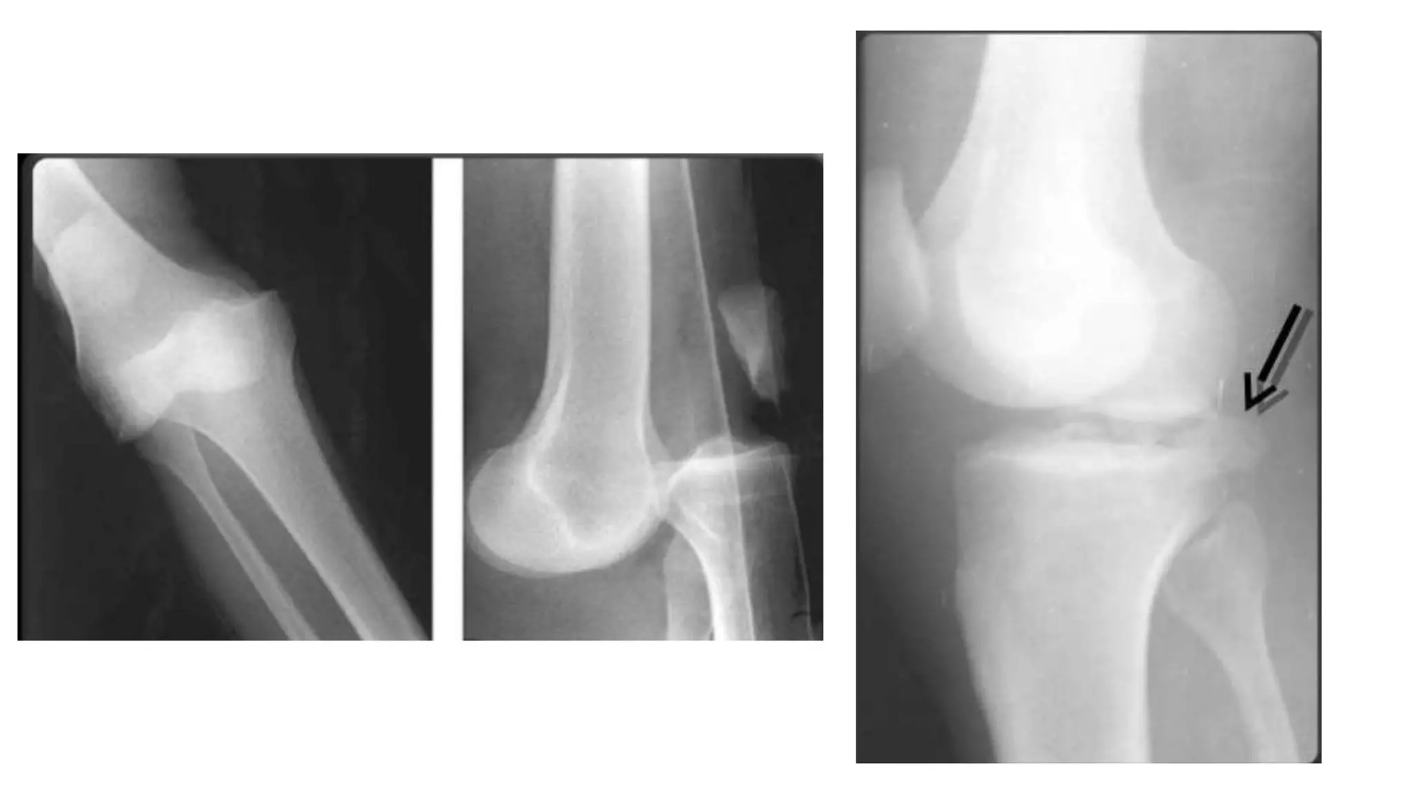
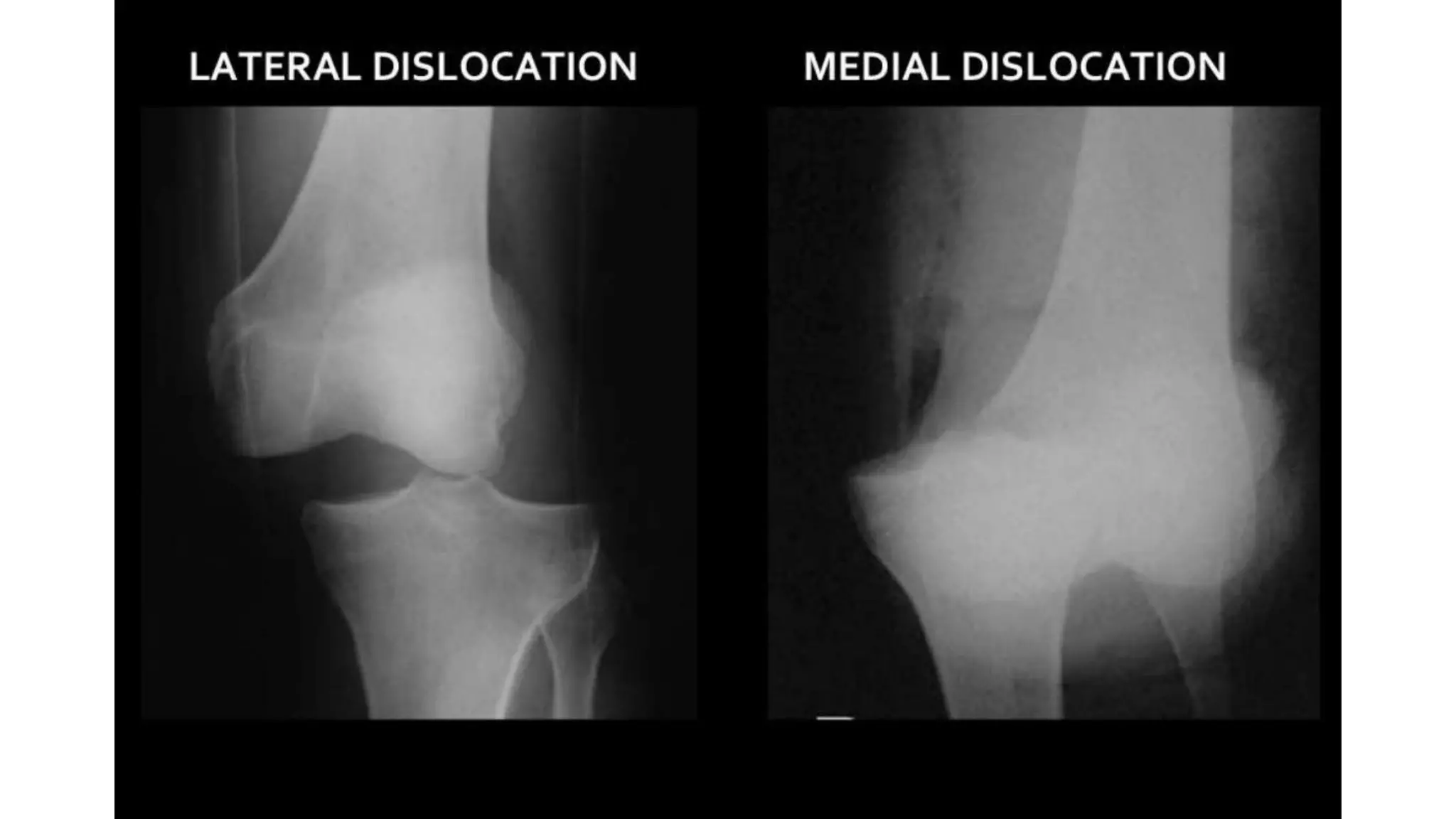
Knee dislocation
DR VIVESH KR SINGH
MS ORTHOPAEDIC
STABILIZERS OF KNEE
JOINTY
• Static
• • Joint Capsule
• • Collateral Ligaments
• • Medial Patellofemoral Ligament • Dynamic
• • Quadriceps
• • Biceps Femoris
• • Pes Anserinus
• • Gastronemius
• • Tensor Fascia Lata
• • Semimembranosus
Mechanism of injury
• Dash board injuries
• Hyperextension injuries
• Fall on flexed knee and plantar flexion
• Anterior blow to the knee
Classification
Kennedy classification:
• Anterior
• Posterior
• Medial
• Lateral
• Rotatory
- Anteromedial
- Anterolateral
- Posteromedial
- Posterolateral (MC ROTATORY)
Clinical features
• May Present with irreducible dislocaton
• Deformity, pain ,cannot ambulate
• Spontaneously reduced- with only effusion
• Normal Knee ,BUT on examination shows instability
• Dislocation + Ipsilateral # lower extremity – Diagnostic Challenge
Associated injuries
• Vascular
• Neurologic
Clinical evaluation
• Measure ankle brachial index
• If ABI >0.9 then monitor with examination
• If ABI <0.9 then arterial duplex ultrasound or CT Angiography.
• If pulses are absent / diminished confirm knee joint is reduced or perform immediate
reduction and reassessment.
• Immediate surgical exploration if pulses are still absent following reduction
• Ischemia time >8 hours has amputation rate as high as 86%
• If pulses present after reduction then measure ABI then observe.
• Neurologic injury - peroneal nerve (10% to 35%):
• Commonly associated with posterolateral dislocation , with varying from
neurapraxia to complete transaction.
• Bracing and tendon transfer may be necessary for treatment of muscular
deficiencies.
PLAIN X-RAYS
• Anteroposterior and
lateral views:
• To evaluate for
fractures and or
dislocation.
• A- PCL intact
knee dislocation
• B- Bicurucate -
Parallel
arrangement of
patella with femur
MRI
• After reduction or suspected spontaneous reduction.
• To assess ligament status
Treatment
• Reduce knee and re-examine vascular status.
• Splint knee in 20-30 degree of flexion
• Confirm reduction is held with repeat radiographs in brace/ splint.
• Reduction maneuvers for specific dislocation :
• Anterior : Axial limb traction is combined with lifting of distal femur.
• Posterior : Axial limb traction is combined with extension and lifting of proximal tibia.
• Medial/ lateral : Axial limb traction is combined with lateral / medial translation of tibia.
• Rotatory : Axial limb traction is combined with derotation of tibia.
• Posterolateral dislocation is believed to be irreducible
owing to buttonholing of the medial femoral Condyle
through the medial capsule, resulting in a Dimple Sign over
the medial aspect of the limb.
•Non operative :
•Immobilization in. Extension for 6 weeks
•External Fixation
-Unstable / subluxation in brace
-Obese patient
-Head injury
-Vascular repair
-Fasciotomy or open wounds
-Removal of fixator under Anesthesia
•Arthroscopy
-Manipulation for flexion
-Assessment of residual laxity
• Indication for operative treatment of knee dislocation:
- Unsuccessful closed reduction.
- Residual soft tissue interposition
- Open injuries
- Vascular injuries
Treatment Indication Advantages Disadvantages
Early open repair (1st week)
Avulsion with large bony
fragments
Secure fixation and maintain
native ligaments
1.Most injuries are either midsubstance
tears or are avulsions without boneand
repair is not possible
2.Wound healing problems due to soft tissue
envelope injury
3.Increased risk of stiffness
Acute (2-4 week) reconstruction
of all ligament
Knee dislocation with bicruciate
injury and no large bony
fragments
Early restoration of ligament
Allows early rehabilitation
Length of surgery
Acute (2-4 week) reconstruction
cruciates staged with delayed (6
weeks) ACL reconstruction
Knee dislocation with bicruciate
injury and no large bony
fragments
Shorter initial procedure,
Return to OR at 6 weeks allows
manipulation to increase motion,
Allows early rehabilitation
1.Requires one additional surgery
2.Rehabilitation in the first 6 weeks
as in an ACL-deficient knee
Delayed (>1 month)
reconstruction after motion is
reestablished and ipsilateral
injuries are healed
Knee dislocation in poor
rehabilitation candidate
Avoid lengthy procedure with
significant complication until
rehabilitation is clarified ,
Fewer complication
1.Difficult to obtain functionalresult equivalent
to early reconstruction 2.Delayed recovery
3.Staged procedure that may require additional
surgeries
Thank you

Knee dislocation

  • 1.
    Knee dislocation DR VIVESHKR SINGH MS ORTHOPAEDIC
  • 2.
    STABILIZERS OF KNEE JOINTY •Static • • Joint Capsule • • Collateral Ligaments • • Medial Patellofemoral Ligament • Dynamic • • Quadriceps • • Biceps Femoris • • Pes Anserinus • • Gastronemius • • Tensor Fascia Lata • • Semimembranosus
  • 3.
    Mechanism of injury •Dash board injuries • Hyperextension injuries • Fall on flexed knee and plantar flexion • Anterior blow to the knee
  • 4.
    Classification Kennedy classification: • Anterior •Posterior • Medial • Lateral • Rotatory - Anteromedial - Anterolateral - Posteromedial - Posterolateral (MC ROTATORY)
  • 5.
    Clinical features • MayPresent with irreducible dislocaton • Deformity, pain ,cannot ambulate • Spontaneously reduced- with only effusion • Normal Knee ,BUT on examination shows instability • Dislocation + Ipsilateral # lower extremity – Diagnostic Challenge
  • 7.
  • 8.
    Clinical evaluation • Measureankle brachial index • If ABI >0.9 then monitor with examination • If ABI <0.9 then arterial duplex ultrasound or CT Angiography. • If pulses are absent / diminished confirm knee joint is reduced or perform immediate reduction and reassessment. • Immediate surgical exploration if pulses are still absent following reduction • Ischemia time >8 hours has amputation rate as high as 86% • If pulses present after reduction then measure ABI then observe.
  • 9.
    • Neurologic injury- peroneal nerve (10% to 35%): • Commonly associated with posterolateral dislocation , with varying from neurapraxia to complete transaction. • Bracing and tendon transfer may be necessary for treatment of muscular deficiencies.
  • 10.
    PLAIN X-RAYS • Anteroposteriorand lateral views: • To evaluate for fractures and or dislocation.
  • 12.
    • A- PCLintact knee dislocation • B- Bicurucate - Parallel arrangement of patella with femur
  • 15.
    MRI • After reductionor suspected spontaneous reduction. • To assess ligament status
  • 16.
    Treatment • Reduce kneeand re-examine vascular status. • Splint knee in 20-30 degree of flexion • Confirm reduction is held with repeat radiographs in brace/ splint. • Reduction maneuvers for specific dislocation : • Anterior : Axial limb traction is combined with lifting of distal femur. • Posterior : Axial limb traction is combined with extension and lifting of proximal tibia. • Medial/ lateral : Axial limb traction is combined with lateral / medial translation of tibia. • Rotatory : Axial limb traction is combined with derotation of tibia.
  • 17.
    • Posterolateral dislocationis believed to be irreducible owing to buttonholing of the medial femoral Condyle through the medial capsule, resulting in a Dimple Sign over the medial aspect of the limb.
  • 18.
    •Non operative : •Immobilizationin. Extension for 6 weeks •External Fixation -Unstable / subluxation in brace -Obese patient -Head injury -Vascular repair -Fasciotomy or open wounds -Removal of fixator under Anesthesia •Arthroscopy -Manipulation for flexion -Assessment of residual laxity
  • 19.
    • Indication foroperative treatment of knee dislocation: - Unsuccessful closed reduction. - Residual soft tissue interposition - Open injuries - Vascular injuries
  • 20.
    Treatment Indication AdvantagesDisadvantages Early open repair (1st week) Avulsion with large bony fragments Secure fixation and maintain native ligaments 1.Most injuries are either midsubstance tears or are avulsions without boneand repair is not possible 2.Wound healing problems due to soft tissue envelope injury 3.Increased risk of stiffness Acute (2-4 week) reconstruction of all ligament Knee dislocation with bicruciate injury and no large bony fragments Early restoration of ligament Allows early rehabilitation Length of surgery Acute (2-4 week) reconstruction cruciates staged with delayed (6 weeks) ACL reconstruction Knee dislocation with bicruciate injury and no large bony fragments Shorter initial procedure, Return to OR at 6 weeks allows manipulation to increase motion, Allows early rehabilitation 1.Requires one additional surgery 2.Rehabilitation in the first 6 weeks as in an ACL-deficient knee Delayed (>1 month) reconstruction after motion is reestablished and ipsilateral injuries are healed Knee dislocation in poor rehabilitation candidate Avoid lengthy procedure with significant complication until rehabilitation is clarified , Fewer complication 1.Difficult to obtain functionalresult equivalent to early reconstruction 2.Delayed recovery 3.Staged procedure that may require additional surgeries
  • 21.