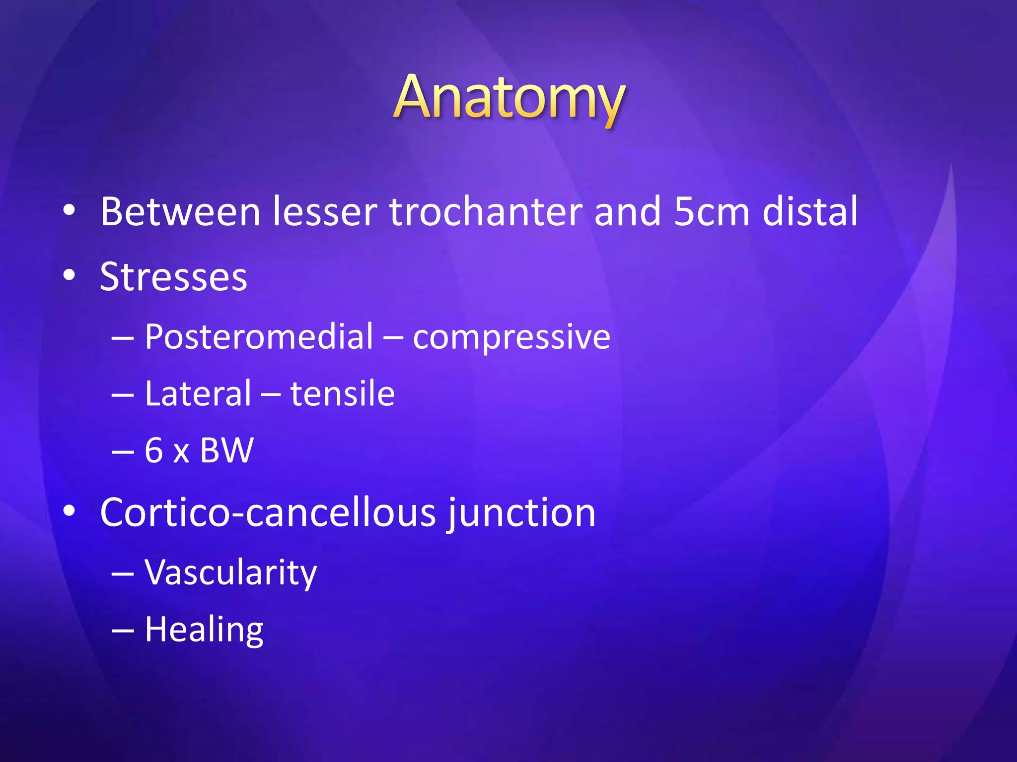
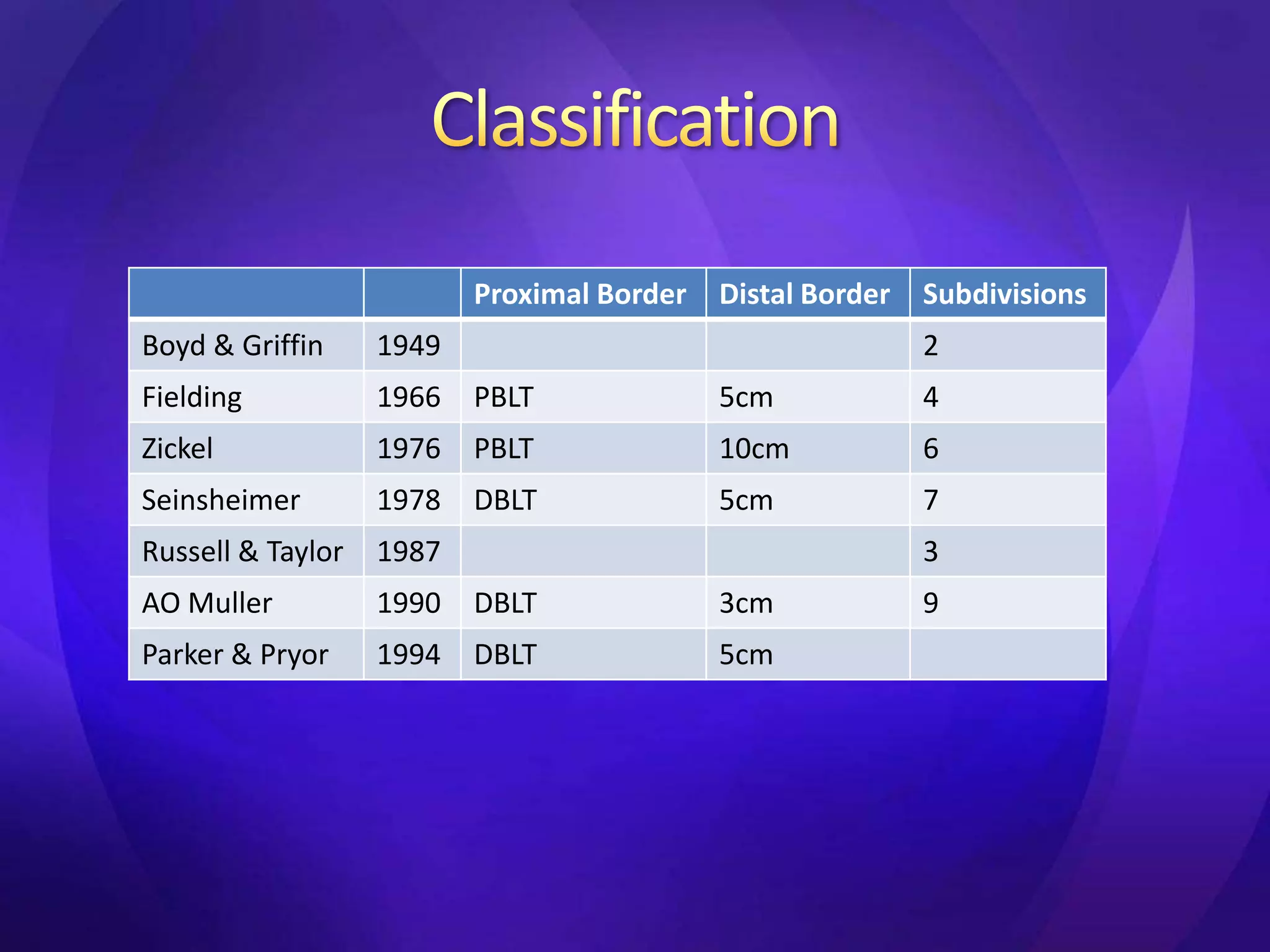
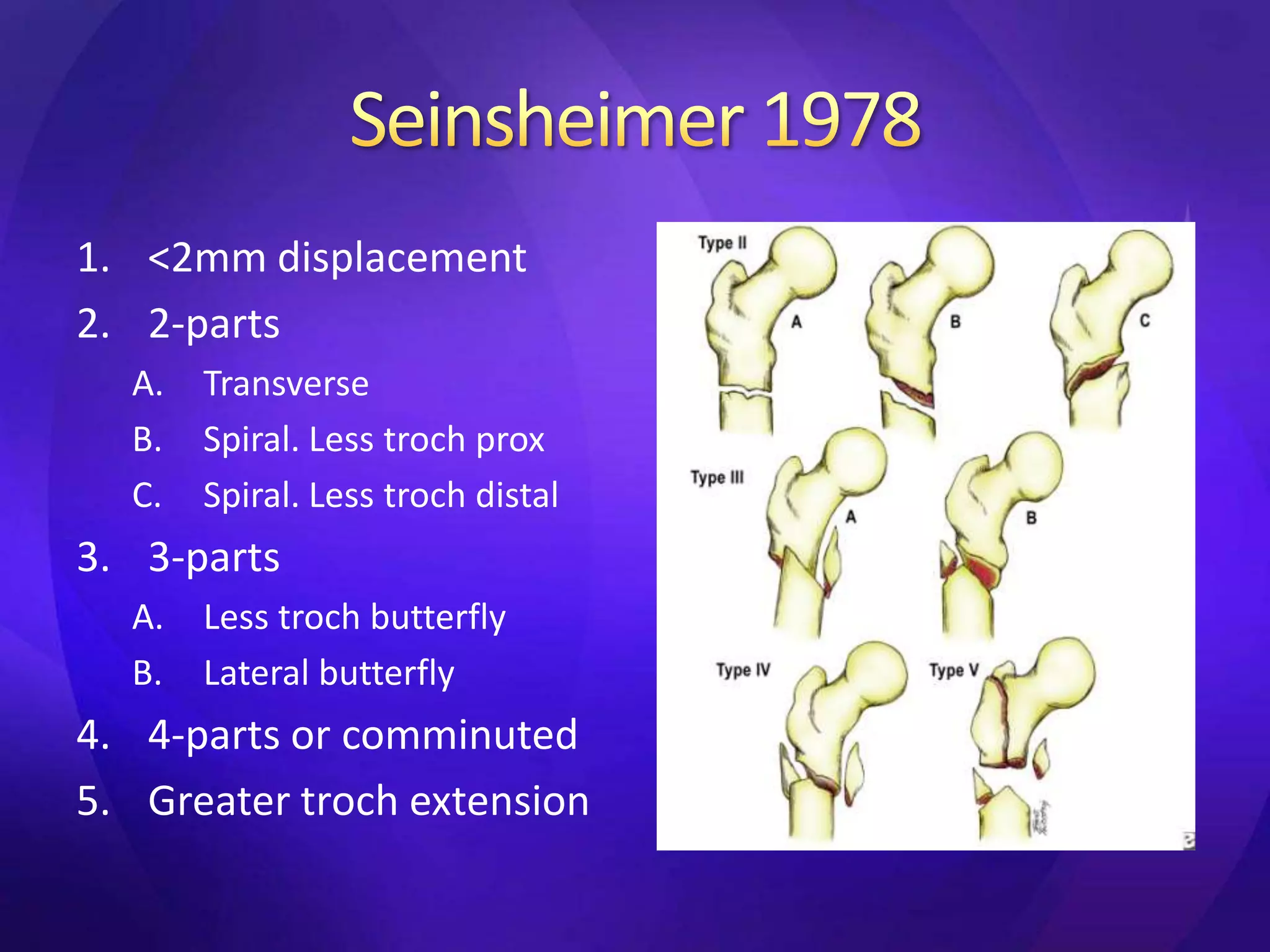
1. This document discusses hip fractures, specifically subtrochanteric fractures. It notes that 10-30% of hip fractures are subtrochanteric and they have a bimodal age distribution in those 20-40 years old from high-energy injuries and those over 60 from low-energy falls.
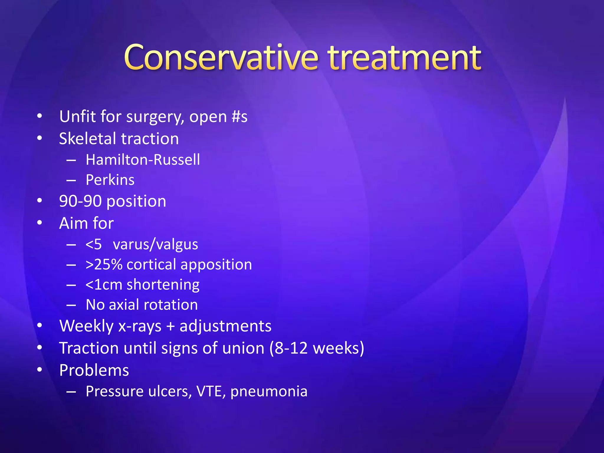
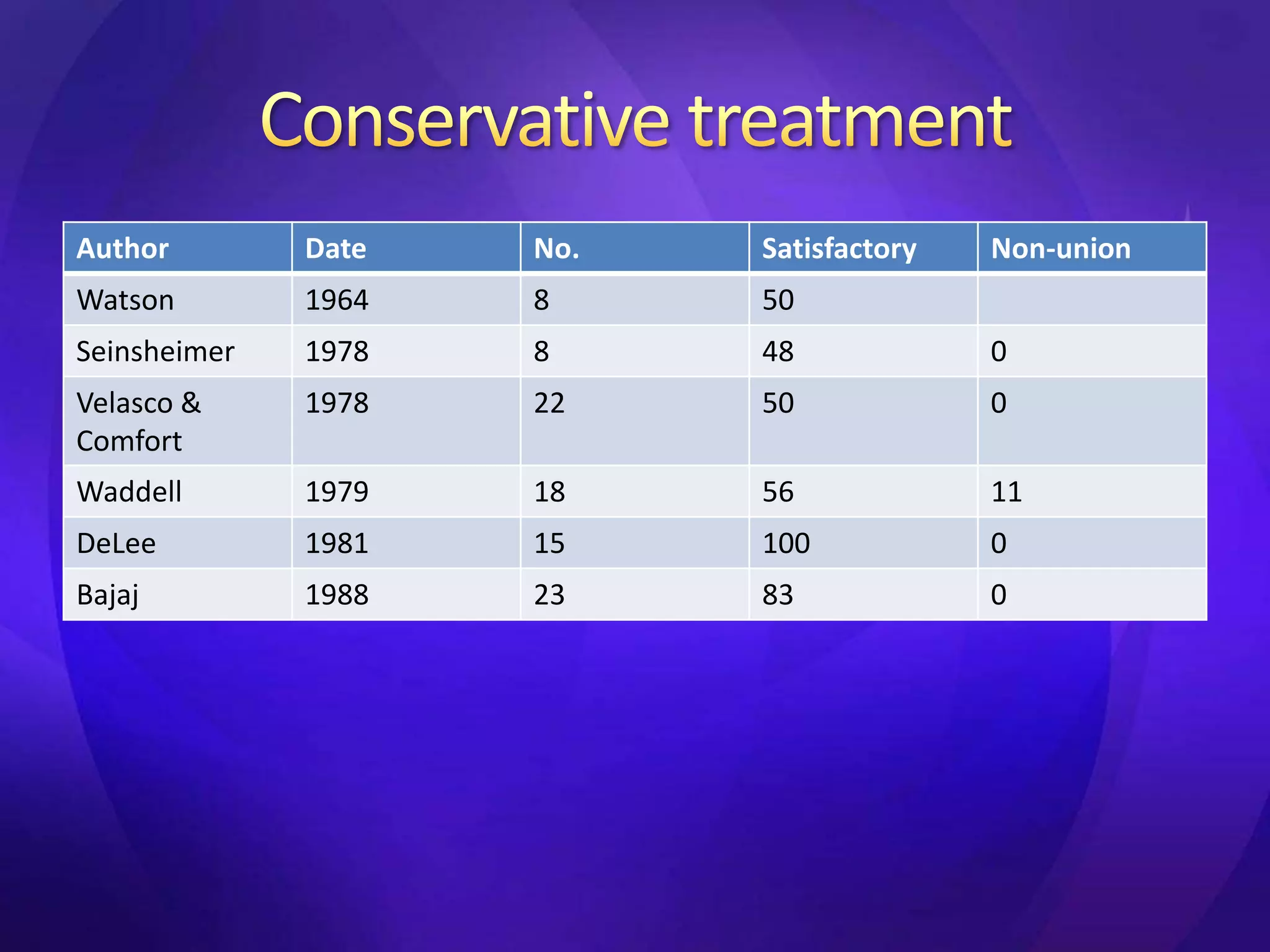
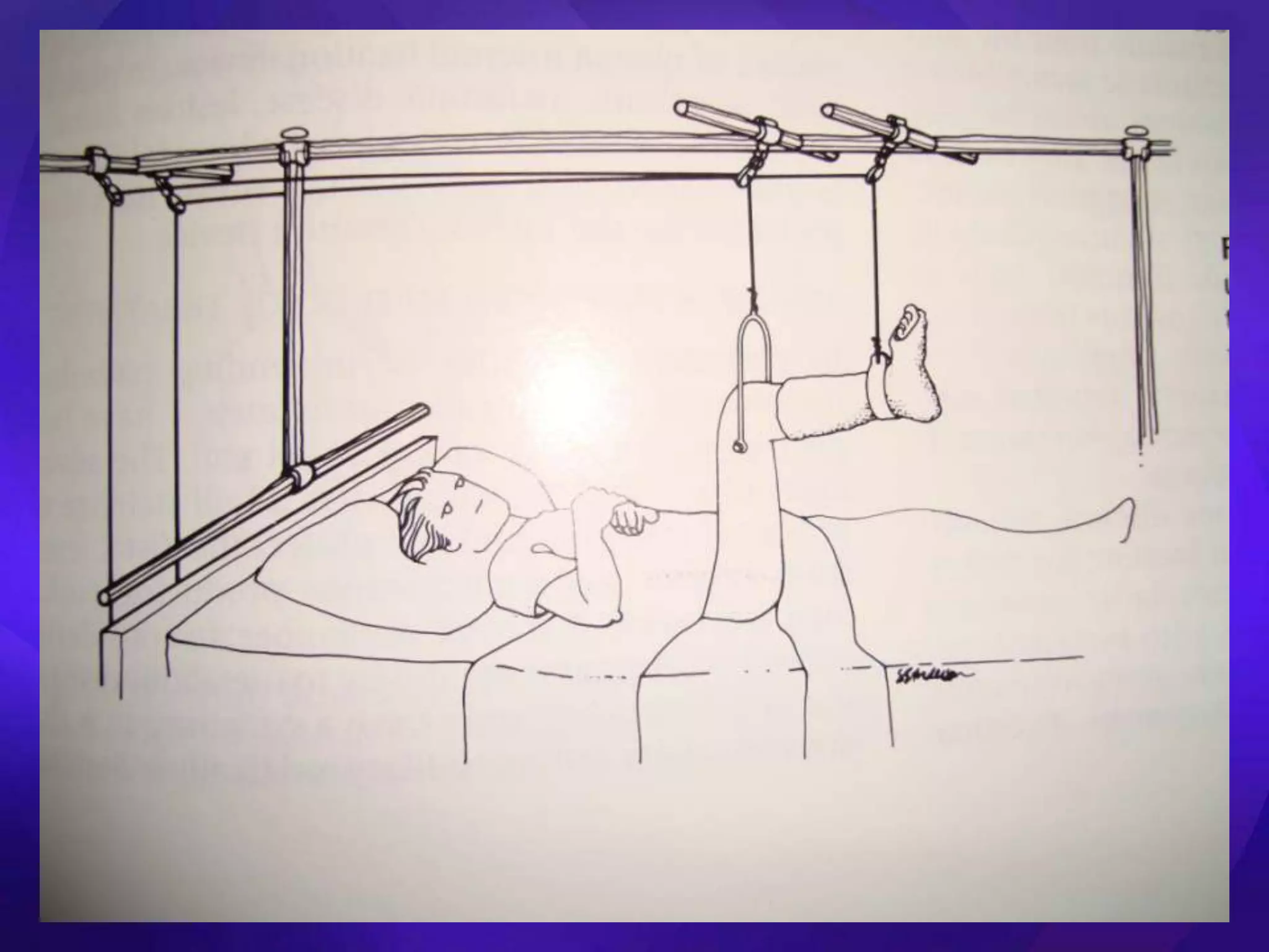
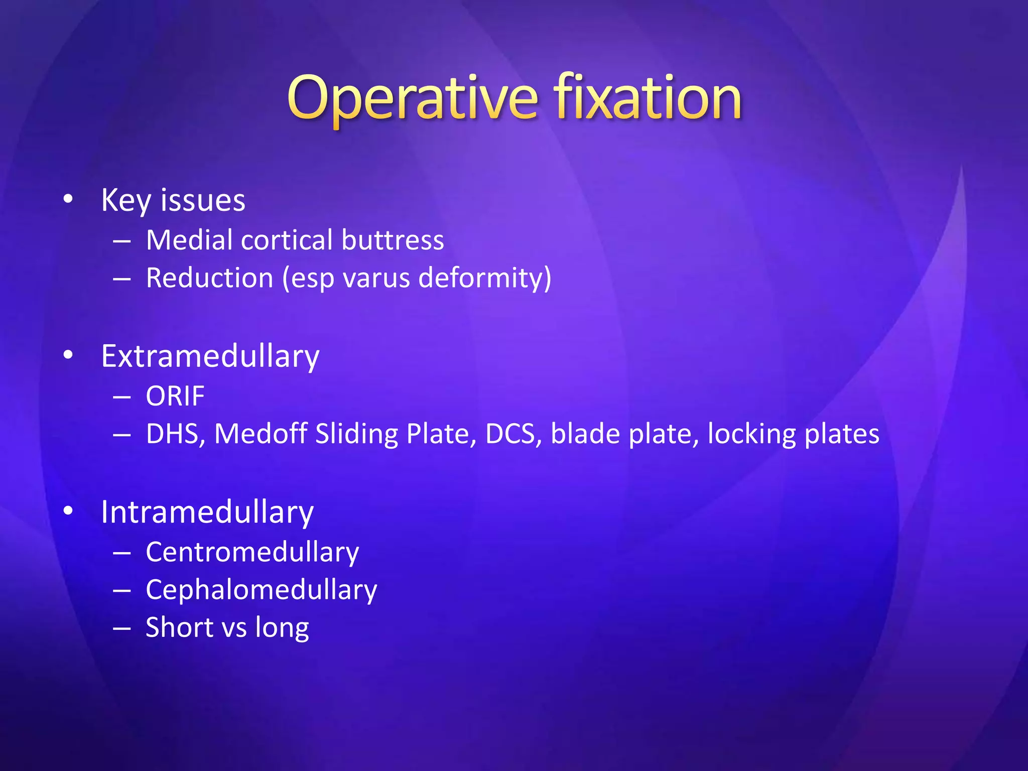
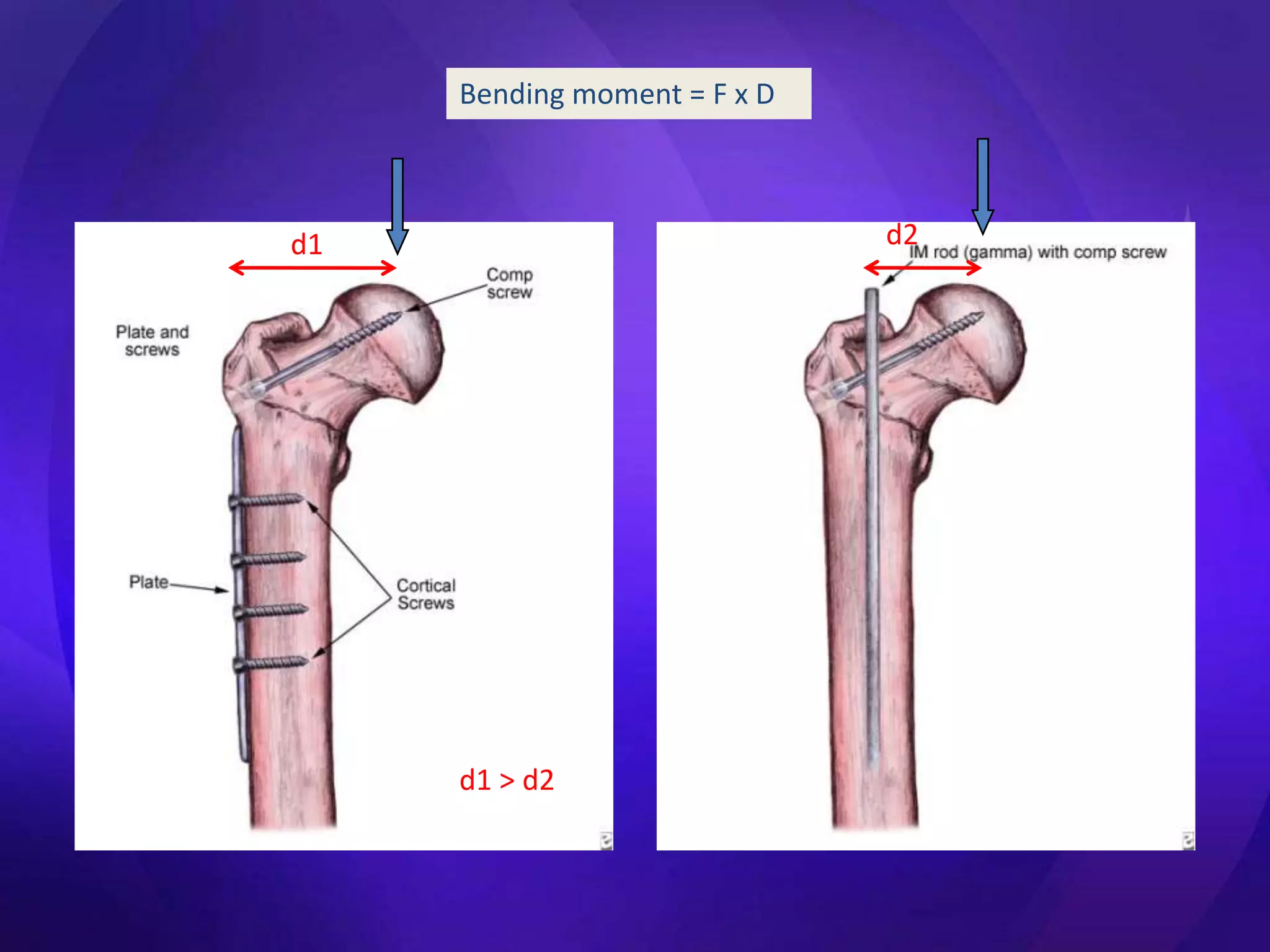
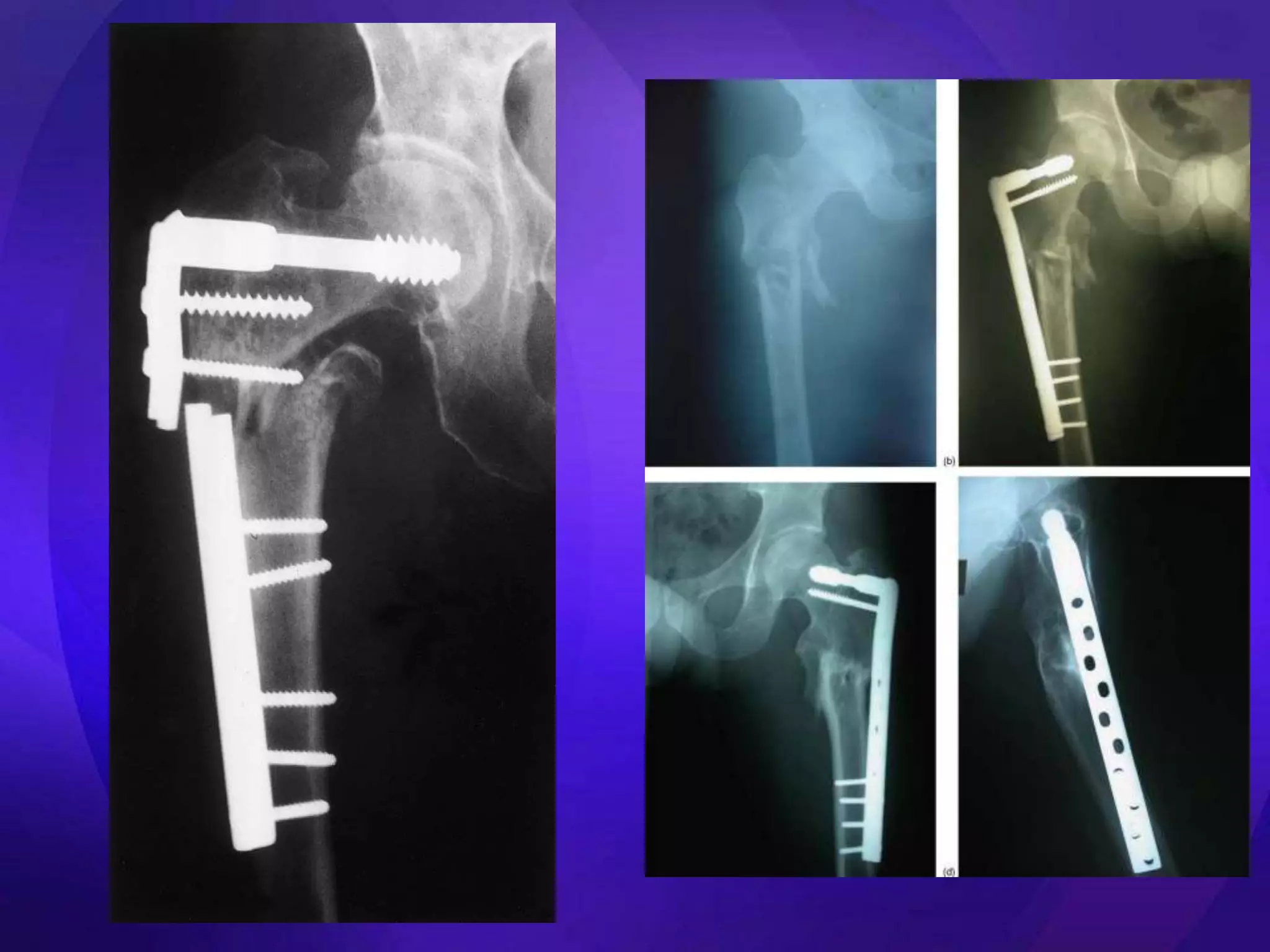
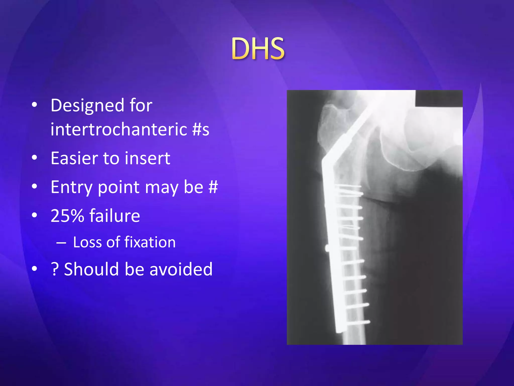
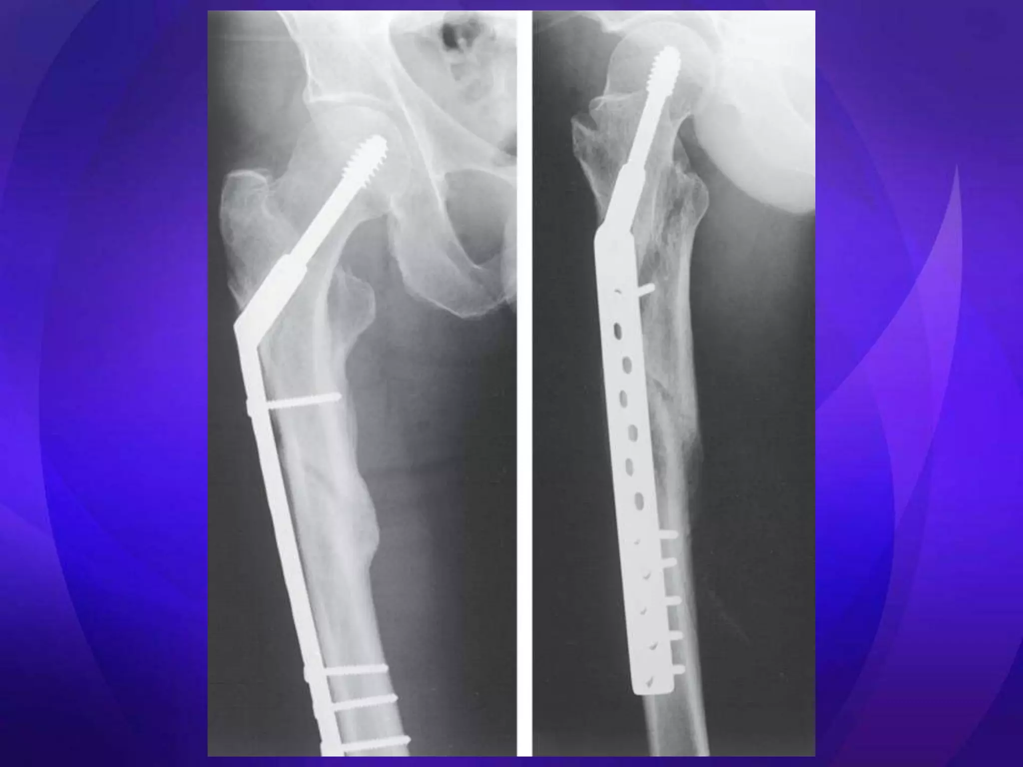
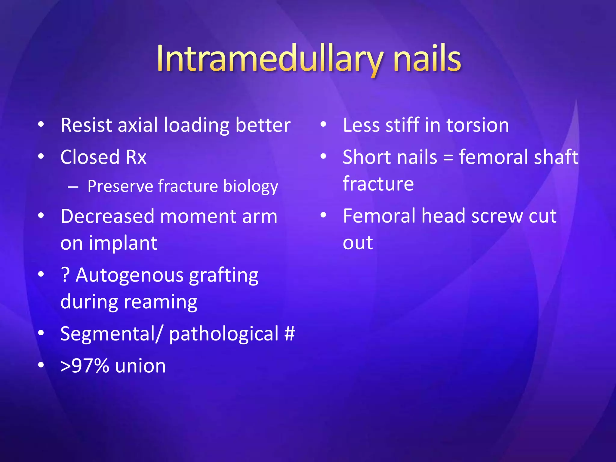
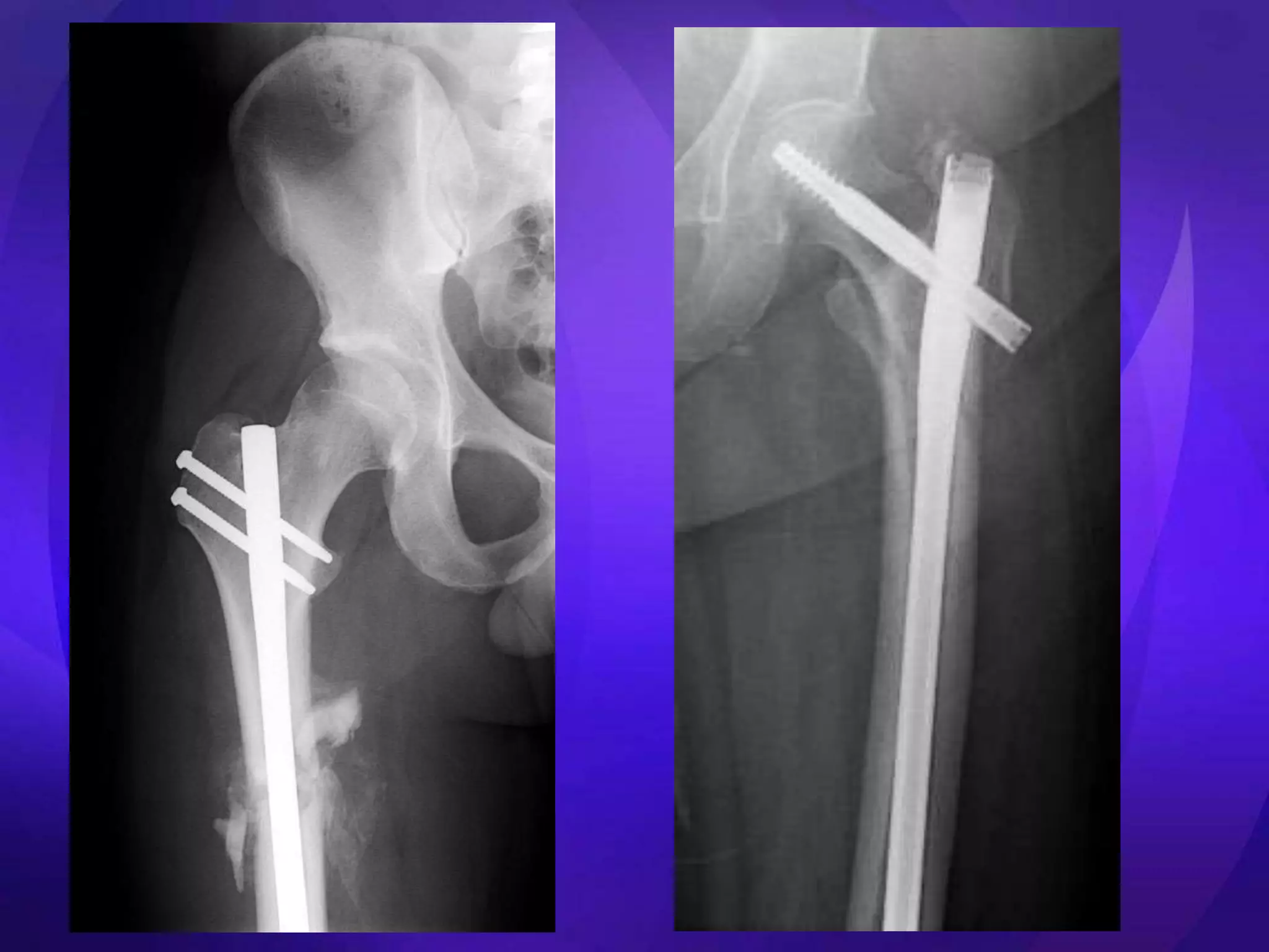
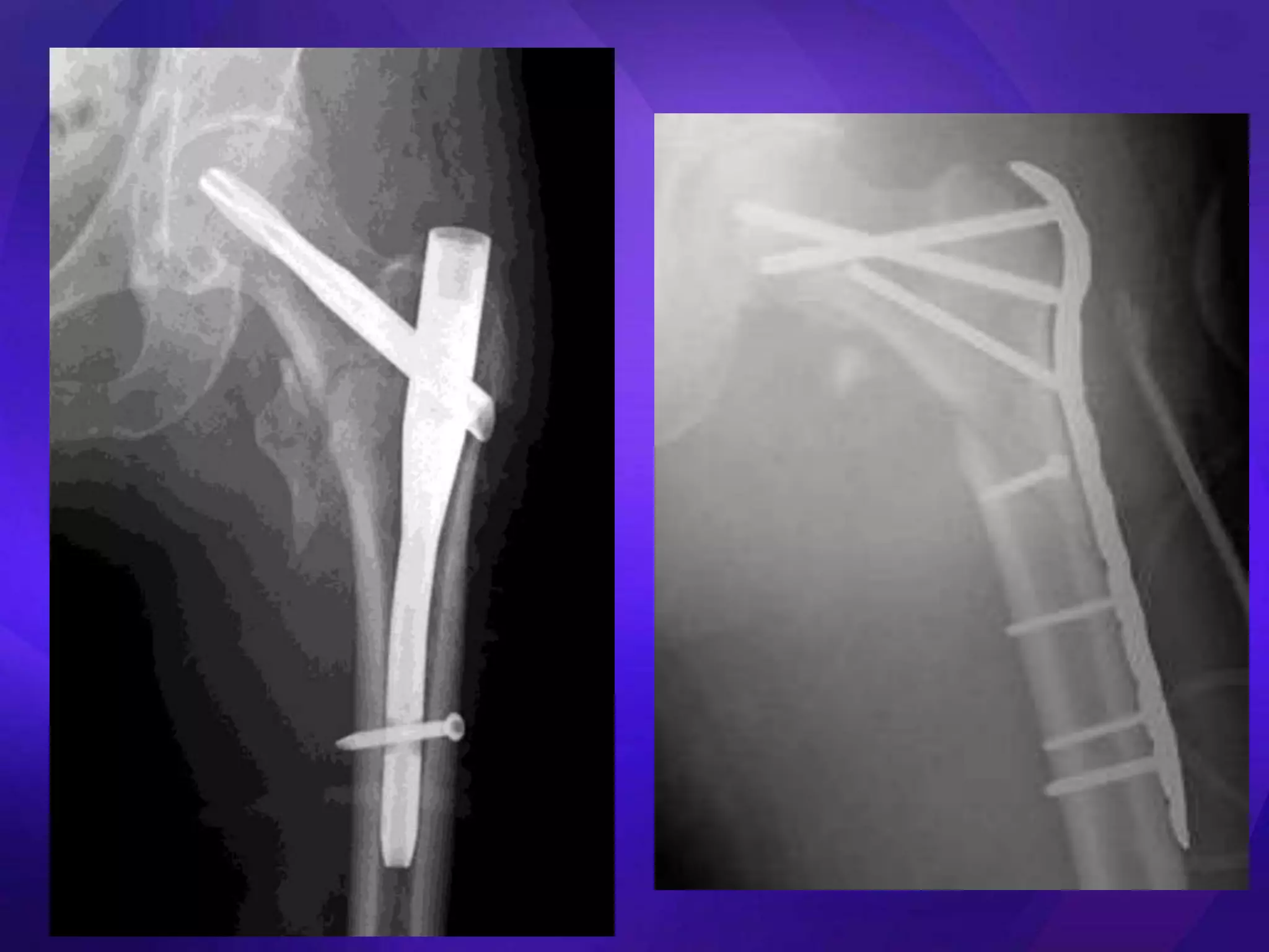
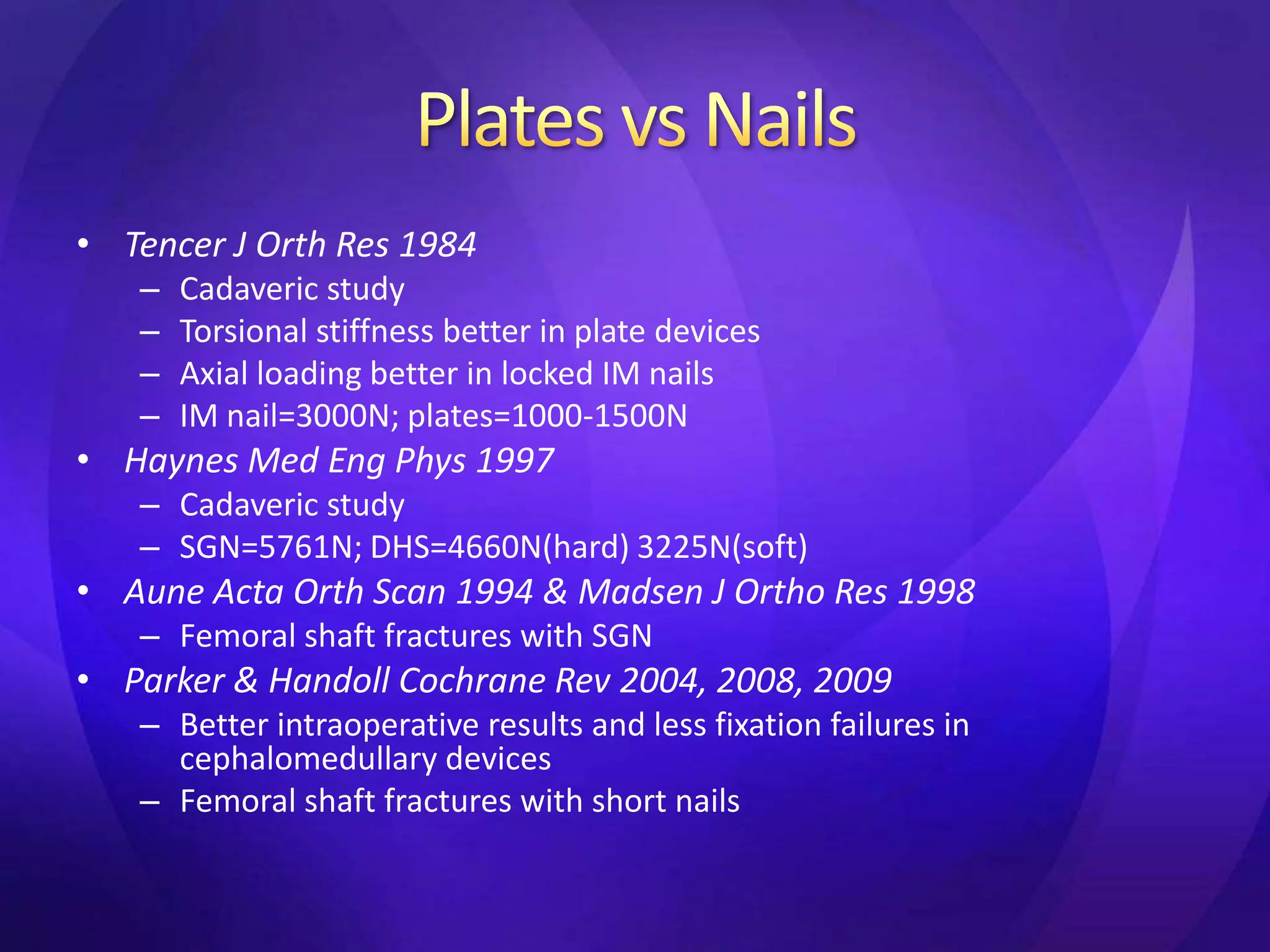
2. It reviews treatment options for subtrochanteric fractures including traction, extramedullary fixation with plates, and intramedullary fixation with nails. Intramedullary nails are preferred as they better resist axial loads and torsion compared to plates.
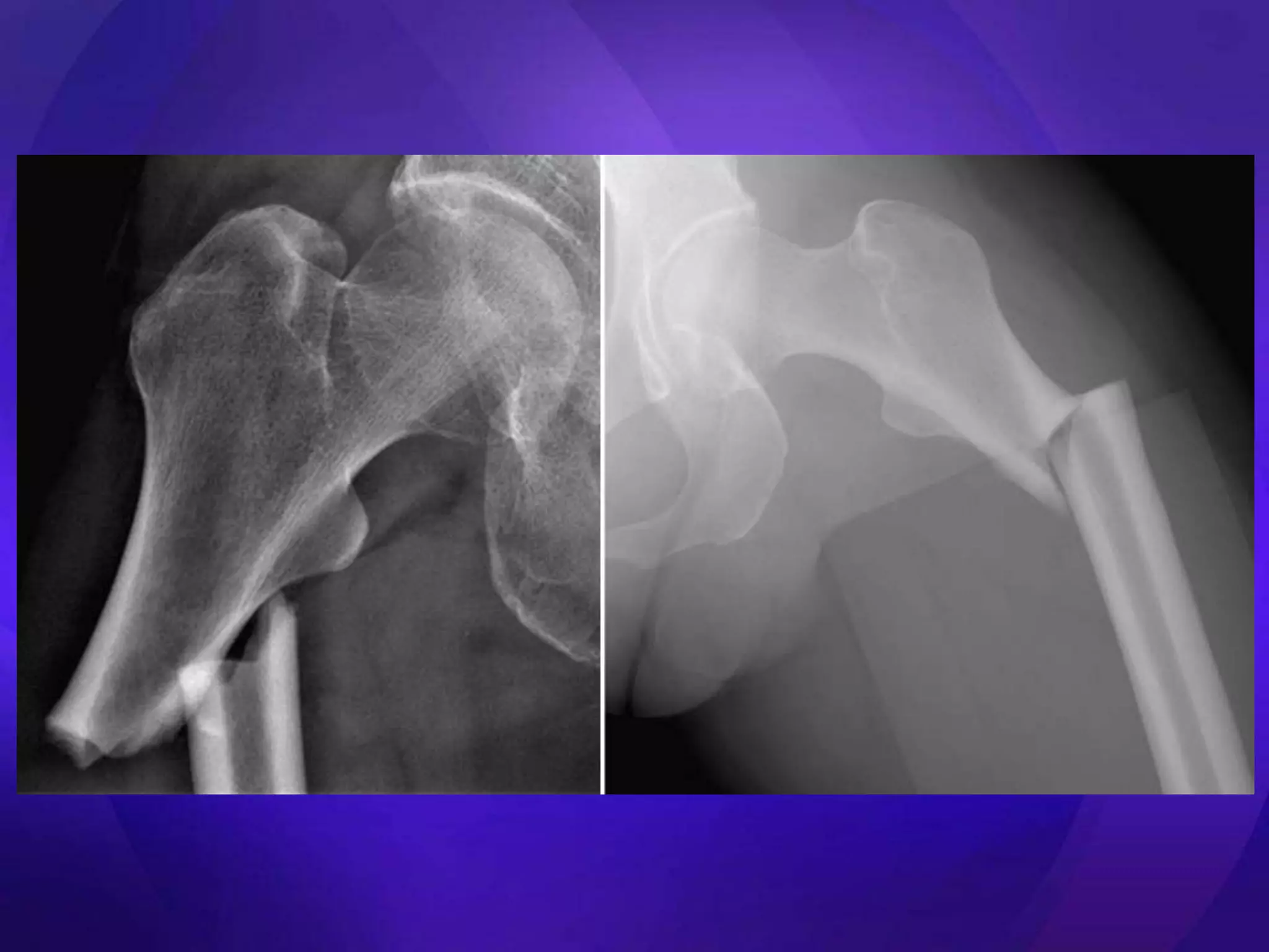
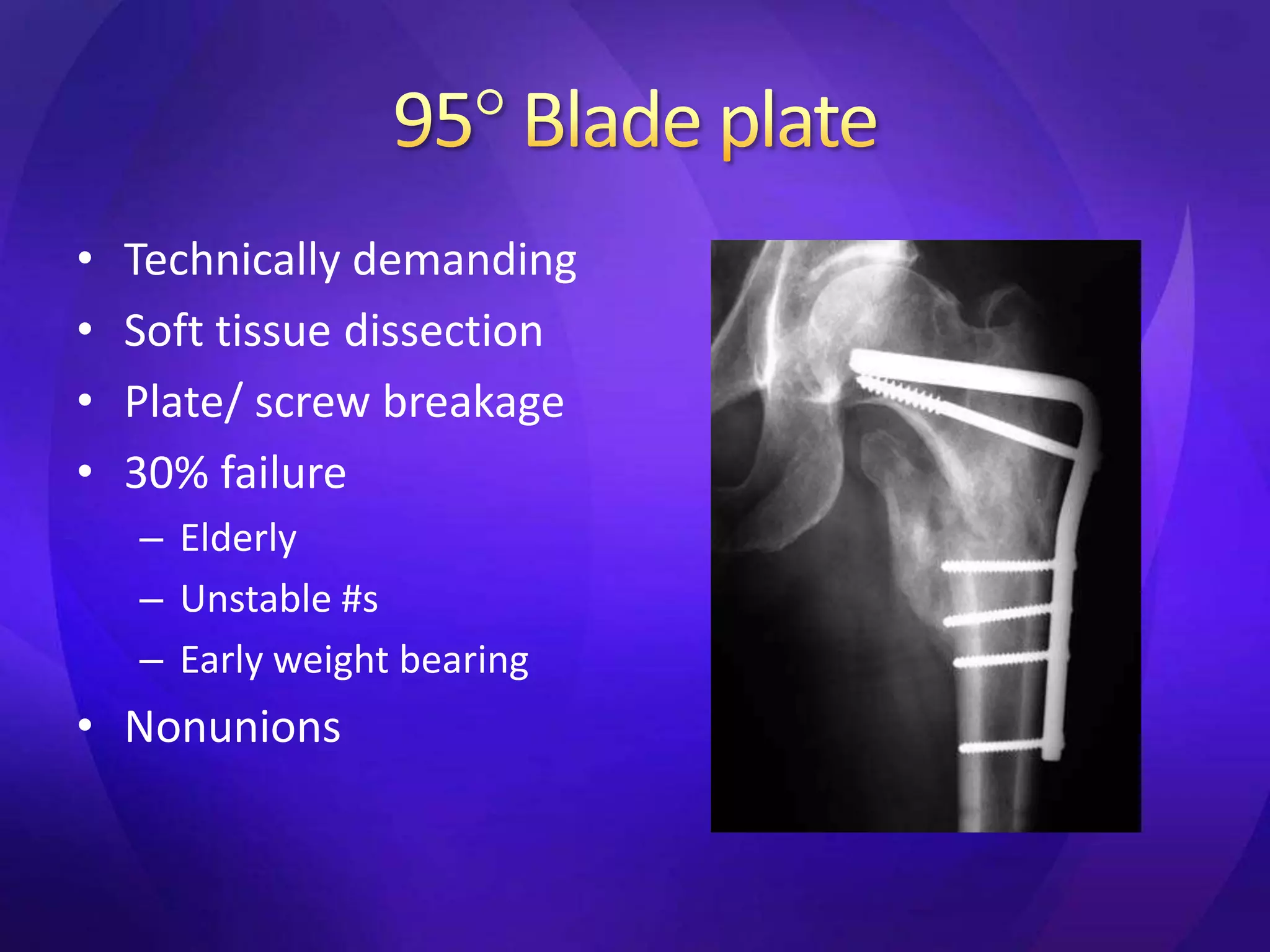
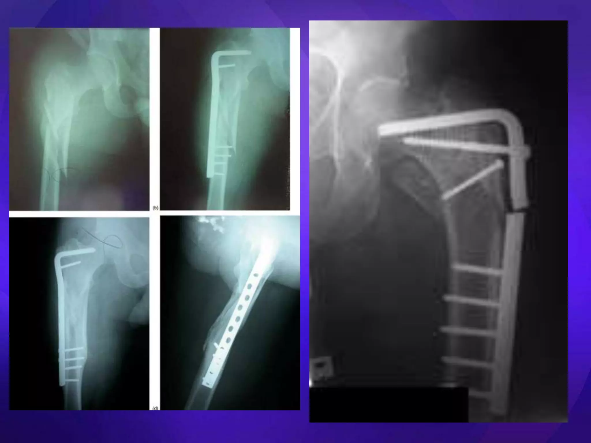
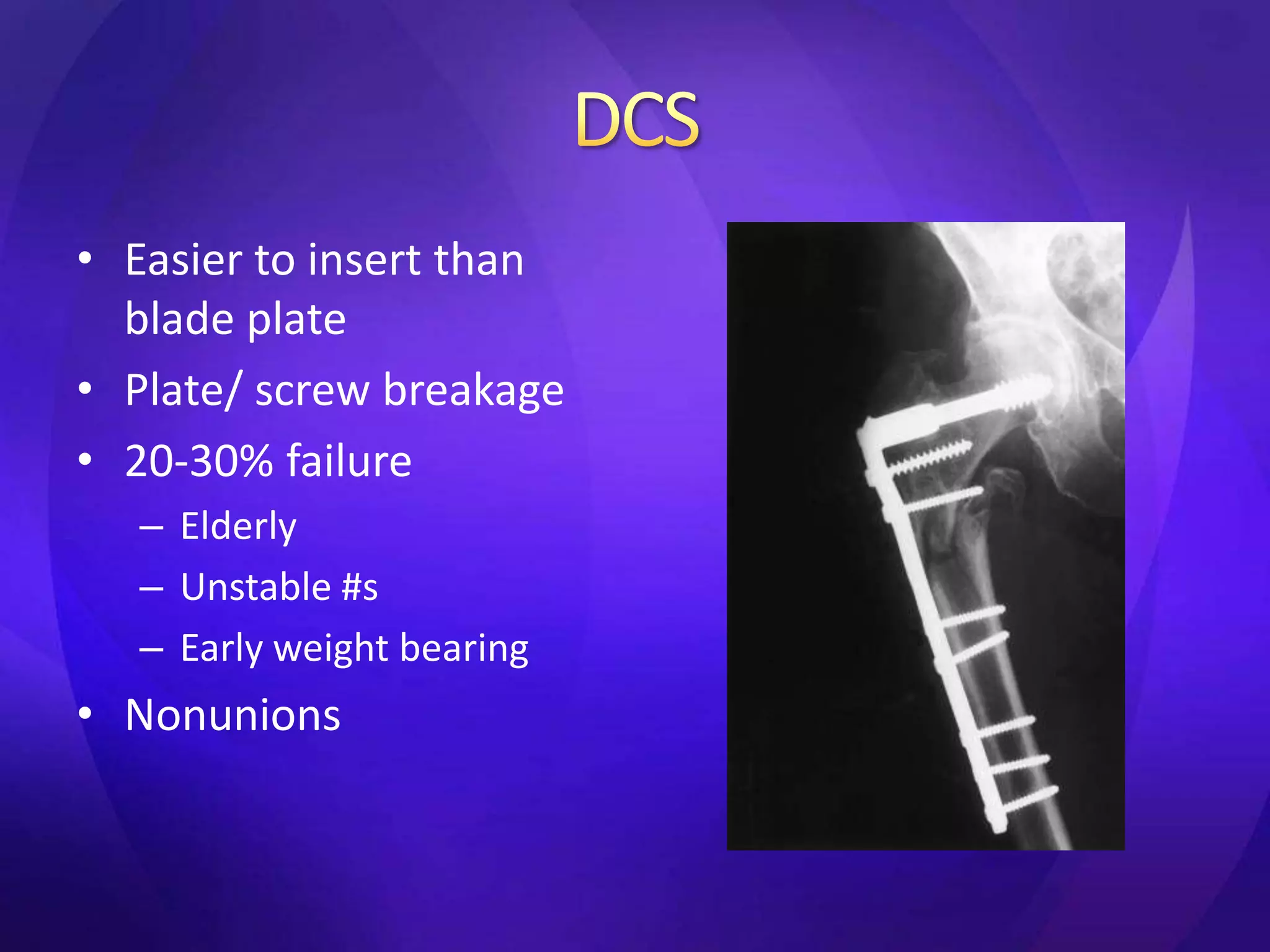
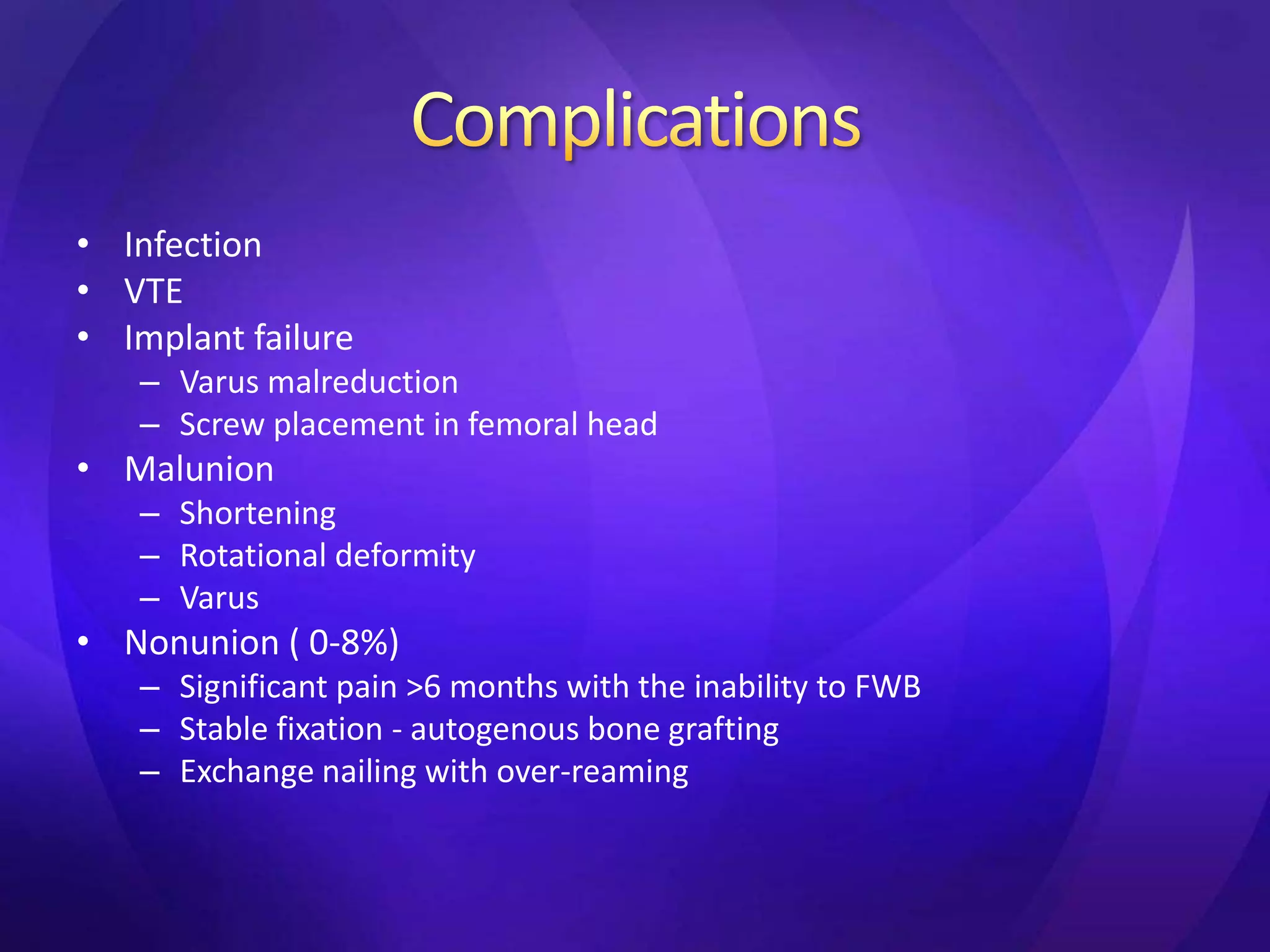
3. Complications of treatment include infection, malunion, nonunion, and implant failure. Proper reduction and fixation are important to
Arthroscopic ACL Reconstruction By Dr Shekhar ShrivastavDelhiArthroscopy
Arthroscopic Acl Reconstruction By Dr Shekhar Shrivastav.
HOW NORMAL KNEE WORKS ?
The knee is the largest joint in the body, and one of the most easily injured. It is made up of the lower end of the thigh bone(femur), the upper end of the shin bone (tibia), and the knee cap (patella), which slides in a groove on the end of the femur. Four bands of tissue, the anterior and posterior cruciate ligaments, and the medial and lateral collateral ligaments connect the femur and the tibia and provide joint stability. The surfaces where the femur, tibia and patella touch are covered with articular cartilage, a smooth substance that cushions the bones and enables them to glide freely. Semicircular rings of tough fibrous-cartilage tissue called the lateral and medial menisci act as shock absorbers and stabilizers.
WHAT IS THE ROLE OF ACL ?
ACL along with other ligaments of the knee joint and meniscus provides stability to the knee joint.
WHAT IS LIGAMENT RECONSTRUCTION ( ACL ) ?
Ligament reconstruction involves replacing the torn ligament with a tendon (graft) from your knee and fixing the graft in place with screws. This procedure is performed with the use of the arthroscope. The anterior cruciate ligament (ACL) is the most common ligament requiring reconstruction procedures. The torn ligament is excised arthroscopically and new ligament is prepared by ligament grafts taken from your own body. Bony tunnels are prepared in femur and tibia using specialized instruments through which the new ligament is passed and fixed with special screws. This procedure requires relative rest or leave from your work or studies for about 2-3 weeks after which you will be allowed normal day to day activities.
WHEN CAN THE PATIENT BE AMBULATED AFTER SURGERY ?
The patient can walk from the same evening of the surgery. Initially the patient is advised to walk with a brace and a walking cane. Strengthening and range of motion exercises for the knee are started from the next day. The patient is discharged from the hospital 2nd or 3rd day after surgery. The patient can walk without support by 10-14 days depending on muscle strengthening. Slow Jogging and other strenuous activities are permitted after 3 months and the patient can return to active sports only 8-9 months after surgery.
Torn ACL Reconstructed ACL
For Further Queries contact your Orthopedic Surgeon at
+ 91 9971192233
Fracture of shaft and distal part of Femoral bone by Dr. Ammar AlsabaeAmmar Alsbae
This ppt show the fracture of shaft and distal part ( condylar and supracondylar ) of femuarl bone which include anatomy , classification , clinical picture , diagnosis , treatment and complications .
This PPT prepared by Ammar Alsabae , A medical student , faculity of medicine , Taiz university . Yemen .
Objectives:
-Recognize the anatomy of the proximal tibia
-Describe initial evaluation and management
-Identify common fracture patterns
-Apply treatment principles and strategies for Partial articular fractures and Complete articular fractures
-Discuss rehabilitation and complications
-Learn Management in selected tibial plateau case scenarios
Pulmonary Thromboembolism - etilogy, types, medical- Surgical and nursing man...VarunMahajani
Disruption of blood supply to lung alveoli due to blockage of one or more pulmonary blood vessels is called as Pulmonary thromboembolism. In this presentation we will discuss its causes, types and its management in depth.
Title: Sense of Smell
Presenter: Dr. Faiza, Assistant Professor of Physiology
Qualifications:
MBBS (Best Graduate, AIMC Lahore)
FCPS Physiology
ICMT, CHPE, DHPE (STMU)
MPH (GC University, Faisalabad)
MBA (Virtual University of Pakistan)
Learning Objectives:
Describe the primary categories of smells and the concept of odor blindness.
Explain the structure and location of the olfactory membrane and mucosa, including the types and roles of cells involved in olfaction.
Describe the pathway and mechanisms of olfactory signal transmission from the olfactory receptors to the brain.
Illustrate the biochemical cascade triggered by odorant binding to olfactory receptors, including the role of G-proteins and second messengers in generating an action potential.
Identify different types of olfactory disorders such as anosmia, hyposmia, hyperosmia, and dysosmia, including their potential causes.
Key Topics:
Olfactory Genes:
3% of the human genome accounts for olfactory genes.
400 genes for odorant receptors.
Olfactory Membrane:
Located in the superior part of the nasal cavity.
Medially: Folds downward along the superior septum.
Laterally: Folds over the superior turbinate and upper surface of the middle turbinate.
Total surface area: 5-10 square centimeters.
Olfactory Mucosa:
Olfactory Cells: Bipolar nerve cells derived from the CNS (100 million), with 4-25 olfactory cilia per cell.
Sustentacular Cells: Produce mucus and maintain ionic and molecular environment.
Basal Cells: Replace worn-out olfactory cells with an average lifespan of 1-2 months.
Bowman’s Gland: Secretes mucus.
Stimulation of Olfactory Cells:
Odorant dissolves in mucus and attaches to receptors on olfactory cilia.
Involves a cascade effect through G-proteins and second messengers, leading to depolarization and action potential generation in the olfactory nerve.
Quality of a Good Odorant:
Small (3-20 Carbon atoms), volatile, water-soluble, and lipid-soluble.
Facilitated by odorant-binding proteins in mucus.
Membrane Potential and Action Potential:
Resting membrane potential: -55mV.
Action potential frequency in the olfactory nerve increases with odorant strength.
Adaptation Towards the Sense of Smell:
Rapid adaptation within the first second, with further slow adaptation.
Psychological adaptation greater than receptor adaptation, involving feedback inhibition from the central nervous system.
Primary Sensations of Smell:
Camphoraceous, Musky, Floral, Pepperminty, Ethereal, Pungent, Putrid.
Odor Detection Threshold:
Examples: Hydrogen sulfide (0.0005 ppm), Methyl-mercaptan (0.002 ppm).
Some toxic substances are odorless at lethal concentrations.
Characteristics of Smell:
Odor blindness for single substances due to lack of appropriate receptor protein.
Behavioral and emotional influences of smell.
Transmission of Olfactory Signals:
From olfactory cells to glomeruli in the olfactory bulb, involving lateral inhibition.
Primitive, less old, and new olfactory systems with different path
The prostate is an exocrine gland of the male mammalian reproductive system
It is a walnut-sized gland that forms part of the male reproductive system and is located in front of the rectum and just below the urinary bladder
Function is to store and secrete a clear, slightly alkaline fluid that constitutes 10-30% of the volume of the seminal fluid that along with the spermatozoa, constitutes semen
A healthy human prostate measures (4cm-vertical, by 3cm-horizontal, 2cm ant-post ).
It surrounds the urethra just below the urinary bladder. It has anterior, median, posterior and two lateral lobes
It’s work is regulated by androgens which are responsible for male sex characteristics
Generalised disease of the prostate due to hormonal derangement which leads to non malignant enlargement of the gland (increase in the number of epithelial cells and stromal tissue)to cause compression of the urethra leading to symptoms (LUTS
TEST BANK for Operations Management, 14th Edition by William J. Stevenson, Ve...kevinkariuki227
TEST BANK for Operations Management, 14th Edition by William J. Stevenson, Verified Chapters 1 - 19, Complete Newest Version.pdf
These simplified slides by Dr. Sidra Arshad present an overview of the non-respiratory functions of the respiratory tract.
Learning objectives:
1. Enlist the non-respiratory functions of the respiratory tract
2. Briefly explain how these functions are carried out
3. Discuss the significance of dead space
4. Differentiate between minute ventilation and alveolar ventilation
5. Describe the cough and sneeze reflexes
Study Resources:
1. Chapter 39, Guyton and Hall Textbook of Medical Physiology, 14th edition
2. Chapter 34, Ganong’s Review of Medical Physiology, 26th edition
3. Chapter 17, Human Physiology by Lauralee Sherwood, 9th edition
4. Non-respiratory functions of the lungs https://academic.oup.com/bjaed/article/13/3/98/278874
Prix Galien International 2024 Forum ProgramLevi Shapiro
June 20, 2024, Prix Galien International and Jerusalem Ethics Forum in ROME. Detailed agenda including panels:
- ADVANCES IN CARDIOLOGY: A NEW PARADIGM IS COMING
- WOMEN’S HEALTH: FERTILITY PRESERVATION
- WHAT’S NEW IN THE TREATMENT OF INFECTIOUS,
ONCOLOGICAL AND INFLAMMATORY SKIN DISEASES?
- ARTIFICIAL INTELLIGENCE AND ETHICS
- GENE THERAPY
- BEYOND BORDERS: GLOBAL INITIATIVES FOR DEMOCRATIZING LIFE SCIENCE TECHNOLOGIES AND PROMOTING ACCESS TO HEALTHCARE
- ETHICAL CHALLENGES IN LIFE SCIENCES
- Prix Galien International Awards Ceremony
Ethanol (CH3CH2OH), or beverage alcohol, is a two-carbon alcohol
that is rapidly distributed in the body and brain. Ethanol alters many
neurochemical systems and has rewarding and addictive properties. It
is the oldest recreational drug and likely contributes to more morbidity,
mortality, and public health costs than all illicit drugs combined. The
5th edition of the Diagnostic and Statistical Manual of Mental Disorders
(DSM-5) integrates alcohol abuse and alcohol dependence into a single
disorder called alcohol use disorder (AUD), with mild, moderate,
and severe subclassifications (American Psychiatric Association, 2013).
In the DSM-5, all types of substance abuse and dependence have been
combined into a single substance use disorder (SUD) on a continuum
from mild to severe. A diagnosis of AUD requires that at least two of
the 11 DSM-5 behaviors be present within a 12-month period (mild
AUD: 2–3 criteria; moderate AUD: 4–5 criteria; severe AUD: 6–11 criteria).
The four main behavioral effects of AUD are impaired control over
drinking, negative social consequences, risky use, and altered physiological
effects (tolerance, withdrawal). This chapter presents an overview
of the prevalence and harmful consequences of AUD in the U.S.,
the systemic nature of the disease, neurocircuitry and stages of AUD,
comorbidities, fetal alcohol spectrum disorders, genetic risk factors, and
pharmacotherapies for AUD.
Couples presenting to the infertility clinic- Do they really have infertility...Sujoy Dasgupta
Dr Sujoy Dasgupta presented the study on "Couples presenting to the infertility clinic- Do they really have infertility? – The unexplored stories of non-consummation" in the 13th Congress of the Asia Pacific Initiative on Reproduction (ASPIRE 2024) at Manila on 24 May, 2024.
ARTIFICIAL INTELLIGENCE IN HEALTHCARE.pdfAnujkumaranit
Artificial intelligence (AI) refers to the simulation of human intelligence processes by machines, especially computer systems. It encompasses tasks such as learning, reasoning, problem-solving, perception, and language understanding. AI technologies are revolutionizing various fields, from healthcare to finance, by enabling machines to perform tasks that typically require human intelligence.
Report Back from SGO 2024: What’s the Latest in Cervical Cancer?bkling
Are you curious about what’s new in cervical cancer research or unsure what the findings mean? Join Dr. Emily Ko, a gynecologic oncologist at Penn Medicine, to learn about the latest updates from the Society of Gynecologic Oncology (SGO) 2024 Annual Meeting on Women’s Cancer. Dr. Ko will discuss what the research presented at the conference means for you and answer your questions about the new developments.
These lecture slides, by Dr Sidra Arshad, offer a quick overview of physiological basis of a normal electrocardiogram.
Learning objectives:
1. Define an electrocardiogram (ECG) and electrocardiography
2. Describe how dipoles generated by the heart produce the waveforms of the ECG
3. Describe the components of a normal electrocardiogram of a typical bipolar leads (limb II)
4. Differentiate between intervals and segments
5. Enlist some common indications for obtaining an ECG
Study Resources:
1. Chapter 11, Guyton and Hall Textbook of Medical Physiology, 14th edition
2. Chapter 9, Human Physiology - From Cells to Systems, Lauralee Sherwood, 9th edition
3. Chapter 29, Ganong’s Review of Medical Physiology, 26th edition
4. Electrocardiogram, StatPearls - https://www.ncbi.nlm.nih.gov/books/NBK549803/
5. ECG in Medical Practice by ABM Abdullah, 4th edition
6. ECG Basics, http://www.nataliescasebook.com/tag/e-c-g-basics
Anti ulcer drugs and their Advance pharmacology ||
Anti-ulcer drugs are medications used to prevent and treat ulcers in the stomach and upper part of the small intestine (duodenal ulcers). These ulcers are often caused by an imbalance between stomach acid and the mucosal lining, which protects the stomach lining.
||Scope: Overview of various classes of anti-ulcer drugs, their mechanisms of action, indications, side effects, and clinical considerations.
2. • 10 -30% of hip fractures
• Bimodal age distribution
– 20-40yrs
• HIGH ENERGY
• RTA, fall from height, GSW (10%)
– >60yrs
• low energy
• Simple fall, osteoporotic, pathological (17-35%)
• Cann. screws below lesser troch
• Bisphosphonate Rx
3. • Screws must not enter lateral femoral cortex
below level of lesser troch
– Kloen et al JOT 2003
• Triangular configurations
– Highest load to failure
– Apex superior – higher risk of subtroch #
– Selvan et al Injury 2004, Lichtblau et al Bull NYU
Hosp Jt Dis 2008
11. 1. <2mm displacement
2. 2-parts
A. Transverse
B. Spiral. Less troch prox
C. Spiral. Less troch distal
3. 3-parts
A. Less troch butterfly
B. Lateral butterfly
4. 4-parts or comminuted
5. Greater troch extension
12. • History
• Pain, NWB, deformity of thigh
• Open/ closed injury
• Neurovascular status
• Full ATLS
– Haemorrhagic shock
– Cranial, thoracic, abdominal injuries (Waddell’s
triad)
13. • Analgesia, femoral nerve block???
– Significantly reduced pain scores and use of opiates
– AK Fletcher et al. Ann Emerg Med. 2003;41:227-233
• Thomas splint
• X-rays
– AP pelvis
– full length femur views
– Contralateral femur (bisphosphonate Rx)
• Pathological
– Full assessment
– Bloods – myeloma screen, bone profile, LFTs
– Bone scan, MRI
14. • Unfit for surgery, open #s
• Skeletal traction
– Hamilton-Russell
– Perkins
• 90-90 position
• Aim for
– <5 varus/valgus
– >25% cortical apposition
– <1cm shortening
– No axial rotation
• Weekly x-rays + adjustments
• Traction until signs of union (8-12 weeks)
• Problems
– Pressure ulcers, VTE, pneumonia
21. • Easier to insert than
blade plate
• Plate/ screw breakage
• 20-30% failure
– Elderly
– Unstable #s
– Early weight bearing
• Nonunions
22.
23. • Designed for
intertrochanteric #s
• Easier to insert
• Entry point may be #
• 25% failure
– Loss of fixation
• ? Should be avoided
24.
25. • Resist axial loading better • Less stiff in torsion
• Closed Rx • Short nails = femoral shaft
– Preserve fracture biology fracture
• Decreased moment arm • Femoral head screw cut
on implant out
• ? Autogenous grafting
during reaming
• Segmental/ pathological #
• >97% union
26.
27.
28. • Tencer J Orth Res 1984
– Cadaveric study
– Torsional stiffness better in plate devices
– Axial loading better in locked IM nails
– IM nail=3000N; plates=1000-1500N
• Haynes Med Eng Phys 1997
– Cadaveric study
– SGN=5761N; DHS=4660N(hard) 3225N(soft)
• Aune Acta Orth Scan 1994 & Madsen J Ortho Res 1998
– Femoral shaft fractures with SGN
• Parker & Handoll Cochrane Rev 2004, 2008, 2009
– Better intraoperative results and less fixation failures in
cephalomedullary devices
– Femoral shaft fractures with short nails
29. • Infection
• VTE
• Implant failure
– Varus malreduction
– Screw placement in femoral head
• Malunion
– Shortening
– Rotational deformity
– Varus
• Nonunion ( 0-8%)
– Significant pain >6 months with the inability to FWB
– Stable fixation - autogenous bone grafting
– Exchange nailing with over-reaming
30. • Fletcher AK et al. Three-in-One Femoral Nerve
Block as Analgesia for Fractured Neck of
Femur in the Emergency Department: A
Randomized, Controlled Trial. Ann Emerg
Med. 2003;41:227-233
• Craig N et al. Subtrochanteric fractures. Bull
Hosp Joint Dis 2001;60:35-46
• Lundy DW. Subtrochanteric femoral fractures.
AAOS 2007;15:663-671
31. 1. Kloen P et al. Subtrochanteric fracture after
cannulated screw fixation of femoral neck
fractures: a report of four cases. J Orthop
Trauma. 2003;17(3):225-9
2. Lichtblau S et al. A biomechanical
comparison of two patterns of screw
insertion. Bull NYU Hosp Jt Dis.
2008;66(4):269-71
32. 1. Goh et al. Subtrochanteric insufficiency fractures in
patients on alendronate therapy. JBJS 89-B(3); 349-
353
2. Lenart et al. Atypical Fractures of the Femoral
Diaphysis in Postmenopausal Women Taking
Alendronate. NEJM 2008 358;12:1304-6
3. Neviaser et al. Low-Energy Femoral Shaft Fractures
Associated With Alendronate Use. J Orth Trauma
2008; 22(5): 346-350
4. Kweka et al. An emerging pattern of subtrochanteric
stress fractures: A long-term complication of
alendronate therapy. Injury 2008 39; 224—231