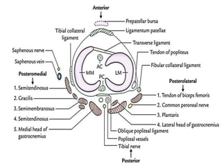
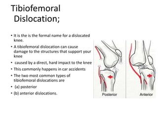
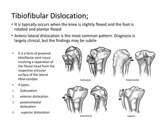
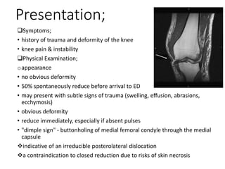
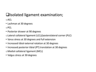
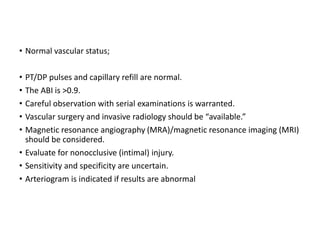
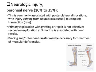
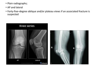
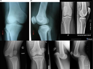
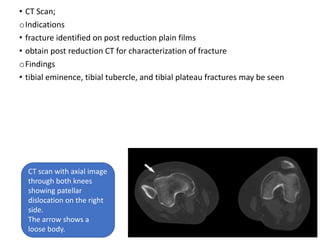
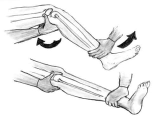
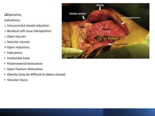
Knee Dislocation
Presenter:- Dr JISHITH REDDY
Introduction;
• Knee dislocations are high energy traumatic injuries characterized by a high
rate of neurovascular injury.
• Traumatic knee dislocation is an uncommon injury that may be limb
threatening; it should therefore be treated as an orthopedic emergency.
• Most knee dislocations are the result of high-energy injuries, such as motor
vehicle or industrial accidents.
• They can also occur with low-energy injuries, such as those that occur in
sports.
• Anterior dislocation is the most common type, resulting from severe
hyperextension.
• the knee may experience up to five times body weight per step.
• Significant soft tissue injury is necessary for knee dislocation, including
ruptures of at least three of four major ligamentous structures of the knee
• This rare injury results from severe violence to the knee so that all of its
supporting ligaments are torn.
• It is a major damage to the joint, and is often associated with injury to the
popliteal artery.
• Joint Laxity - it is presumed that excessive passive motion in the knee joint
automatically leads to instability during dynamic and functional activities.
• Joint Instability - it is the sensation of the knee twisting or moving from side
to side when doing basic activities.
• Dislocation - is defined as complete displacement of the tibia with respect
to the femur with disruption of three or more of the stabilizing
ligaments. Small avulsion fractures from the ligaments and capsular
insertions may be present.
• Subluxation - means that the kneecap has briefly slid out of its normal place
in that groove
Anatomy;
Range of motion;
•It is from 0 degrees of extension to
140 degrees of flexion
•8 to 12 degrees of rotation through
the flexion–extension arc
Osteology;
The ginglymoid (hinge joint) consists
of three articulations;
1. Patellofemoral
2. Tibiofemoral
3. Tibiofibular
• Ligaments;
• Cruciate Ligaments
1. Anterior Cruciate ligament
2. Posterior Cruciate ligament
• Collateral Ligaments
1. Lateral collateral ligaments
2. Medial collateral ligaments
• Meniscal Ligaments
1. Medial meniscus
2. Lateral meniscus
• Ligamentum Patellae
• Stability of the Knee Joint;
1. By strength and actions of the surrounding muscles and
tendons
2. Medial and lateral collateral ligaments maintain side-to-side
stability.
3. Cruciate ligaments maintain anteroposterior stability.
4. Iliotibial tract helps in stabilizing a partly flexed knee.
• Blood Supply;
1. Genicular branches of popliteal artery
2. Descending genicular branch of femoral artery
3. Descending branch of lateral circumflex femoral artery
4. Recurrent branches of anterior tibial artery
5. Circumflex fibular of posterior tibial artery
• Movements;
• Main/active movements;
1. Flexion
2. extension
• Conjunct movements;
1. Medial rotation
2. Lateral rotation
•Relations of the Knee Joint-
Anteriorly -Tendon of quadriceps femoris, patella, ligamentum patellae, patellar
plexus of the nerves, and prepatellar synovial bursa.
• Anteromedially - Medial patellar retinaculum
• Anterolaterally -Lateral patellar retinaculum, iliotibial tract
• Posteriorly- Popliteal vessels, tibial nerve, oblique popliteal ligament
• Postero-laterally- Biceps femoris tendon, common peroneal nerve, lateral
head of gastrocnemius, plantaris
• Postero-medially- Sartorius, gracilis, semimembranosus, semitendinosus,
medial head of gastrocnemius, popliteus
Mechanism of injury;
• High energy;
A motor vehicle accident with a “dashboard” injury involves axial loading to the tibia
of a flexed knee
• Low energy;
This includes athletic injuries and falls in an obese patient
• Hyperextension with or without varus/valgus leads to anterior dislocation.
• Flexion plus posterior force leads to posterior dislocation
• Rotary knee dislocation are caused by rotary forces.
• Most frequently (93% of the time), the kneecap pops out laterally, to the side of the
groove. Patellar dislocation is usually an acute injury caused by impact or by a sudden
turn and twist.
• Posterolateral dislocation is caused when the leg is in valgus stress with internal tibial
rotation
TYPES OF DISLOCATIONS
• PATELLOFEMORAL DISLOCATION
• TIBIOFEMORAL DISLOCATION
• TIBIOFIBULAR DISLOCATION
Patellofemoral Dislocation;
• Occurs when the foot is planted and a rapid change of direction or
twisting occurs.
• Usually a pre-existence ligamentous laxity is required to allow a
dislocation to occur in this manner
• Direct blows to a knee can cause dislocations as well.
Tibiofemoral
Dislocation;
• It is the is the formal name for a dislocated
knee.
• A tibiofemoral dislocation can cause
damage to the structures that support your
knee
• caused by a direct, hard impact to the knee
• This commonly happens in car accidents
• The two most common types of
tibiofemoral dislocations are
• (a) posterior
• (b) anterior dislocations.
Tibiofibular Dislocation;
• It is typically occurs when the knee is slightly flexed and the foot is
rotated and plantar flexed
• Antero lateral dislocation is the most common pattern. Diagnosis is
largely clinical, but the findings may be subtle
 It is a form of proximal
tibiofibular joint injury
involving a separation of
the fibular head from the
respective articular
surface of the lateral
tibial condyle.
 4 types;
1. Subluxation
2. anterior dislocation
3. posteromedial
dislocation
4. superior dislocation
CLINICAL EVALUATION;
• Patients present with gross knee distortion unless the knee underwent
spontaneous reduction.
• Immediate reduction should be undertaken without waiting for radiographs.
The arterial supply is of paramount importance. Secondary consideration
should be given to neurologic status.
• Patients who sustain a knee dislocation that spontaneously reduces may
have a relatively normal appearing knee. Subtle signs of injury such as mild
abrasions, or a minimal effusion, or complaints of knee pain may be the only
abnormalities.
• The extent of ligamentous injury is related to the degree of displacement,
with injury occurring with displacement greater than 10% to 25% of the
resting length of the ligament. Gross instability may be realized after
reduction. These injuries may be difficult to assess acutely.
Presentation;
Symptoms;
• history of trauma and deformity of the knee
• knee pain & instability
Physical Examination;
oappearance
• no obvious deformity
• 50% spontaneously reduce before arrival to ED
• may present with subtle signs of trauma (swelling, effusion, abrasions,
ecchymosis)
• obvious deformity
• reduce immediately, especially if absent pulses
• "dimple sign" - buttonholing of medial femoral condyle through the medial
capsule
indicative of an irreducible posterolateral dislocation
a contraindication to closed reduction due to risks of skin necrosis
Isolated ligament examination;
oACL
• Lachman at 30 degrees
oPCL
• Posterior drawer at 90 degrees
oLateral collateral ligament (LCL)/posterolateral corner (PLC)
• Varus stress at 30 degrees and full extension
• Increased tibial external rotation at 30 degrees
• Increased posterior tibial (PT) translation at 30 degrees
oMedial collateral ligament (MCL)
• Valgus stress at 30 degrees
Combined ligament examination;
oLCL/PLC and cruciate
• Increased varus in full extension and at 30 degrees
oMCL and cruciate
• Increased valgus in full extension and at 30 degrees
oPLC and PCL
• Increased tibial external rotation at 30 and 90 degrees
• Increased PT translation at 30 and 90 degrees
oStability in full extension
• Excludes significant PCL or capsular injury
• A careful neurovascular examination is critical, both before and after
reduction, and serially thereafter, because vasospasm or thrombosis
resulting from an unsuspected intimal tear may cause delayed ischemia
hours or even days after reduction.
• Vascular injury—popliteal artery disruption (20% to 60%):
• The popliteal artery is at risk during traumatic dislocations of the knee owing
to the bowstring effect across the popliteal fossa secondary to proximal and
distal tethering.
• The mechanism of arterial injury varies with the type of dislocation. When
anterior dislocations injure the artery, it is usually by traction, resulting in an
intimal tear.
• In contrast, vascular injuries associated with posterior dislocations are
frequently complete arterial tears.
Vascular examination;
 Dorsalis pedis (DP) and posterior tibial (PT) artery pulses should be evaluated
 Pulse absent;
• Consider immediate closed reduction.
• If still absent, proceed to the operating room for exploration.
• If pulse returns, consider angiogram versus observation.
• The maximum ischemic time should be 6 to 8 hours.
 Pulse present;
• If the ankle–brachial index (ABI) is >0.9, observe the patient.
• If the ABI is <0.9, proceed with angiogram and/or exploration.
Vascular injuries;
oPrinciples;
• Evaluate and document the vascular status (DP/PT pulses and capillary
refill) in any patient with a proven or suspected knee dislocation.
• Once the dislocation is reduced, the circulation should be reevaluated.
• Revascularization should be performed within 8 hours.
• Arteriography should not delay surgical reanastomosis.
• It is unacceptable to suggest spasm as a cause for decreased or absent
pulses in an attempt to justify observation.
• If arterial insufficiency or abnormality is present, there is a vascular
injury.
• Arterial injury is treated with excision of the damaged segment and
reanastomosis with a reverse saphenous vein graft.
• An experienced vascular surgeon should be consulted to verify clinical
findings and to interpret studies.
Vascular injuries;
oRecommendations;
 Ischemic limb after reduction
• Immediate surgical exploration is indicated.
• Injury and location are predictable.
• Arteriogram is indicated only if an additional associated proximal injury is
present.
 Abnormal vascular status; viable limb
• Diminished pulses are noted.
• Decreased capillary refill is seen.
• The ABI is <0.9.
• An “urgent” arteriogram is indicated.
• Normal vascular status;
• PT/DP pulses and capillary refill are normal.
• The ABI is >0.9.
• Careful observation with serial examinations is warranted.
• Vascular surgery and invasive radiology should be “available.”
• Magnetic resonance angiography (MRA)/magnetic resonance imaging (MRI)
should be considered.
• Evaluate for nonocclusive (intimal) injury.
• Sensitivity and specificity are uncertain.
• Arteriogram is indicated if results are abnormal
Neurologic injury;
peroneal nerve (10% to 35%):
• This is commonly associated with posterolateral dislocations,
with injury varying from neurapraxia (usual) to complete
transection (rare).
• Primary exploration with grafting or repair is not effective;
secondary exploration at 3 months is associated with poor
results.
• Bracing and/or tendon transfer may be necessary for treatment
of muscular deficiencies.
RADIOGRAPHIC EVALUATION;
• A knee dislocation is a potentially limb-threatening condition.
• Because of the high incidence of neurovascular compromise, immediate
reduction is recommended before radiographic evaluation.
• Following reduction, anteroposterior (AP) and lateral views of the knee should be
obtained to assess the reduction and evaluate associated injuries.
• Widened knee joint spaces may indicate soft tissue interposition and the need
for open reduction.
• Plain radiographs;
• AP and lateral
• Forty-five–degree oblique and/or plateau views if an associated fracture is
suspected
• Findings that can be seen;
oObvious dislocation
oIrregular/asymmetric joint space
oLateral capsular sign (Segond)
oAvulsions
oOsteochondral defects
• The use of angiography in every case of knee dislocation is controversial.
• Vascular compromise is an indication for operative intervention. Identifying
intimal tears in a neurovascularly intact limb may be unnecessary because
most do not result in thrombosis and vascular occlusion.
• Some authors advocate selective arteriography only if the ABI is <0.9.
• Regardless, the patient should be closely observed for evidence of vascular
insufficiency.
•MRI;
• Acts as a valuable diagnostic tool
• Used in preoperative planning
• For the identification of ligament avulsions
• MCL: injury location (femur, tibia, midsubstance)
• Lateral structures: popliteus, LCL, biceps
• Meniscal pathology;
Displaced in notch, an indication for early surgery
Limited arthroscopy secondary to extravasation
• Articular cartilage lesions might be seen
MRI of Normal Knee
• CT Scan;
oIndications
• fracture identified on post reduction plain films
• obtain post reduction CT for characterization of fracture
oFindings
• tibial eminence, tibial tubercle, and tibial plateau fractures may be seen
CT scan with axial image
through both knees
showing patellar
dislocation on the right
side.
The arrow shows a
loose body.
CLASSIFICATION;
• Kennedy classification
Kennedy classification
(based on the direction of displacement of the tibia)
Anterior (30-50%) most commondue to hyperextension injury
usually involves tear of PCL
an arterial injury is generally an intimal
tear due to traction
the highest rate of peroneal nerve injury
Posterior (30-40%) 2nd most common
due to axial load to the flexed knee
(dashboard injury)
the highest rate of vascular injury based on
Kennedy classification has highest incidence
of a complete tear of the popliteal artery
Lateral (13%) due to a varus or valgus force
usually involves tears of both ACL
and PCL
Medial (3%) varus or valgus force
usually disrupted PLC and PCL
Rotational (4%) usually irreducible
posterolateral is most common
rotational dislocation
buttonholing of femoral condyle through
the capsule
• Kennedy classification
• Schenck Classification
Schenck Classification
(based on the number of ruptured ligaments)
KD I Multiligamentous injury with the
involvement of the ACL or PCL
KD II Injury to ACL and PCL only (2
ligaments)
KD III Injury to ACL, PCL, and PMC or PLC
(3 ligaments).KDIIIM (ACL, PCL,
MCL) and KDIIIL (ACL, PCL, PLC, LCL).
KD IV Injury to ACL, PCL, PMC, and PLC (4
ligaments)Has the highest rate of
vascular injury (5-15%%)
KD V Multiligamentous injury with
periarticular fracture
Treatment;
• Immediate closed reduction is essential, even in the field and especially in the
compromised limb.
• Direct pressure on the popliteal space should be avoided during or after
reduction. Reduction maneuvers for specific dislocations.
• Anterior: Axial limb traction is combined with lifting of the distal femur.
• Posterior: Axial limb traction is combined with extension and lifting of the
proximal tibia.
• Medial/lateral: Axial limb traction is combined with lateral/medial
translation of the tibia.
• Rotatory: Axial limb traction is combined with derotation of the tibia.
• The posterolateral dislocation is believed to be “irreducible” owing to
buttonholing of the medial femoral condyle through the medial capsule,
resulting in a dimple sign over the medial aspect of the limb; it requires open
reduction.
• The knee should be splinted at 20 to 30 degrees of flexion. The knee must be
perfectly reduced in the splint.
• External fixation;
• This approach is better for the grossly unstable knee.
• It protects vascular repair.
• It permits skin care for open injuries
General Treatment Considerations;
• Mostly it is recommended to repair of the torn structures.
• Nonoperative treatment has been associated with poor results.
• Period of immobilization
• A shorter period leads to improved motion and residual laxity.
• A longer period leads to improved stability and limited motion.
• Recent clinical series have reported better results with operative treatment.
There are high rates of heterotopic ossification.
• No prospective, controlled, randomized trials of comparable injuries have
been reported.
• Once stiffness occurs, it is very difficult to treat.
• Complete PLC disruption is best treated with early open repair.
• Late reconstruction is difficult.
• Reconstitution of the PCL is important.
• It allows tibiofemoral positioning.
• Collateral and ACL surgery evolves around PCL reconstitution.
• ACL reconstruction before PCL treatment is never indicated.
Nonoperative;
Nonoperative treatment has been associated with poor results.
oEmergent closed reduction followed by vascular assessment/consult
• Indications:
• considered an orthopedic emergency
• Vascular consult indicated if:
• pulses are absent or diminished following reduction
• if arterial injury confirmed by arterial duplex ultrasound or CT angiography
oImmobilization as definitive management (6
weeks)
• Indications (rare)
• Successful closed reduction without vacular compromise
• Most cases require some form of surgical stabilization following reduction
• Outcomes
• worse outcomes are seen with nonoperative management
• prolonged immobilization will lead to loss of ROM with persistent
instability
• Technique;
Approach
• anterior dislocation - traction and anterior translation of the
femur
• posterior dislocation - traction, extension, and anterior
translation of the tibia
• medial/lateral - traction and medial or lateral translation
• rotatory - axial limb traction and rotation in the opposite
direction of deformity
Splinting
• 20 to 30 degrees of flexion
Operative;
Indications;
o Unsuccessful closed reduction
o Residual soft issue interposition
o Open injuries
o Vascular injuries
Open reduction;
• Indications
• Irreducible knee
• Posterolateral dislocation
• Open fracture-dislocation
• Obesity (may be difficult to obtain closed)
• Vascular injury
• Technique;
approach
• midline incision with a medial parapatellar
arthrotomy
soft tissue
• the medial capsule may need to be pulled over
medial condyle if buttonholed
• acute associated soft tissue injuries (patellar tendon
rupture, periarticular avulsion, or displaced menisci)
may benefit from acute repair
bone work
• periarticular fractures may be fixed acutely or
spanned with external fixator depending on
surgeon preference
instrumentation
• place knee-spanning external fixator in 20-30
degrees of flexion with knee reduced in AP and
sagittal planes
external fixation
o indications
• vascular repair (takes precedence)
• open fracture-dislocation
• compartment syndrome
• obese (if difficult to maintain reduction)
• polytrauma patient
Delayed ligamentous reconstruction/repair
Indications
• instability will require some kind of ligamentous
repair or fixation
• patients can be placed in a knee immobilizer until
treated operatively
• improved outcomes with early treatment (within 3
weeks)
o Technique
approach
arthroscopic versus open
• arthroscopic may not be possible if large capsular
injury and creates a risk of fluid extravasation and
compartment syndrome
• PLC and PMC require open reconstruction given
subcutaneous nature and proximity to
neurovascular structures
soft tissue work
• arthroscopic reconstruction of ACL and/or PCL
• address intraarticular pathology (menisci, cartilage
defects, capsular injury)
• open repair versus reconstruction of collateral
ligaments
Outcomes
• acute (< 3 weeks) and staged reconstruction have
equivalent outcomes
Treatment Recommendations of Specific Patterns
1. ACL + MCL (class I knee dislocation)
 MCL: Predictable healing
 Cylinder cast or knee immobilization in extension
for 2 weeks
 Hinged brace permitting to range of motion
 Delayed ACL reconstruction
• Motion restored
• Residual laxity and desired activity level
2. ACL + LCL/PLC (class I knee dislocation)
• Delayed surgery at 14 days
• Capsular healing
• Identification of lateral structures
• Arthroscopic ACL: femoral fixation
• Instruments and experience with open techniques
• Femoral fixation
• Tibial fixation/ACL tensioned after LCL/PLC
• Open posterolateral repair/reconstruction
3. ACL + PLC (class II knee dislocation)
• Collateral ligaments intact
• Hinged brace and early range of motion
• Extension stop at 0 degrees
• Arthroscopic reconstruction after 6 weeks
• PCL only in most cases
• ACL/PCL limited to high-demand patient
• Sedentary individuals: No surgery
4. ACL + PLC + MCL (class IIIM knee dislocation)
• Immobilization in extension
• Early surgery (2 weeks)
• Examination under anesthesia and limited
diagnostic arthroscopy (MRI)
• Single straight medial parapatellar incision
• Open PCL reconstruction or repair
• MCL repair
• 5. ACL + PLC + LCL/PLC (class IIIL knee dislocation)
• Immobilization in extension
• Delayed surgery at 14 days
• Diagnostic arthroscopy
• Arthroscopic or open PCL
• Open LCL/PLC
• Incisions critical: Avoidance of the midline
• PCL: medial (open or arthroscopic)
• Straight posterolateral
Complications;
1. Vascular compromise
o incidence
• 5-15% in all dislocations
• 40-50% in anterior or posterior dislocations
o risk factors-KD IV injuries have the highest rate of
vascular injuries
o treatment
• emergent vascular repair and prophylactic fasciotomies
2. Stiffness (arthrofibrosis)
o Incidence- most common complication (38%)
o risk factors-more common with delayed mobilization
o treatment
• avoid stiffness with early motion
• arthroscopic lysis of adhesion
• manipulation under anesthesia
3. Laxity and instability
oincidence
• 37% of some instability, however, redislocation is uncommon
otreatment
• bracing
• revision reconstruction
4. Peroneal nerve injury
oIncidence
• 25% occurrence of a peroneal nerve injury
• 50% recover partially
orisk factors -anterior dislocations
oTreatment
• AFO to prevent equinus contracture
• neurolysis or exploration at the time of reconstruction
• nerve repair or reconstruction or tendon transfers if chronic nerve palsy
persists
• dynamic tendon transfer involves transferring the posterior tibial tendon
(PTT) to the foot
THANK YOU
Next topic –
Journal – pediatric tuberculosis
by – DR AJINKYA MORE

Knee dislocation powerpoint for medical students

  • 1.
  • 2.
    Introduction; • Knee dislocationsare high energy traumatic injuries characterized by a high rate of neurovascular injury. • Traumatic knee dislocation is an uncommon injury that may be limb threatening; it should therefore be treated as an orthopedic emergency. • Most knee dislocations are the result of high-energy injuries, such as motor vehicle or industrial accidents. • They can also occur with low-energy injuries, such as those that occur in sports. • Anterior dislocation is the most common type, resulting from severe hyperextension. • the knee may experience up to five times body weight per step. • Significant soft tissue injury is necessary for knee dislocation, including ruptures of at least three of four major ligamentous structures of the knee
  • 3.
    • This rareinjury results from severe violence to the knee so that all of its supporting ligaments are torn. • It is a major damage to the joint, and is often associated with injury to the popliteal artery. • Joint Laxity - it is presumed that excessive passive motion in the knee joint automatically leads to instability during dynamic and functional activities. • Joint Instability - it is the sensation of the knee twisting or moving from side to side when doing basic activities. • Dislocation - is defined as complete displacement of the tibia with respect to the femur with disruption of three or more of the stabilizing ligaments. Small avulsion fractures from the ligaments and capsular insertions may be present. • Subluxation - means that the kneecap has briefly slid out of its normal place in that groove
  • 4.
    Anatomy; Range of motion; •Itis from 0 degrees of extension to 140 degrees of flexion •8 to 12 degrees of rotation through the flexion–extension arc Osteology; The ginglymoid (hinge joint) consists of three articulations; 1. Patellofemoral 2. Tibiofemoral 3. Tibiofibular
  • 5.
    • Ligaments; • CruciateLigaments 1. Anterior Cruciate ligament 2. Posterior Cruciate ligament • Collateral Ligaments 1. Lateral collateral ligaments 2. Medial collateral ligaments • Meniscal Ligaments 1. Medial meniscus 2. Lateral meniscus • Ligamentum Patellae
  • 6.
    • Stability ofthe Knee Joint; 1. By strength and actions of the surrounding muscles and tendons 2. Medial and lateral collateral ligaments maintain side-to-side stability. 3. Cruciate ligaments maintain anteroposterior stability. 4. Iliotibial tract helps in stabilizing a partly flexed knee. • Blood Supply; 1. Genicular branches of popliteal artery 2. Descending genicular branch of femoral artery 3. Descending branch of lateral circumflex femoral artery 4. Recurrent branches of anterior tibial artery 5. Circumflex fibular of posterior tibial artery
  • 8.
    • Movements; • Main/activemovements; 1. Flexion 2. extension • Conjunct movements; 1. Medial rotation 2. Lateral rotation
  • 9.
    •Relations of theKnee Joint- Anteriorly -Tendon of quadriceps femoris, patella, ligamentum patellae, patellar plexus of the nerves, and prepatellar synovial bursa. • Anteromedially - Medial patellar retinaculum • Anterolaterally -Lateral patellar retinaculum, iliotibial tract • Posteriorly- Popliteal vessels, tibial nerve, oblique popliteal ligament • Postero-laterally- Biceps femoris tendon, common peroneal nerve, lateral head of gastrocnemius, plantaris • Postero-medially- Sartorius, gracilis, semimembranosus, semitendinosus, medial head of gastrocnemius, popliteus
  • 11.
    Mechanism of injury; •High energy; A motor vehicle accident with a “dashboard” injury involves axial loading to the tibia of a flexed knee • Low energy; This includes athletic injuries and falls in an obese patient • Hyperextension with or without varus/valgus leads to anterior dislocation. • Flexion plus posterior force leads to posterior dislocation • Rotary knee dislocation are caused by rotary forces. • Most frequently (93% of the time), the kneecap pops out laterally, to the side of the groove. Patellar dislocation is usually an acute injury caused by impact or by a sudden turn and twist. • Posterolateral dislocation is caused when the leg is in valgus stress with internal tibial rotation
  • 12.
    TYPES OF DISLOCATIONS •PATELLOFEMORAL DISLOCATION • TIBIOFEMORAL DISLOCATION • TIBIOFIBULAR DISLOCATION
  • 13.
    Patellofemoral Dislocation; • Occurswhen the foot is planted and a rapid change of direction or twisting occurs. • Usually a pre-existence ligamentous laxity is required to allow a dislocation to occur in this manner • Direct blows to a knee can cause dislocations as well.
  • 14.
    Tibiofemoral Dislocation; • It isthe is the formal name for a dislocated knee. • A tibiofemoral dislocation can cause damage to the structures that support your knee • caused by a direct, hard impact to the knee • This commonly happens in car accidents • The two most common types of tibiofemoral dislocations are • (a) posterior • (b) anterior dislocations.
  • 15.
    Tibiofibular Dislocation; • Itis typically occurs when the knee is slightly flexed and the foot is rotated and plantar flexed • Antero lateral dislocation is the most common pattern. Diagnosis is largely clinical, but the findings may be subtle  It is a form of proximal tibiofibular joint injury involving a separation of the fibular head from the respective articular surface of the lateral tibial condyle.  4 types; 1. Subluxation 2. anterior dislocation 3. posteromedial dislocation 4. superior dislocation
  • 16.
    CLINICAL EVALUATION; • Patientspresent with gross knee distortion unless the knee underwent spontaneous reduction. • Immediate reduction should be undertaken without waiting for radiographs. The arterial supply is of paramount importance. Secondary consideration should be given to neurologic status. • Patients who sustain a knee dislocation that spontaneously reduces may have a relatively normal appearing knee. Subtle signs of injury such as mild abrasions, or a minimal effusion, or complaints of knee pain may be the only abnormalities. • The extent of ligamentous injury is related to the degree of displacement, with injury occurring with displacement greater than 10% to 25% of the resting length of the ligament. Gross instability may be realized after reduction. These injuries may be difficult to assess acutely.
  • 17.
    Presentation; Symptoms; • history oftrauma and deformity of the knee • knee pain & instability Physical Examination; oappearance • no obvious deformity • 50% spontaneously reduce before arrival to ED • may present with subtle signs of trauma (swelling, effusion, abrasions, ecchymosis) • obvious deformity • reduce immediately, especially if absent pulses • "dimple sign" - buttonholing of medial femoral condyle through the medial capsule indicative of an irreducible posterolateral dislocation a contraindication to closed reduction due to risks of skin necrosis
  • 18.
    Isolated ligament examination; oACL •Lachman at 30 degrees oPCL • Posterior drawer at 90 degrees oLateral collateral ligament (LCL)/posterolateral corner (PLC) • Varus stress at 30 degrees and full extension • Increased tibial external rotation at 30 degrees • Increased posterior tibial (PT) translation at 30 degrees oMedial collateral ligament (MCL) • Valgus stress at 30 degrees
  • 20.
    Combined ligament examination; oLCL/PLCand cruciate • Increased varus in full extension and at 30 degrees oMCL and cruciate • Increased valgus in full extension and at 30 degrees oPLC and PCL • Increased tibial external rotation at 30 and 90 degrees • Increased PT translation at 30 and 90 degrees oStability in full extension • Excludes significant PCL or capsular injury
  • 21.
    • A carefulneurovascular examination is critical, both before and after reduction, and serially thereafter, because vasospasm or thrombosis resulting from an unsuspected intimal tear may cause delayed ischemia hours or even days after reduction. • Vascular injury—popliteal artery disruption (20% to 60%): • The popliteal artery is at risk during traumatic dislocations of the knee owing to the bowstring effect across the popliteal fossa secondary to proximal and distal tethering. • The mechanism of arterial injury varies with the type of dislocation. When anterior dislocations injure the artery, it is usually by traction, resulting in an intimal tear. • In contrast, vascular injuries associated with posterior dislocations are frequently complete arterial tears.
  • 22.
    Vascular examination;  Dorsalispedis (DP) and posterior tibial (PT) artery pulses should be evaluated  Pulse absent; • Consider immediate closed reduction. • If still absent, proceed to the operating room for exploration. • If pulse returns, consider angiogram versus observation. • The maximum ischemic time should be 6 to 8 hours.  Pulse present; • If the ankle–brachial index (ABI) is >0.9, observe the patient. • If the ABI is <0.9, proceed with angiogram and/or exploration.
  • 24.
    Vascular injuries; oPrinciples; • Evaluateand document the vascular status (DP/PT pulses and capillary refill) in any patient with a proven or suspected knee dislocation. • Once the dislocation is reduced, the circulation should be reevaluated. • Revascularization should be performed within 8 hours. • Arteriography should not delay surgical reanastomosis. • It is unacceptable to suggest spasm as a cause for decreased or absent pulses in an attempt to justify observation. • If arterial insufficiency or abnormality is present, there is a vascular injury. • Arterial injury is treated with excision of the damaged segment and reanastomosis with a reverse saphenous vein graft. • An experienced vascular surgeon should be consulted to verify clinical findings and to interpret studies.
  • 25.
    Vascular injuries; oRecommendations;  Ischemiclimb after reduction • Immediate surgical exploration is indicated. • Injury and location are predictable. • Arteriogram is indicated only if an additional associated proximal injury is present.  Abnormal vascular status; viable limb • Diminished pulses are noted. • Decreased capillary refill is seen. • The ABI is <0.9. • An “urgent” arteriogram is indicated.
  • 26.
    • Normal vascularstatus; • PT/DP pulses and capillary refill are normal. • The ABI is >0.9. • Careful observation with serial examinations is warranted. • Vascular surgery and invasive radiology should be “available.” • Magnetic resonance angiography (MRA)/magnetic resonance imaging (MRI) should be considered. • Evaluate for nonocclusive (intimal) injury. • Sensitivity and specificity are uncertain. • Arteriogram is indicated if results are abnormal
  • 27.
    Neurologic injury; peroneal nerve(10% to 35%): • This is commonly associated with posterolateral dislocations, with injury varying from neurapraxia (usual) to complete transection (rare). • Primary exploration with grafting or repair is not effective; secondary exploration at 3 months is associated with poor results. • Bracing and/or tendon transfer may be necessary for treatment of muscular deficiencies.
  • 28.
    RADIOGRAPHIC EVALUATION; • Aknee dislocation is a potentially limb-threatening condition. • Because of the high incidence of neurovascular compromise, immediate reduction is recommended before radiographic evaluation. • Following reduction, anteroposterior (AP) and lateral views of the knee should be obtained to assess the reduction and evaluate associated injuries. • Widened knee joint spaces may indicate soft tissue interposition and the need for open reduction.
  • 29.
    • Plain radiographs; •AP and lateral • Forty-five–degree oblique and/or plateau views if an associated fracture is suspected
  • 31.
    • Findings thatcan be seen; oObvious dislocation oIrregular/asymmetric joint space oLateral capsular sign (Segond) oAvulsions oOsteochondral defects • The use of angiography in every case of knee dislocation is controversial. • Vascular compromise is an indication for operative intervention. Identifying intimal tears in a neurovascularly intact limb may be unnecessary because most do not result in thrombosis and vascular occlusion. • Some authors advocate selective arteriography only if the ABI is <0.9. • Regardless, the patient should be closely observed for evidence of vascular insufficiency.
  • 33.
    •MRI; • Acts asa valuable diagnostic tool • Used in preoperative planning • For the identification of ligament avulsions • MCL: injury location (femur, tibia, midsubstance) • Lateral structures: popliteus, LCL, biceps • Meniscal pathology; Displaced in notch, an indication for early surgery Limited arthroscopy secondary to extravasation • Articular cartilage lesions might be seen
  • 34.
  • 35.
    • CT Scan; oIndications •fracture identified on post reduction plain films • obtain post reduction CT for characterization of fracture oFindings • tibial eminence, tibial tubercle, and tibial plateau fractures may be seen CT scan with axial image through both knees showing patellar dislocation on the right side. The arrow shows a loose body.
  • 36.
    CLASSIFICATION; • Kennedy classification Kennedyclassification (based on the direction of displacement of the tibia) Anterior (30-50%) most commondue to hyperextension injury usually involves tear of PCL an arterial injury is generally an intimal tear due to traction the highest rate of peroneal nerve injury Posterior (30-40%) 2nd most common due to axial load to the flexed knee (dashboard injury) the highest rate of vascular injury based on Kennedy classification has highest incidence of a complete tear of the popliteal artery Lateral (13%) due to a varus or valgus force usually involves tears of both ACL and PCL Medial (3%) varus or valgus force usually disrupted PLC and PCL Rotational (4%) usually irreducible posterolateral is most common rotational dislocation buttonholing of femoral condyle through the capsule
  • 37.
  • 38.
    • Schenck Classification SchenckClassification (based on the number of ruptured ligaments) KD I Multiligamentous injury with the involvement of the ACL or PCL KD II Injury to ACL and PCL only (2 ligaments) KD III Injury to ACL, PCL, and PMC or PLC (3 ligaments).KDIIIM (ACL, PCL, MCL) and KDIIIL (ACL, PCL, PLC, LCL). KD IV Injury to ACL, PCL, PMC, and PLC (4 ligaments)Has the highest rate of vascular injury (5-15%%) KD V Multiligamentous injury with periarticular fracture
  • 39.
    Treatment; • Immediate closedreduction is essential, even in the field and especially in the compromised limb. • Direct pressure on the popliteal space should be avoided during or after reduction. Reduction maneuvers for specific dislocations. • Anterior: Axial limb traction is combined with lifting of the distal femur. • Posterior: Axial limb traction is combined with extension and lifting of the proximal tibia. • Medial/lateral: Axial limb traction is combined with lateral/medial translation of the tibia. • Rotatory: Axial limb traction is combined with derotation of the tibia.
  • 40.
    • The posterolateraldislocation is believed to be “irreducible” owing to buttonholing of the medial femoral condyle through the medial capsule, resulting in a dimple sign over the medial aspect of the limb; it requires open reduction. • The knee should be splinted at 20 to 30 degrees of flexion. The knee must be perfectly reduced in the splint. • External fixation; • This approach is better for the grossly unstable knee. • It protects vascular repair. • It permits skin care for open injuries
  • 41.
    General Treatment Considerations; •Mostly it is recommended to repair of the torn structures. • Nonoperative treatment has been associated with poor results. • Period of immobilization • A shorter period leads to improved motion and residual laxity. • A longer period leads to improved stability and limited motion. • Recent clinical series have reported better results with operative treatment. There are high rates of heterotopic ossification.
  • 42.
    • No prospective,controlled, randomized trials of comparable injuries have been reported. • Once stiffness occurs, it is very difficult to treat. • Complete PLC disruption is best treated with early open repair. • Late reconstruction is difficult. • Reconstitution of the PCL is important. • It allows tibiofemoral positioning. • Collateral and ACL surgery evolves around PCL reconstitution. • ACL reconstruction before PCL treatment is never indicated.
  • 43.
    Nonoperative; Nonoperative treatment hasbeen associated with poor results. oEmergent closed reduction followed by vascular assessment/consult • Indications: • considered an orthopedic emergency • Vascular consult indicated if: • pulses are absent or diminished following reduction • if arterial injury confirmed by arterial duplex ultrasound or CT angiography
  • 45.
    oImmobilization as definitivemanagement (6 weeks) • Indications (rare) • Successful closed reduction without vacular compromise • Most cases require some form of surgical stabilization following reduction • Outcomes • worse outcomes are seen with nonoperative management • prolonged immobilization will lead to loss of ROM with persistent instability
  • 46.
    • Technique; Approach • anteriordislocation - traction and anterior translation of the femur • posterior dislocation - traction, extension, and anterior translation of the tibia • medial/lateral - traction and medial or lateral translation • rotatory - axial limb traction and rotation in the opposite direction of deformity Splinting • 20 to 30 degrees of flexion
  • 47.
    Operative; Indications; o Unsuccessful closedreduction o Residual soft issue interposition o Open injuries o Vascular injuries Open reduction; • Indications • Irreducible knee • Posterolateral dislocation • Open fracture-dislocation • Obesity (may be difficult to obtain closed) • Vascular injury
  • 48.
    • Technique; approach • midlineincision with a medial parapatellar arthrotomy soft tissue • the medial capsule may need to be pulled over medial condyle if buttonholed • acute associated soft tissue injuries (patellar tendon rupture, periarticular avulsion, or displaced menisci) may benefit from acute repair bone work • periarticular fractures may be fixed acutely or spanned with external fixator depending on surgeon preference instrumentation • place knee-spanning external fixator in 20-30 degrees of flexion with knee reduced in AP and sagittal planes
  • 50.
    external fixation o indications •vascular repair (takes precedence) • open fracture-dislocation • compartment syndrome • obese (if difficult to maintain reduction) • polytrauma patient
  • 51.
    Delayed ligamentous reconstruction/repair Indications •instability will require some kind of ligamentous repair or fixation • patients can be placed in a knee immobilizer until treated operatively • improved outcomes with early treatment (within 3 weeks) o Technique approach arthroscopic versus open • arthroscopic may not be possible if large capsular injury and creates a risk of fluid extravasation and compartment syndrome • PLC and PMC require open reconstruction given subcutaneous nature and proximity to neurovascular structures
  • 52.
    soft tissue work •arthroscopic reconstruction of ACL and/or PCL • address intraarticular pathology (menisci, cartilage defects, capsular injury) • open repair versus reconstruction of collateral ligaments Outcomes • acute (< 3 weeks) and staged reconstruction have equivalent outcomes
  • 53.
    Treatment Recommendations ofSpecific Patterns 1. ACL + MCL (class I knee dislocation)  MCL: Predictable healing  Cylinder cast or knee immobilization in extension for 2 weeks  Hinged brace permitting to range of motion  Delayed ACL reconstruction • Motion restored • Residual laxity and desired activity level
  • 54.
    2. ACL +LCL/PLC (class I knee dislocation) • Delayed surgery at 14 days • Capsular healing • Identification of lateral structures • Arthroscopic ACL: femoral fixation • Instruments and experience with open techniques • Femoral fixation • Tibial fixation/ACL tensioned after LCL/PLC • Open posterolateral repair/reconstruction
  • 55.
    3. ACL +PLC (class II knee dislocation) • Collateral ligaments intact • Hinged brace and early range of motion • Extension stop at 0 degrees • Arthroscopic reconstruction after 6 weeks • PCL only in most cases • ACL/PCL limited to high-demand patient • Sedentary individuals: No surgery
  • 56.
    4. ACL +PLC + MCL (class IIIM knee dislocation) • Immobilization in extension • Early surgery (2 weeks) • Examination under anesthesia and limited diagnostic arthroscopy (MRI) • Single straight medial parapatellar incision • Open PCL reconstruction or repair • MCL repair
  • 57.
    • 5. ACL+ PLC + LCL/PLC (class IIIL knee dislocation) • Immobilization in extension • Delayed surgery at 14 days • Diagnostic arthroscopy • Arthroscopic or open PCL • Open LCL/PLC • Incisions critical: Avoidance of the midline • PCL: medial (open or arthroscopic) • Straight posterolateral
  • 58.
    Complications; 1. Vascular compromise oincidence • 5-15% in all dislocations • 40-50% in anterior or posterior dislocations o risk factors-KD IV injuries have the highest rate of vascular injuries o treatment • emergent vascular repair and prophylactic fasciotomies 2. Stiffness (arthrofibrosis) o Incidence- most common complication (38%) o risk factors-more common with delayed mobilization o treatment • avoid stiffness with early motion • arthroscopic lysis of adhesion • manipulation under anesthesia
  • 59.
    3. Laxity andinstability oincidence • 37% of some instability, however, redislocation is uncommon otreatment • bracing • revision reconstruction
  • 60.
    4. Peroneal nerveinjury oIncidence • 25% occurrence of a peroneal nerve injury • 50% recover partially orisk factors -anterior dislocations oTreatment • AFO to prevent equinus contracture • neurolysis or exploration at the time of reconstruction • nerve repair or reconstruction or tendon transfers if chronic nerve palsy persists • dynamic tendon transfer involves transferring the posterior tibial tendon (PTT) to the foot
  • 61.
    THANK YOU Next topic– Journal – pediatric tuberculosis by – DR AJINKYA MORE