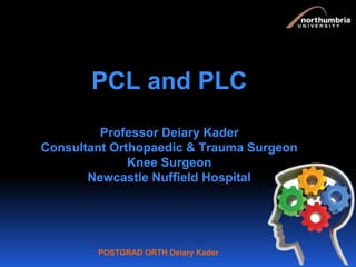
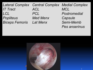
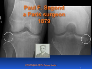
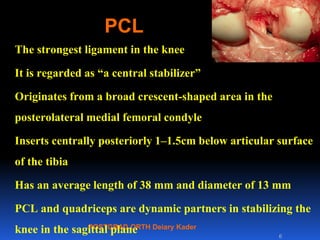
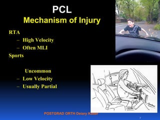
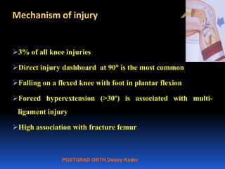
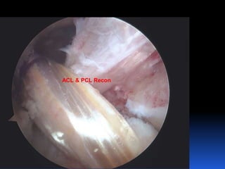
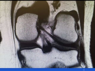
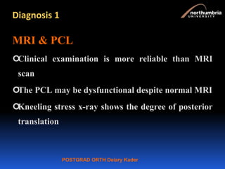
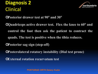
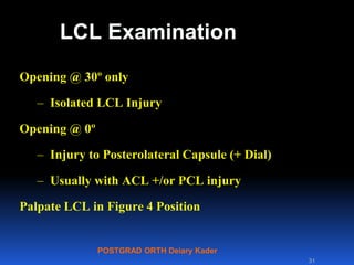
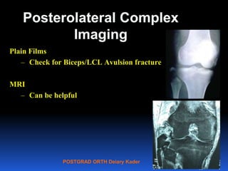
This document contains lecture slides from Professor Deiary Kader on various topics related to orthopaedics of the knee. It discusses the anatomy and classification of knee stabilizers. It provides details on the posterior cruciate ligament anatomy, mechanisms of injury, grading of instability, and treatment options including rehabilitation and reconstruction. The document also discusses the posterolateral corner complex, its anatomy, mechanisms of injury, examination techniques, and treatment including non-surgical and surgical options. Finally, it covers knee dislocations, classification, examination, management principles, and surgical approaches.
Arthroscopic ACL Reconstruction By Dr Shekhar ShrivastavDelhiArthroscopy
Arthroscopic Acl Reconstruction By Dr Shekhar Shrivastav.
HOW NORMAL KNEE WORKS ?
The knee is the largest joint in the body, and one of the most easily injured. It is made up of the lower end of the thigh bone(femur), the upper end of the shin bone (tibia), and the knee cap (patella), which slides in a groove on the end of the femur. Four bands of tissue, the anterior and posterior cruciate ligaments, and the medial and lateral collateral ligaments connect the femur and the tibia and provide joint stability. The surfaces where the femur, tibia and patella touch are covered with articular cartilage, a smooth substance that cushions the bones and enables them to glide freely. Semicircular rings of tough fibrous-cartilage tissue called the lateral and medial menisci act as shock absorbers and stabilizers.
WHAT IS THE ROLE OF ACL ?
ACL along with other ligaments of the knee joint and meniscus provides stability to the knee joint.
WHAT IS LIGAMENT RECONSTRUCTION ( ACL ) ?
Ligament reconstruction involves replacing the torn ligament with a tendon (graft) from your knee and fixing the graft in place with screws. This procedure is performed with the use of the arthroscope. The anterior cruciate ligament (ACL) is the most common ligament requiring reconstruction procedures. The torn ligament is excised arthroscopically and new ligament is prepared by ligament grafts taken from your own body. Bony tunnels are prepared in femur and tibia using specialized instruments through which the new ligament is passed and fixed with special screws. This procedure requires relative rest or leave from your work or studies for about 2-3 weeks after which you will be allowed normal day to day activities.
WHEN CAN THE PATIENT BE AMBULATED AFTER SURGERY ?
The patient can walk from the same evening of the surgery. Initially the patient is advised to walk with a brace and a walking cane. Strengthening and range of motion exercises for the knee are started from the next day. The patient is discharged from the hospital 2nd or 3rd day after surgery. The patient can walk without support by 10-14 days depending on muscle strengthening. Slow Jogging and other strenuous activities are permitted after 3 months and the patient can return to active sports only 8-9 months after surgery.
Torn ACL Reconstructed ACL
For Further Queries contact your Orthopedic Surgeon at
+ 91 9971192233
Surgical Approaches to Acetabulum and PelvisBijay Mehta
Important surgical approaches to acetabulum and pelvis are described.
Ilioinguinal approach, Modified Stoppa Approach, Kocher lagenbeck Approach, Ilifemoral approach and extensile approaches are well illustrated and described.
arthroscopy of the knee joint is a relatively common orthopedic procedure to treat a host of sports injuries and other knee diseases. Commonly a 4 mm size scope is used via two standard arthroscopy portals. Arthroscopic examination of the knee confirms MRI findings. Synovial fluid and biopsy can be taken to confirm diagnosis.
Posterolateral corner injuries of knee joint Samir Dwidmuthe
Missed posterolateral corner injuries of knee joint is a common cause for failure of ACL and PCL reconstruction only next to malpositioned tunnels.
Isolated PLC injuries are uncommon, making up <2% of all acute knee ligamentous injuries. Covey JBJS 2001
Incidence of PLC injuries associated with concomitant ACL and PCL disruptions are much more common (43% to 80%). Ranawat JAAOS 2008
A recent (MRI) analysis of surgical tibialplateau fractures demonstrated an incidence of PLC injuries in 68% of cases. Gardner JOT 2005
Take home message
PLC injuries to be ruled out in every case of ACL& PCL rupture.
Neurovascular integrity to be checked in every case.
Grade I & II can be managed conservatively.
Grade III Acute- Repair.
Grade III Chronic- Anatomic PLC recon.
Beware of varus knee alignment.
We have a moral responsibility to care for the wellbeing of our staff
Incivility cannot be tackled by increasing awareness and education alone
Governing bodies, surgeons and healthcare managers must play a bigger role in setting the tone for professionalism
The BOA needs to build systems that serve as a check against some of our human impulses
NVBDCP.pptx Nation vector borne disease control programSapna Thakur
NVBDCP was launched in 2003-2004 . Vector-Borne Disease: Disease that results from an infection transmitted to humans and other animals by blood-feeding arthropods, such as mosquitoes, ticks, and fleas. Examples of vector-borne diseases include Dengue fever, West Nile Virus, Lyme disease, and malaria.
Couples presenting to the infertility clinic- Do they really have infertility...Sujoy Dasgupta
Dr Sujoy Dasgupta presented the study on "Couples presenting to the infertility clinic- Do they really have infertility? – The unexplored stories of non-consummation" in the 13th Congress of the Asia Pacific Initiative on Reproduction (ASPIRE 2024) at Manila on 24 May, 2024.
Title: Sense of Taste
Presenter: Dr. Faiza, Assistant Professor of Physiology
Qualifications:
MBBS (Best Graduate, AIMC Lahore)
FCPS Physiology
ICMT, CHPE, DHPE (STMU)
MPH (GC University, Faisalabad)
MBA (Virtual University of Pakistan)
Learning Objectives:
Describe the structure and function of taste buds.
Describe the relationship between the taste threshold and taste index of common substances.
Explain the chemical basis and signal transduction of taste perception for each type of primary taste sensation.
Recognize different abnormalities of taste perception and their causes.
Key Topics:
Significance of Taste Sensation:
Differentiation between pleasant and harmful food
Influence on behavior
Selection of food based on metabolic needs
Receptors of Taste:
Taste buds on the tongue
Influence of sense of smell, texture of food, and pain stimulation (e.g., by pepper)
Primary and Secondary Taste Sensations:
Primary taste sensations: Sweet, Sour, Salty, Bitter, Umami
Chemical basis and signal transduction mechanisms for each taste
Taste Threshold and Index:
Taste threshold values for Sweet (sucrose), Salty (NaCl), Sour (HCl), and Bitter (Quinine)
Taste index relationship: Inversely proportional to taste threshold
Taste Blindness:
Inability to taste certain substances, particularly thiourea compounds
Example: Phenylthiocarbamide
Structure and Function of Taste Buds:
Composition: Epithelial cells, Sustentacular/Supporting cells, Taste cells, Basal cells
Features: Taste pores, Taste hairs/microvilli, and Taste nerve fibers
Location of Taste Buds:
Found in papillae of the tongue (Fungiform, Circumvallate, Foliate)
Also present on the palate, tonsillar pillars, epiglottis, and proximal esophagus
Mechanism of Taste Stimulation:
Interaction of taste substances with receptors on microvilli
Signal transduction pathways for Umami, Sweet, Bitter, Sour, and Salty tastes
Taste Sensitivity and Adaptation:
Decrease in sensitivity with age
Rapid adaptation of taste sensation
Role of Saliva in Taste:
Dissolution of tastants to reach receptors
Washing away the stimulus
Taste Preferences and Aversions:
Mechanisms behind taste preference and aversion
Influence of receptors and neural pathways
Impact of Sensory Nerve Damage:
Degeneration of taste buds if the sensory nerve fiber is cut
Abnormalities of Taste Detection:
Conditions: Ageusia, Hypogeusia, Dysgeusia (parageusia)
Causes: Nerve damage, neurological disorders, infections, poor oral hygiene, adverse drug effects, deficiencies, aging, tobacco use, altered neurotransmitter levels
Neurotransmitters and Taste Threshold:
Effects of serotonin (5-HT) and norepinephrine (NE) on taste sensitivity
Supertasters:
25% of the population with heightened sensitivity to taste, especially bitterness
Increased number of fungiform papillae
Pulmonary Thromboembolism - etilogy, types, medical- Surgical and nursing man...VarunMahajani
Disruption of blood supply to lung alveoli due to blockage of one or more pulmonary blood vessels is called as Pulmonary thromboembolism. In this presentation we will discuss its causes, types and its management in depth.
New Directions in Targeted Therapeutic Approaches for Older Adults With Mantl...i3 Health
i3 Health is pleased to make the speaker slides from this activity available for use as a non-accredited self-study or teaching resource.
This slide deck presented by Dr. Kami Maddocks, Professor-Clinical in the Division of Hematology and
Associate Division Director for Ambulatory Operations
The Ohio State University Comprehensive Cancer Center, will provide insight into new directions in targeted therapeutic approaches for older adults with mantle cell lymphoma.
STATEMENT OF NEED
Mantle cell lymphoma (MCL) is a rare, aggressive B-cell non-Hodgkin lymphoma (NHL) accounting for 5% to 7% of all lymphomas. Its prognosis ranges from indolent disease that does not require treatment for years to very aggressive disease, which is associated with poor survival (Silkenstedt et al, 2021). Typically, MCL is diagnosed at advanced stage and in older patients who cannot tolerate intensive therapy (NCCN, 2022). Although recent advances have slightly increased remission rates, recurrence and relapse remain very common, leading to a median overall survival between 3 and 6 years (LLS, 2021). Though there are several effective options, progress is still needed towards establishing an accepted frontline approach for MCL (Castellino et al, 2022). Treatment selection and management of MCL are complicated by the heterogeneity of prognosis, advanced age and comorbidities of patients, and lack of an established standard approach for treatment, making it vital that clinicians be familiar with the latest research and advances in this area. In this activity chaired by Michael Wang, MD, Professor in the Department of Lymphoma & Myeloma at MD Anderson Cancer Center, expert faculty will discuss prognostic factors informing treatment, the promising results of recent trials in new therapeutic approaches, and the implications of treatment resistance in therapeutic selection for MCL.
Target Audience
Hematology/oncology fellows, attending faculty, and other health care professionals involved in the treatment of patients with mantle cell lymphoma (MCL).
Learning Objectives
1.) Identify clinical and biological prognostic factors that can guide treatment decision making for older adults with MCL
2.) Evaluate emerging data on targeted therapeutic approaches for treatment-naive and relapsed/refractory MCL and their applicability to older adults
3.) Assess mechanisms of resistance to targeted therapies for MCL and their implications for treatment selection
Report Back from SGO 2024: What’s the Latest in Cervical Cancer?bkling
Are you curious about what’s new in cervical cancer research or unsure what the findings mean? Join Dr. Emily Ko, a gynecologic oncologist at Penn Medicine, to learn about the latest updates from the Society of Gynecologic Oncology (SGO) 2024 Annual Meeting on Women’s Cancer. Dr. Ko will discuss what the research presented at the conference means for you and answer your questions about the new developments.
TEST BANK for Operations Management, 14th Edition by William J. Stevenson, Ve...kevinkariuki227
TEST BANK for Operations Management, 14th Edition by William J. Stevenson, Verified Chapters 1 - 19, Complete Newest Version.pdf
Title: Sense of Smell
Presenter: Dr. Faiza, Assistant Professor of Physiology
Qualifications:
MBBS (Best Graduate, AIMC Lahore)
FCPS Physiology
ICMT, CHPE, DHPE (STMU)
MPH (GC University, Faisalabad)
MBA (Virtual University of Pakistan)
Learning Objectives:
Describe the primary categories of smells and the concept of odor blindness.
Explain the structure and location of the olfactory membrane and mucosa, including the types and roles of cells involved in olfaction.
Describe the pathway and mechanisms of olfactory signal transmission from the olfactory receptors to the brain.
Illustrate the biochemical cascade triggered by odorant binding to olfactory receptors, including the role of G-proteins and second messengers in generating an action potential.
Identify different types of olfactory disorders such as anosmia, hyposmia, hyperosmia, and dysosmia, including their potential causes.
Key Topics:
Olfactory Genes:
3% of the human genome accounts for olfactory genes.
400 genes for odorant receptors.
Olfactory Membrane:
Located in the superior part of the nasal cavity.
Medially: Folds downward along the superior septum.
Laterally: Folds over the superior turbinate and upper surface of the middle turbinate.
Total surface area: 5-10 square centimeters.
Olfactory Mucosa:
Olfactory Cells: Bipolar nerve cells derived from the CNS (100 million), with 4-25 olfactory cilia per cell.
Sustentacular Cells: Produce mucus and maintain ionic and molecular environment.
Basal Cells: Replace worn-out olfactory cells with an average lifespan of 1-2 months.
Bowman’s Gland: Secretes mucus.
Stimulation of Olfactory Cells:
Odorant dissolves in mucus and attaches to receptors on olfactory cilia.
Involves a cascade effect through G-proteins and second messengers, leading to depolarization and action potential generation in the olfactory nerve.
Quality of a Good Odorant:
Small (3-20 Carbon atoms), volatile, water-soluble, and lipid-soluble.
Facilitated by odorant-binding proteins in mucus.
Membrane Potential and Action Potential:
Resting membrane potential: -55mV.
Action potential frequency in the olfactory nerve increases with odorant strength.
Adaptation Towards the Sense of Smell:
Rapid adaptation within the first second, with further slow adaptation.
Psychological adaptation greater than receptor adaptation, involving feedback inhibition from the central nervous system.
Primary Sensations of Smell:
Camphoraceous, Musky, Floral, Pepperminty, Ethereal, Pungent, Putrid.
Odor Detection Threshold:
Examples: Hydrogen sulfide (0.0005 ppm), Methyl-mercaptan (0.002 ppm).
Some toxic substances are odorless at lethal concentrations.
Characteristics of Smell:
Odor blindness for single substances due to lack of appropriate receptor protein.
Behavioral and emotional influences of smell.
Transmission of Olfactory Signals:
From olfactory cells to glomeruli in the olfactory bulb, involving lateral inhibition.
Primitive, less old, and new olfactory systems with different path
These simplified slides by Dr. Sidra Arshad present an overview of the non-respiratory functions of the respiratory tract.
Learning objectives:
1. Enlist the non-respiratory functions of the respiratory tract
2. Briefly explain how these functions are carried out
3. Discuss the significance of dead space
4. Differentiate between minute ventilation and alveolar ventilation
5. Describe the cough and sneeze reflexes
Study Resources:
1. Chapter 39, Guyton and Hall Textbook of Medical Physiology, 14th edition
2. Chapter 34, Ganong’s Review of Medical Physiology, 26th edition
3. Chapter 17, Human Physiology by Lauralee Sherwood, 9th edition
4. Non-respiratory functions of the lungs https://academic.oup.com/bjaed/article/13/3/98/278874
- Video recording of this lecture in English language: https://youtu.be/lK81BzxMqdo
- Video recording of this lecture in Arabic language: https://youtu.be/Ve4P0COk9OI
- Link to download the book free: https://nephrotube.blogspot.com/p/nephrotube-nephrology-books.html
- Link to NephroTube website: www.NephroTube.com
- Link to NephroTube social media accounts: https://nephrotube.blogspot.com/p/join-nephrotube-on-social-media.html
New Drug Discovery and Development .....NEHA GUPTA
The "New Drug Discovery and Development" process involves the identification, design, testing, and manufacturing of novel pharmaceutical compounds with the aim of introducing new and improved treatments for various medical conditions. This comprehensive endeavor encompasses various stages, including target identification, preclinical studies, clinical trials, regulatory approval, and post-market surveillance. It involves multidisciplinary collaboration among scientists, researchers, clinicians, regulatory experts, and pharmaceutical companies to bring innovative therapies to market and address unmet medical needs.
ARTIFICIAL INTELLIGENCE IN HEALTHCARE.pdfAnujkumaranit
Artificial intelligence (AI) refers to the simulation of human intelligence processes by machines, especially computer systems. It encompasses tasks such as learning, reasoning, problem-solving, perception, and language understanding. AI technologies are revolutionizing various fields, from healthcare to finance, by enabling machines to perform tasks that typically require human intelligence.
Ethanol (CH3CH2OH), or beverage alcohol, is a two-carbon alcohol
that is rapidly distributed in the body and brain. Ethanol alters many
neurochemical systems and has rewarding and addictive properties. It
is the oldest recreational drug and likely contributes to more morbidity,
mortality, and public health costs than all illicit drugs combined. The
5th edition of the Diagnostic and Statistical Manual of Mental Disorders
(DSM-5) integrates alcohol abuse and alcohol dependence into a single
disorder called alcohol use disorder (AUD), with mild, moderate,
and severe subclassifications (American Psychiatric Association, 2013).
In the DSM-5, all types of substance abuse and dependence have been
combined into a single substance use disorder (SUD) on a continuum
from mild to severe. A diagnosis of AUD requires that at least two of
the 11 DSM-5 behaviors be present within a 12-month period (mild
AUD: 2–3 criteria; moderate AUD: 4–5 criteria; severe AUD: 6–11 criteria).
The four main behavioral effects of AUD are impaired control over
drinking, negative social consequences, risky use, and altered physiological
effects (tolerance, withdrawal). This chapter presents an overview
of the prevalence and harmful consequences of AUD in the U.S.,
the systemic nature of the disease, neurocircuitry and stages of AUD,
comorbidities, fetal alcohol spectrum disorders, genetic risk factors, and
pharmacotherapies for AUD.
6. POSTGRAD ORTH Deiary Kader
PCL
The strongest ligament in the knee
It is regarded as “a central stabilizer”
Originates from a broad crescent-shaped area in the
posterolateral medial femoral condyle
Inserts centrally posteriorly 1–1.5cm below articular surface
of the tibia
Has an average length of 38 mm and diameter of 13 mm
PCL and quadriceps are dynamic partners in stabilizing the
knee in the sagittal plane 6
7. POSTGRAD ORTH Deiary Kader
PCL
Mechanism of Injury
RTA
– High Velocity
– Often MLI
Sports
Uncommon
– Low Velocity
– Usually Partial
7
8. POSTGRAD ORTH Deiary Kader
Mechanism of injury
3% of all knee injuries
Direct injury dashboard at 90 is the most common
Falling on a flexed knee with foot in plantar flexion
Forced hyperextension (>30º) is associated with multi-
ligament injury
High association with fracture femur
9. POSTGRAD ORTH Deiary Kader
PCL Injury Diagnosis
9
Physical Exam
– Posterior Drawer
– Step off sign
Plain Radiographs
– Look for bony avulsions
– Standing films for chronic injuries (Arthritis)
– Stress Radiographs helpful
MRI
– Not Sensitive
– MLI (common)
10. POSTGRAD ORTH Deiary Kader
PCL
Three components:
AL: Antero-lateral: long and thick part, twice the size
of the posteromedial bundle; tightens in flexion
PM: Posteromedial: tight in extension
Meniscofemoral ligaments: mechanically very strong
Anterior: Humphrey’s ligament
Posterior: Wrisberg’s ligament
15. POSTGRAD ORTH Deiary Kader
Diagnosis 1
MRI & PCL
Clinical examination is more reliable than MRI
scan
The PCL may be dysfunctional despite normal MRI
Kneeling stress x-ray shows the degree of posterior
translation
16. POSTGRAD ORTH Deiary Kader
Diagnosis 2
Clinical
Posterior drawer test at 90 and 30
Quadriceps active drawer test. Flex the knee to 60 and
control the foot then ask the patient to contract the
quads. The test is positive when the tibia reduces.
Posterior sag sign (step-off)
Posterolateral rotatory instability (Dial test prone)
External rotation recurvatum test
17. POSTGRAD ORTH Deiary Kader
Grading of PCL instability
Normal tibia step-off is 10 mm at 90 flexion
Instability could be mild, moderate or severe
Grade I instability is when there is a 5-mm step-off
Grade II instability is when there is no step-off (flush)
Grade III instability is when there is –5 mm step-off
There is a high association between Grade III PCL
injury and posterolateral corner injury.
18. POSTGRAD ORTH Deiary Kader
Management
In isolation, it often causes little long-term
instability. However, it may lead to medial
or PFJ pain (OA) at a later date.
More troublesome in soccer players due to
difficulty in deceleration.
19. POSTGRAD ORTH Deiary Kader
Management 2
Presentation
Acute isolated PCL injury is commonly missed
Present with very little pain in the knee without hemarthrosis
There may be only bruising at the popliteal fossa.
Chronic PCL injury on the other hand may present with pain in
the medial compartment or anterior knee pain.
20. POSTGRAD ORTH Deiary Kader
Treatment
Treat acute, isolated PCL injury conservatively.
Extension brace with calf support (Posterior Tibial Support,
PTS Brace) until the pain subsides (4-6 weeks) with quadriceps
rehabilitation
Start early passive motion only in prone position to maintain
anterior tibia translation.
22. POSTGRAD ORTH Deiary Kader
Surgical reconstruction
Indications
Acute combined injuries
Acute bony avulsion
Symptomatic chronic PCL injuries that failed rehabilitation.
There is no difference in clinical outcome between single and
double bundle PCL reconstruction.
23. POSTGRAD ORTH Deiary Kader
Complications
Immediate
Neurovascular injury popliteal vessels
Infection
Technical error → tunnel placement, graft tensioning, insecure fixation
Delayed
Loss of motion
Avascular necrosis (medial femoral condyle)
Recurrent or persistent laxity (common) when a combined injury is not
adequately addressed
24. POSTGRAD ORTH Deiary Kader
Posterolateral Complex
Components:
– Biceps, ITB, Popliteus,
Popliteofibular ligament, arcuate
ligament, LCL
Function
– Resists External and Varus rotation
Mechanism of Injury
– Direct blow to anteromedial tibia
– Hyperextension/varus
24
arcuate
25. POSTGRAD ORTH Deiary Kader
The Posterolateral Corner
The LCL is a cord like structure 5-7 cm in length
Is the primary static restraint to varus opening of the knee
Secondary restraint to posterolateral rotation
The popliteus is a static and dynamic external rotation stabiliser.
The popletiofibular ligament acts as
a primary restraint to external rotation of
the tibia on the femur at 30º of flexion
25
26. POSTGRAD ORTH Deiary Kader
The Posterolateral Corner
(PLC)
They are the primary stabilisers of external tibial rotation at
all knee flexion angles and the secondary restraints to
anterior and posterior translation
Isolated PLC sectioning produce a maximal average increase of
13° of ER at 30° of knee flexion and only an average increase of
5.3° at 90°.
Isolated PCL sectioning has no effect on external tibial rotation
Combined injury to the PCL and PLC leads to ER of 20.9° at
90° of knee flexion
26
28. POSTGRAD ORTH Deiary Kader
28
Fib
Pop
Extension
The popliteus tendon inserted
10 mm distal
5 mm posterior to the lateral epicondyle
The LCL inserted
1-2 mm proximal
4-5 mm posterior to the lateral epicondyle
31. POSTGRAD ORTH Deiary Kader
LCL Examination
Opening @ 30º only
– Isolated LCL Injury
Opening @ 0º
– Injury to Posterolateral Capsule (+ Dial)
– Usually with ACL +/or PCL injury
Palpate LCL in Figure 4 Position
31
32. POSTGRAD ORTH Deiary Kader
Posterolateral Complex
Imaging
Plain Films
– Check for Biceps/LCL Avulsion fracture
MRI
– Can be helpful
32
33. POSTGRAD ORTH Deiary Kader
Posterolateral Complex
Injury--Treatment
Partial
– Grade I & II Instability with a good end
point
– Nonsurgical Treatment
– 3 week immobilization in extension
Complete Acute
– Primary repair best
– Augment with allo/auto graft
Complete Chronic
– Reconstruct Popliteus and LCL
33
35. POSTGRAD ORTH Deiary Kader
Posterolateral Complex
Injury--Treatment
Partial
– Grade I & II Instability with a good end point
– Nonsurgical Treatment
– 3 week immobilization in extension
Complete Acute
– Primary repair best
– Augment with allo/auto graft
Complete Chronic
– Reconstruct Popliteus and LCL
35
36. POSTGRAD ORTH Deiary Kader
PLC Reconstruction
The reconstruction can be fibula based such as
modified Larson’s technique or combined tibia and
fibula based such as LaPrade’s anatomical
reconstruction.
36
37. POSTGRAD ORTH Deiary Kader
The principles of surgery
Early repair (within 3 weeks) of torn and detached ligaments,
tendons and capsule in acute injuries. A combination of early
repair and reconstruction has been shown to provide better
results.
Late reconstruction of the two or three of the main stabilisers of
the posterolateral corner of the knee i.e. the lateral collateral
ligament, Popliteus tendon, and popliteofibular ligament in
chronic cases.
Combined ACL/PCL and PLC injury must be treated by
reconstruction of all injured ligaments.
37
38. POSTGRAD ORTH Deiary Kader
Knee dislocation
Any triple-ligament knee injury constitutes a
frank dislocation. This is relatively rare but
a severe and potentially limb-threatening
injury.
High-energy injury such as RTA.
Sporting accident.
May be missed on initial assessment.
38
39. POSTGRAD ORTH Deiary Kader
Vascular Injuries
Previously it was thought there was a 50%
incidence of vascular compromise Now 3.3-18%
20%–30% incidence of nerve injury.
Fracture incidence may be as high as 60%.
39
41. POSTGRAD ORTH Deiary Kader
Classification
Classified on the basis on tibial displacement in respect to the femur
Closed or open
High or low energy
Dislocation or subluxation
Neurovascular involvement
Anterior (common: 30-50% of dislocations, associated with intimal tears)
Posterior; also medial, lateral (highest rate of peroneal nerve injury) and
rotatory (usually irreducible) or combined
Hyperextension leads to anterior dislocation
Dashboard injury leads to posterior dislocation
41
42. POSTGRAD ORTH Deiary Kader
Examination
Valgus and varus laxity
Anteroposterior translation
Recurvatum
>10º hyperextension suggests ACL injury
>30º hyperextension indicates PCL injury
Rotation indicates MCL and LCL injury
42
43. POSTGRAD ORTH Deiary Kader
Management
Surgical emergency
Deal with life-threatening injuries first
Circulation in A&E
Serial examination for 48 hours.
Ankle brachial Index (ABI)
ABI <0.9 is suggestive of significant arterial injury
Involve the vascular surgeon
Radiography before manipulation
– (assess direction and associated fracture)
Reduction as soon as possible in the emergency/operating Room
43
44. POSTGRAD ORTH Deiary Kader
Management
Immobilization in an extension knee splint
Check radiograph to confirm congruity, if not,
consider external fixator
Conservative management out of favour
Early surgical reconstruction and/or repair, is
currently recommended by the Knee Dislocation
Study Group
44
45. POSTGRAD ORTH Deiary Kader
Management
Surgery as soon as the vascular surgeon allows
Most ACL/PCL/MCL can be treated with bracing the MCL followed by
combined ACL/PCL reconstruction once range of movement is
restarted, usually after 6 weeks.
ACL/PCL/posterolateral corner can be treated by repairing the
posterolateral corner acutely (within three weeks) and delayed ACL/PCL
reconstruction 8 weeks later.
Open dislocation, fracture dislocation and vascular compromise require
staged procedures.
45