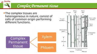
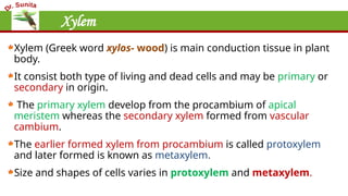
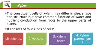
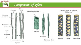
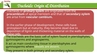
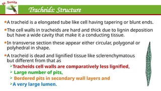
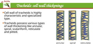
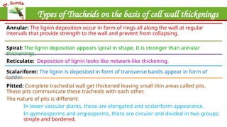
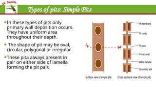
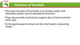
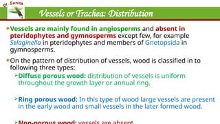
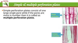
Plant Anatomy
By: Dr. Sunita Sangwan
Assistant Prof.
Deptt. of Botany
MNSGC Bhiwani
Complex Permanent tissue
The complex tissues are
heterogeneous in nature, consist of
cells of common origin performing
different functions.
Complex
Permanent
tissue
Xylem
Phloem
Xylem
Xylem (Greek word xylos- wood) is main conduction tissue in plant
body.
It consist both type of living and dead cells and may be primary or
secondary in origin.
The primary xylem develop from the procambium of apical
meristem whereas the secondary xylem formed from vascular
cambium.
The earlier formed xylem from procambium is called protoxylem
and later formed is known as metaxylem.
Size and shapes of cells varies in protoxylem and metaxylem.
Xylem
The constituent cells of xylem may differ in size, shape
and structure but have common function of water and
nutrient conduction from roots to the upper parts of
plants.
It consists of four kinds of cells:
1.Tracheids, 2. vessels
3. Xylem
fibres
4. Xylem
parenchym
a
Components of xylem
Xylem Tracheids
Tracheids: Origin & Distribution
Tracheids of primary xylem are originated from the
procambium of apical meristem and that of secondary xylem
are arise from vascular cambium.
In the earlier phase of development, these cells have
protoplasm but at maturity, they become dead due to
deposition of lignin and thickening material on the walls of
these cells.
The tracheids are the basic cell of xylem found in pteridophytes,
gymnosperms and angiosperms.
It act as main conducting tissue in pteridophytes and
gymnosperms where vessels are absent.
They present in both primary and secondary xylem.
Tracheids: Structure
A tracheid is a elongated tube like cell having tapering or blunt ends.
The cell walls in tracheids are hard and thick due to lignin deposition
but have a wide cavity that make it a conducting tissue.
In transverse section these appear either circular, polygonal or
polyhedral in shape.
A tracheid is dead and lignified tissue like sclerenchymatous tissue
but different from that as
Tracheids cell walls are comparatively less lignified,
 Large number of pits,
 Bordered pits in secondary wall layers and
A very large lumen.
Tracheids: cell wall thickenings
Cell wall of tracheids is highly
characteristic and specialized
type.
Tracheids possess various types
of wall thickening like annular,
spiral, scalariform, reticulate
and pitted.
Types of Tracheids on the basis of cell wall thickenings
Annular: The lignin deposition occur in form of rings all along the wall at regular
intervals that provide strength to the wall and prevent from collapsing.
Spiral: The lignin deposition appears spiral in shape. It is stronger than annular
thickenings.
Reticulate: Deposition of lignin looks like network-like thickening.
Scalariform: The lignin is deposited in form of transverse bands appear in form of
ladder.
Pitted: Complete trachedial wall get thickened leaving small thin areas called pits.
These pits communicate these tracheids with each other.
The nature of pits is different:
In lower vascular plants, these are elongated and scalariform appearance.
In gymnosperms and angiosperms, there are circular and divided in two groups:
simple and bordered.
Types of pits: Simple Pits
In these types of pits only
primary wall deposition occurs.
They have uniform area
throughout their depth.
The shape of pit may be oval,
circular, polygonal or irregular.
These pita always present in
pair on either side of lamella
forming the pit pair.
Types of pits: Bordered Pits
In bordered pits secondary walls outgrows
the pit to form a dome-like structure with
a small perforation in the middle.
In the central part primary wall thickened
to form torus.
The bordered pits may occur opposite to
each other to form the bordered pit pairs.
Sometimes in two opposite pits one is
bordered and other is simple pit, such pit
is called half bordered pit pairs.
During conduction of water torus act as a
valve to check the flow.
Functions of Tracheids
The main function of tracheids is to conduct water and
dissolved solutes due to specialised structure.
They also provide mechanical support due to hard and their
thick cells.
In the gymnosperms these are the chief water conducting
tissue.
Vessels or Trachea
Vessels or Trachea: Origin
The origin pattern of vessels is same as that of tracheids but
show variation in development pattern.
Vessel in primary xylem originate from procambial cells and that
of secondary xylem from cambial cells.
The vessels enlarge in size rapidly before deposition of
secondary wall material and deposition of secondary wall
material determined by microfibrils.
Vessels or Trachea: Distribution
Vessels are mainly found in angiosperms and absent in
pteridophytes and gymnosperms except few, for example
Selaginella in pteridophytes and members of Gnetopsida in
gymnosperms.
On the pattern of distribution of vessels, wood is classified in to
following three types:
Diffuse porous wood: distribution of vessels is uniform
throughout the growth layer or annual ring.
Ring porous wood: In this type of wood large vessels are present
in the early wood and small vessels in the later formed wood.

Vessels or Trachea : Structure
Vessel is long tube like structure that has thick walls due to
deposition of lignin and a large lumen.
Vessels are made by the longitudinal arrangement of dead cells
with perforated transverse walls making the entire structure like
a water pipe.
Based on the type of perforation there are main two types of
perforation plates:
Simple perforation plates
Multiple perforation plates.
Simple & multiple perforation plates
Simple perforation plates consist of one
large single pore while if the pores are
more in number then it is called as
multiple perforation plates.
Simple
perforation
Multiple
perforation
Cell wall thickening of xylem Vessels
Depending upon the manner
of pore arrangement these
are reticulate (reticulate
shape), scalariform (elongate
and arranged in series) and
foraminate (circular
perforation arranged in
groups).
Deposition of secondary wall
is similar to the tracheids.
A. T. S. showing prominent reticulate pitting, vessels, and multiseriate
rays.
B. L. S. of corn stem: xylem vessel elements with annular secondary walls
Functions of vessels
Vessels are chief conducting tissue especially in the
angiosperms.
Vessels are more efficient conducting system than tracheids due
to presence of perforation plates.
They also provide mechanical support to the plant body.
Xylem Fibres
Xylem Fibres: Distribution
Xylem fibres are present in both primary and secondary xylem
but their abundance is more in secondary xylem as its main
function is to provide mechanical strength to the plant body.
They originate from the same meristematic tissue from which
other xylem cells developed.
Xylem Fibres : Structure
Fibres are dead cell with thick lignified cell walls and narrow
lumen.
These are usually longer than tracheids.
Based on wall thickening and pits number fibres are of two type:
1. Libriform Fibres
2. Fibre tracheids
Libriform Fibres
These fibres have very highly thickened cell walls, narrow lumen
and very little number of pits.
These are the true fibres having all features of
sclerenchymatous fibres and found more in number in woody
plants.
Their main function is to provide mechanical strength to the
plant.
Fibre tracheids
These are intermediate between the tracheids and fibres.
Their cell walls are thicker than the tracheids but thinner than the true fibres.
They possess the bordered pits but border is not well developed.
These are of different types.
In some fibre tracheids protoplast divides by transverse septa
that remains unlignified. These are called as septate fibre
tracheids.
In secondary xylem of dicotyledons, inner layer of secondary
wall is poor in lignin and contains a lot of α-cellulose due to
which this layer absorbs water and swell up to fill the whole
lumen of fibre. This type of fibre are known as Gelatinous or
Mucilagenous fibres.
Xylem Parenchyma
Xylem Parenchyma
The parenchymatous cells are present in both primary and
secondary xylem but more abundant in primary xylem.
The cell wall of xylem parenchyma in primary xylem is thin and
made up of cellulose but that found in secondary xylem is
thickened, lignified and have pits.
Types of xylem parenchyma
In secondary xylem two types of
parenchyma is found:
Axial or Woody Parenchyma: These
are originated from fusiform initial of
vascular cambium. These are
elongated and arranged in vertical
series.
Ray Parenchyma: These are arise
from the ray initials of vascular
cambium. Cells are much elongated
and arranged in both radial and
transverse series.
Functions of Xylem Parenchyma
Xylem parenchyma store food reserve in form of starch or fats.
Ray parenchyma help in radial conduction of water.
Some parenchymatous cells are structurally modified for solute
transfer termed as transfer cells.
Difference between different xylem component
Features Xylem Tracheids Xylem Vessels Xylem Fibres
Origin Originated from single procambial
cell
Originate from a row of procambial
cells placed end to end.
Developed from provascular or
vascular cambium.
Distribution Found in all vascular plants Only present in angiosperms. Only present in angiosperms.
Size Usually very short about 1 cm in
length rarely reach up to 12 cm.
They are long about 10 cm and
sometimes can reach up to 2-6
meters
These are dead cells longer than
tracheids.
Cell walls Cell walls remain intact but having
many bordered pits.
Cell walls are either absent or
perforated.
Cell wall is perforated with pits .
Arrangement Tracheids are placed one above
the other but separated by cross
walls and are not syncytes.
Vessels are syncytes or tubes. Thick cell wall provide mechanical
support.
Lumen Lumen is not much wide Vessels have wide lumen. Lumen is absent or very narrow.
Primary Xylem
This is differentiated into protoxylem and metaxylem.
Protoxylem is found nearest to central axis in stem (endarch xylem) and away
from centre in roots (exarch xylem).
The protoxylem elements have annular, spiral and sometimes reticulate
secondary thickenings.
Fibers are absent in protoxylem.
Metaxylem appears after protoxylem.
Metaxylem has only pitted secondary walls. Metaxylem is more complex than
protoxylem and its elements are wider.
Secondary Xylem
The vascular cambium (intrafascicular cambium and interfascicular cambium)
producing secondary xylem consists of fusiform and ray initials.
When compared with primary xylem, secondary xylem is more complex and
shows orderly development. Pitted and scalariform secondary thickenings
are developed.
The xylem is called the primary xylem if its elements are developed from
apical meristem. Secondary xylem elements develop from the vascular
cambium during the process of secondary growth.
It is very difficult to differentiate in xylem and phloem in hydrophytes due to
less development of vascular tissues.
Primary Xylem Secondary Xylem
1. Formed by the pro-cambial activity of apical Meristem.
2. Protoxylem and metaxylem are easily distinct.
3. The tracheids and vessels are comparatively narrower and
longer.
4. Axial and radial systems are not distinct.
5. Annual rings are not found.
6. No differentiation of heartwood and sapwood.
7. Tylosis are not found in the vessels.
8. Xylem fibres are either absent or present in very few
numbers
9. Medullary rays consist of only one type of cells so they are
homocellular.
10. Medullary rays found in primary xylem are formed by
histogen of apical meristem.
1. It formed by vascular cambium that is a Lateral Meristem.
2. Protoxylem and metaxylem are not distinct.
3. Both are normally wide and short.
4. Both axial and radial systems are clearly differentiated
from each other.
5. Annual rings are clearly distinct in the wood of temperate
trees and shrubs.
6. Heartwood and sapwood are clearly distinct in Woody
trees.
7. Vessels show tylosis.
8. Xylem fibres are found abundantly
9. Medullary Rays may be homocellular or heterocellular.
10. Secondary medullary rays are originated by the activity of
ray initials of the cambial ring.
Thanks

Xylem- complex permanent tissue in plants

  • 1.
    Plant Anatomy By: Dr.Sunita Sangwan Assistant Prof. Deptt. of Botany MNSGC Bhiwani
  • 2.
    Complex Permanent tissue Thecomplex tissues are heterogeneous in nature, consist of cells of common origin performing different functions. Complex Permanent tissue Xylem Phloem
  • 3.
    Xylem Xylem (Greek wordxylos- wood) is main conduction tissue in plant body. It consist both type of living and dead cells and may be primary or secondary in origin. The primary xylem develop from the procambium of apical meristem whereas the secondary xylem formed from vascular cambium. The earlier formed xylem from procambium is called protoxylem and later formed is known as metaxylem. Size and shapes of cells varies in protoxylem and metaxylem.
  • 4.
    Xylem The constituent cellsof xylem may differ in size, shape and structure but have common function of water and nutrient conduction from roots to the upper parts of plants. It consists of four kinds of cells: 1.Tracheids, 2. vessels 3. Xylem fibres 4. Xylem parenchym a
  • 5.
  • 6.
  • 7.
    Tracheids: Origin &Distribution Tracheids of primary xylem are originated from the procambium of apical meristem and that of secondary xylem are arise from vascular cambium. In the earlier phase of development, these cells have protoplasm but at maturity, they become dead due to deposition of lignin and thickening material on the walls of these cells. The tracheids are the basic cell of xylem found in pteridophytes, gymnosperms and angiosperms. It act as main conducting tissue in pteridophytes and gymnosperms where vessels are absent. They present in both primary and secondary xylem.
  • 8.
    Tracheids: Structure A tracheidis a elongated tube like cell having tapering or blunt ends. The cell walls in tracheids are hard and thick due to lignin deposition but have a wide cavity that make it a conducting tissue. In transverse section these appear either circular, polygonal or polyhedral in shape. A tracheid is dead and lignified tissue like sclerenchymatous tissue but different from that as Tracheids cell walls are comparatively less lignified,  Large number of pits,  Bordered pits in secondary wall layers and A very large lumen.
  • 9.
    Tracheids: cell wallthickenings Cell wall of tracheids is highly characteristic and specialized type. Tracheids possess various types of wall thickening like annular, spiral, scalariform, reticulate and pitted.
  • 10.
    Types of Tracheidson the basis of cell wall thickenings Annular: The lignin deposition occur in form of rings all along the wall at regular intervals that provide strength to the wall and prevent from collapsing. Spiral: The lignin deposition appears spiral in shape. It is stronger than annular thickenings. Reticulate: Deposition of lignin looks like network-like thickening. Scalariform: The lignin is deposited in form of transverse bands appear in form of ladder. Pitted: Complete trachedial wall get thickened leaving small thin areas called pits. These pits communicate these tracheids with each other. The nature of pits is different: In lower vascular plants, these are elongated and scalariform appearance. In gymnosperms and angiosperms, there are circular and divided in two groups: simple and bordered.
  • 11.
    Types of pits:Simple Pits In these types of pits only primary wall deposition occurs. They have uniform area throughout their depth. The shape of pit may be oval, circular, polygonal or irregular. These pita always present in pair on either side of lamella forming the pit pair.
  • 12.
    Types of pits:Bordered Pits In bordered pits secondary walls outgrows the pit to form a dome-like structure with a small perforation in the middle. In the central part primary wall thickened to form torus. The bordered pits may occur opposite to each other to form the bordered pit pairs. Sometimes in two opposite pits one is bordered and other is simple pit, such pit is called half bordered pit pairs. During conduction of water torus act as a valve to check the flow.
  • 13.
    Functions of Tracheids Themain function of tracheids is to conduct water and dissolved solutes due to specialised structure. They also provide mechanical support due to hard and their thick cells. In the gymnosperms these are the chief water conducting tissue.
  • 14.
  • 15.
    Vessels or Trachea:Origin The origin pattern of vessels is same as that of tracheids but show variation in development pattern. Vessel in primary xylem originate from procambial cells and that of secondary xylem from cambial cells. The vessels enlarge in size rapidly before deposition of secondary wall material and deposition of secondary wall material determined by microfibrils.
  • 16.
    Vessels or Trachea:Distribution Vessels are mainly found in angiosperms and absent in pteridophytes and gymnosperms except few, for example Selaginella in pteridophytes and members of Gnetopsida in gymnosperms. On the pattern of distribution of vessels, wood is classified in to following three types: Diffuse porous wood: distribution of vessels is uniform throughout the growth layer or annual ring. Ring porous wood: In this type of wood large vessels are present in the early wood and small vessels in the later formed wood. 
  • 17.
    Vessels or Trachea: Structure Vessel is long tube like structure that has thick walls due to deposition of lignin and a large lumen. Vessels are made by the longitudinal arrangement of dead cells with perforated transverse walls making the entire structure like a water pipe. Based on the type of perforation there are main two types of perforation plates: Simple perforation plates Multiple perforation plates.
  • 18.
    Simple & multipleperforation plates Simple perforation plates consist of one large single pore while if the pores are more in number then it is called as multiple perforation plates. Simple perforation Multiple perforation
  • 19.
    Cell wall thickeningof xylem Vessels Depending upon the manner of pore arrangement these are reticulate (reticulate shape), scalariform (elongate and arranged in series) and foraminate (circular perforation arranged in groups). Deposition of secondary wall is similar to the tracheids. A. T. S. showing prominent reticulate pitting, vessels, and multiseriate rays. B. L. S. of corn stem: xylem vessel elements with annular secondary walls
  • 20.
    Functions of vessels Vesselsare chief conducting tissue especially in the angiosperms. Vessels are more efficient conducting system than tracheids due to presence of perforation plates. They also provide mechanical support to the plant body.
  • 21.
  • 22.
    Xylem Fibres: Distribution Xylemfibres are present in both primary and secondary xylem but their abundance is more in secondary xylem as its main function is to provide mechanical strength to the plant body. They originate from the same meristematic tissue from which other xylem cells developed.
  • 23.
    Xylem Fibres :Structure Fibres are dead cell with thick lignified cell walls and narrow lumen. These are usually longer than tracheids. Based on wall thickening and pits number fibres are of two type: 1. Libriform Fibres 2. Fibre tracheids
  • 24.
    Libriform Fibres These fibreshave very highly thickened cell walls, narrow lumen and very little number of pits. These are the true fibres having all features of sclerenchymatous fibres and found more in number in woody plants. Their main function is to provide mechanical strength to the plant.
  • 25.
    Fibre tracheids These areintermediate between the tracheids and fibres. Their cell walls are thicker than the tracheids but thinner than the true fibres. They possess the bordered pits but border is not well developed. These are of different types. In some fibre tracheids protoplast divides by transverse septa that remains unlignified. These are called as septate fibre tracheids. In secondary xylem of dicotyledons, inner layer of secondary wall is poor in lignin and contains a lot of α-cellulose due to which this layer absorbs water and swell up to fill the whole lumen of fibre. This type of fibre are known as Gelatinous or Mucilagenous fibres.
  • 26.
  • 27.
    Xylem Parenchyma The parenchymatouscells are present in both primary and secondary xylem but more abundant in primary xylem. The cell wall of xylem parenchyma in primary xylem is thin and made up of cellulose but that found in secondary xylem is thickened, lignified and have pits.
  • 28.
    Types of xylemparenchyma In secondary xylem two types of parenchyma is found: Axial or Woody Parenchyma: These are originated from fusiform initial of vascular cambium. These are elongated and arranged in vertical series. Ray Parenchyma: These are arise from the ray initials of vascular cambium. Cells are much elongated and arranged in both radial and transverse series.
  • 29.
    Functions of XylemParenchyma Xylem parenchyma store food reserve in form of starch or fats. Ray parenchyma help in radial conduction of water. Some parenchymatous cells are structurally modified for solute transfer termed as transfer cells.
  • 30.
    Difference between differentxylem component Features Xylem Tracheids Xylem Vessels Xylem Fibres Origin Originated from single procambial cell Originate from a row of procambial cells placed end to end. Developed from provascular or vascular cambium. Distribution Found in all vascular plants Only present in angiosperms. Only present in angiosperms. Size Usually very short about 1 cm in length rarely reach up to 12 cm. They are long about 10 cm and sometimes can reach up to 2-6 meters These are dead cells longer than tracheids. Cell walls Cell walls remain intact but having many bordered pits. Cell walls are either absent or perforated. Cell wall is perforated with pits . Arrangement Tracheids are placed one above the other but separated by cross walls and are not syncytes. Vessels are syncytes or tubes. Thick cell wall provide mechanical support. Lumen Lumen is not much wide Vessels have wide lumen. Lumen is absent or very narrow.
  • 31.
    Primary Xylem This isdifferentiated into protoxylem and metaxylem. Protoxylem is found nearest to central axis in stem (endarch xylem) and away from centre in roots (exarch xylem). The protoxylem elements have annular, spiral and sometimes reticulate secondary thickenings. Fibers are absent in protoxylem. Metaxylem appears after protoxylem. Metaxylem has only pitted secondary walls. Metaxylem is more complex than protoxylem and its elements are wider.
  • 32.
    Secondary Xylem The vascularcambium (intrafascicular cambium and interfascicular cambium) producing secondary xylem consists of fusiform and ray initials. When compared with primary xylem, secondary xylem is more complex and shows orderly development. Pitted and scalariform secondary thickenings are developed. The xylem is called the primary xylem if its elements are developed from apical meristem. Secondary xylem elements develop from the vascular cambium during the process of secondary growth. It is very difficult to differentiate in xylem and phloem in hydrophytes due to less development of vascular tissues.
  • 33.
    Primary Xylem SecondaryXylem 1. Formed by the pro-cambial activity of apical Meristem. 2. Protoxylem and metaxylem are easily distinct. 3. The tracheids and vessels are comparatively narrower and longer. 4. Axial and radial systems are not distinct. 5. Annual rings are not found. 6. No differentiation of heartwood and sapwood. 7. Tylosis are not found in the vessels. 8. Xylem fibres are either absent or present in very few numbers 9. Medullary rays consist of only one type of cells so they are homocellular. 10. Medullary rays found in primary xylem are formed by histogen of apical meristem. 1. It formed by vascular cambium that is a Lateral Meristem. 2. Protoxylem and metaxylem are not distinct. 3. Both are normally wide and short. 4. Both axial and radial systems are clearly differentiated from each other. 5. Annual rings are clearly distinct in the wood of temperate trees and shrubs. 6. Heartwood and sapwood are clearly distinct in Woody trees. 7. Vessels show tylosis. 8. Xylem fibres are found abundantly 9. Medullary Rays may be homocellular or heterocellular. 10. Secondary medullary rays are originated by the activity of ray initials of the cambial ring.
  • 34.