Presented by:- Surender Kaur
B.Sc 3rd sem
Uid-190606001
 Tissue
 1. Meristematic 2. Permanent tissue
tissue
a. Simple b. Complex c. Secretory
1.parenchyma 1.xylem 1. External secretory
2.collenchyma 2.phloem structures
3.sclernchyma 2. Internal secretory
structure
Secretory Tissue
 many substance are released from the cytoplasm
of the cells
 Among these secretion some are beneficial to the
plant some are not
 These substance are called secretions and the
cells/tissues concerned with these secretions are
called secretory tissues
 The structure and chemical composition of
secretions varies
 Secretion may be water, nectar, salt, tannins,
resins, latex, gums, digestive enzymes, hormones
etc.
Types of secretory tissue
1.External secretory tissue
Glandular trichomes
Nectaries
Hydathodes
2.Internal secretory tissue
secretory cells
glands
resin ducts
laticifers / laticiferous tissue
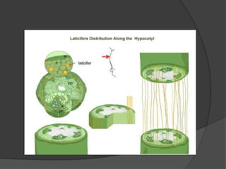
Laticifers / laticiferous tissue
 Specialized parenchyma cells; living
 Fused tube like cells filled with viscous fluid called latex
 White in color / yellow( Papaveraceae and
Cannabinaceae)/ colorless ( Moraceae)
 Suspension contains various substances like waxes,
resins, proteins, essential oils, alkaloids and sugars
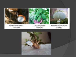
 Alkaloid-Papaver
 Enzymes-Papaya
 Latex-source of rubber (Hevea and Ficus)
 Chicle (Achras); Papain (Corica)
 Monocots - Dracaena
Cell structure of laticifers
 Cells contain living protoplast
 Vacuoles well developed
 Vacuoles store latex
 Cells usually multinucleate
 Latex may be milky white , colored or
colorless
Chemical composition of latex
 Latex is a mixture of chemical compounds
 Contains carbohydrates, organic acid, alkaloids, terpenes,
resins and enzymes
 Latex of Euphorbia mili contains dumbbell shaped starch
grains
 Latex of Papaverm somniferum contain alkaloids (morphine,
codeine)
 Latex of Carica papaya contain digestive enzyme popain
 Latex of Hevea contain rubber
 Latex of Achras saporta contain chicles
 Rubber particles suspended in the latex
Types of laticifers
 On the basis of structure and
development-
Nonarticulate latex ducts / Latex cells
Articulate latex duct/ Latex vessels
- Content and funtion of them are same
and both may occur in same family
Types of laticifers
Nonarticulate latex ducts/ Latex
cells
 These are multinucleated cells of immeasurable length
 Initials originates within the meristematic mass of embryo
 Develops from single cell
 This initials grow more rapidly than neighbouring cells and
form highly branched systems in the intercellular space
 Elongate by intrusive growth
 Occupy intercellular spaces of other cells
 Eg. Apocynaceae
Asclepiadaceae
Euphorbiaceae
Urticaceae
Nonarticulate latex ducts
 They originate as minute structures, elongate quickly and
ramify in all directions of the plant by repeated branching
,but they not fuse together , thus no netted structure are
formed as they are formed in articulate ducts
 The walls of the ducts are soft and very often thick
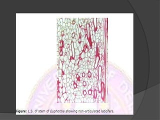
 These may be or may not be branched
 Branched nonarticulated laticifers commonly occur in
leaves. here they travel through the vascular bundles ramify
in the mesophyll and often reach the epidermis
 Unbranched non-articulated laticifres of Vinca and
Cannabis occur in the primary phloem but are apperently
absent in the secondry tissue
Types of Nonarticulate latex ducts /
latex cells
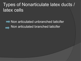
Non articulated unbranched laticifer
Non articulated branched laticifer
a. Non-articulate unbranched
laticifers
 They develop into long ,more or less
straight tubes
 Eg:- Cannabaceae (Humulus , Cannabis)
Apocynaceae (Vinca)
Urticaceae (Urtica)
b. Non articulated branched
laticifers
 They branched repeatedly, each cell thus
forming an immense system of tubes
 Eg:- Asclepiadaeae (Asclepias, Cryptostegia)
Euphorbiaeae (Euphorbia)
Moraceae ( Ficus, Morus)
Apocynaceae( Nerium oleander)
Cyclanthaceae( Cyclanthus bipartitus)
Articulate latex ducts/ Latex
vessels
 Compound in origin : made up of number of cells, joined
end to end to form longitudinal files of cells
 These are living coenocytic cells
 Initiation takes place in the ontogeny of plant
 Resembles with xylem vessels ; joined together with end
walls dissoled
 Eg- Compositae, Euphorbiacece, Papaveraceae ,
Moraceae
 Laticifer show various arrangement and a frequent
association with the phloem
Articulate latex ducts
 In Carica papaya the laticifer apparently
occur not only in the phloem but also in
the xylem
First pic shows both non articulate and articulate laticifers
second shows only articulate laticifers
Types of articulate latex ducts
/vessels
 Articulated nonanastomosing laticifer
Articulated anastomosing laticifer
A. Articulated nonanastomosing laticifer
B. Articulated anastomosing latiifer
a.Articulated nonanastomosing
laticifers
 They consist of a series of long cell chainsor
compound chains or tubes not connected with
each other laterally
 Eg:- Papaveraceae (Chelidonium)
Convolvulaceae(Convolvulus,Dichondra)
Sapotaceae (Achrassaota, Manilkarazapota)
Musaceae (Musa)
Liliaceae (Allium)
b. Articulated anastomosing
laticifers
 They form lateral anastomes with similar
cell chains or tubes, all united into a
netlike structure or reticulum
 Eg:-Caricaceae (Carica papaya)
Euphorbiaceae (Hevea, Manihot)
Papaveraceae (Argemone,Papaver)
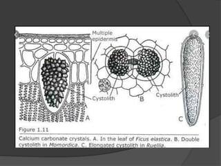
Lithocysts
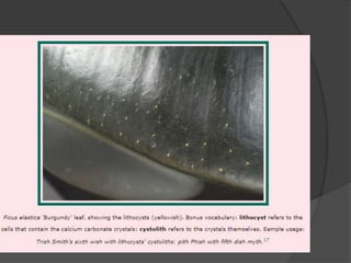
Cystolith which is a botanical term for outgrowths
of the epidermal cell wall , usually of calcium
carbonate , formed in a cellulose matrix in
special cells called Lithocysts , generally in the
leaf of plants
it includes in genera of Acanthaceae
plants in the family Urticaceae also form leaf
cystolith , but only during their later flowering
seed setting stages
also in plants of family Cannabaceae- Cannabis
and Moraceae – Ficus elastica
Laticifers
Laticifers
Laticifers
Laticifers