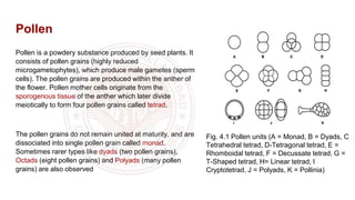
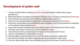
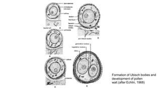
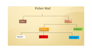
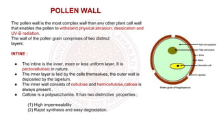
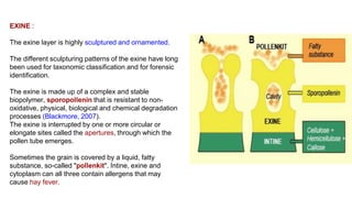
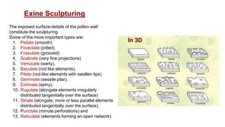
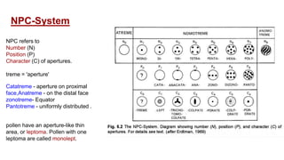
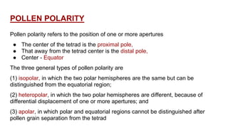
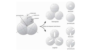
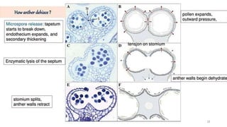
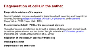
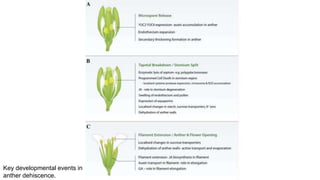
Vascular Cambium & Seasonal activity & its Role in Stem & RootFatima Ramay
Vascular Cambium & Seasonal activity & its Role in Stem & Root:
The vascular cambium (pl. cambia or cambiums) is a lateral meristem in the vascular tissue of plants.
The vascular cambium is a cylindrical layer of cambium that runs through the stem of a plant that undergoes secondary growth.
In Dicots:
The vascular cambium is in dicot stems and roots, located between the xylem and the phloem in the stem and root of a vascular plant, and is the source of both the secondary xylem growth (inwards, towards the pith) and the secondary phloem growth (outwards).
In Monocots:
Monocot stems, such as corn, palms and bamboos, do not have a vascular cambium and do not exhibit secondary growth by the production of concentric annual rings. They cannot increase in girth by adding lateral layers of cells as in conifers and woody dicots.
Cambium of some plants remains active for the entire period of their life, i.e., cambial cells divide and resulting cells mature to form xylem and phloem elements.
This type of seasonal activity usually found in the plants present in the tropical regions, and not all plants show cambial activity.
Percentage of ringless trees in the rain forests of;India : 75%Amazon : 43%Malaysia : 15%
In regions with definite seasonal climate; seasonal activity of cambium ceased with onset of unfavorable conditions; In Autumn, it enters the dormant state and lasts for the end of summer; In Spring, cambium again becomes active.
Duration of cambial activity is also affected by day-length, e.g., In Robinia pseudoacacia, cambium is dormant under short-day condition.
The cambium cells formed in circular in cross section from the beginning onwards.
The cambial ring is partially primary (fascicular cambium) and partially secondary (interfascicular cambium).
Periderm originates from the cortical cells (extra stelar in origin).
In Dicot stem, for mechanical support xylem is with comparatively smaller vessels, greater fibers and less parenchyma.
More amount of cork is produces for protection.
Lenticels on periderm are very prominent.
The cambial ring formed is wavy in the beginning and later becomes circular.
The cambium ring is completely secondary in origin.
Periderm originates from the pericycle (intra stelar in origin).
In Dicot root, xylem is with big thin walled vessels with few fibers and more parenchyma.
Less amount of cork is produced as root is underground.
Lenticels on periderm are not very prominent.
The Shoot apex is also known as the terminal bud of plants that grows from 0.1-1.0 mm and consists of the apical meristem, developing leaves and the immediate surrounding leaf primordial. The shoot apex is present in both dicot and monocot plants.
The "Telome theory" of Walter Zimmermann (1930, 1952) is the most accepted theory that is based on fossil record and synthesizes the major steps in the evolution of vascular plants.
It describes how the primitive type of vascular plants developed from Rhynia like plants.
The ovules is also known as megasporongia which are borne on a cushion-like tissue called placenta in the ovary. One or more than one ovules are present inside the ovary.
Taxus baccata commonly known as ‘Yew’, is an evergreen tree attaining a height of 9-20 metres with a massive trunk. Taxus is usually dioecious, but occasionally monoecious trees are also reported.
The reproductive structures become prominent on the plant in February-March. The male and female plants do not show any distinction in their vegetative organization. The differentiation between them can be made only when the plants are in the flowering or fruiting stage. Vegetative reproduction in Taxus is not known.
Class 12||Chapter 2|| Sexual Reproduction in flowering plantsPrathamBiology
This chapter includes flowers, their detailed structure and developmental processess which took place durin sexual reproduction. Helpful for Board and NEET students.
Fell free for any query or suggestion
Mail us on: biologypratham@gmail.com
Website : www.prathambiology.in
Seminar of U.V. Spectroscopy by SAMIR PANDASAMIR PANDA
Spectroscopy is a branch of science dealing the study of interaction of electromagnetic radiation with matter.
Ultraviolet-visible spectroscopy refers to absorption spectroscopy or reflect spectroscopy in the UV-VIS spectral region.
Ultraviolet-visible spectroscopy is an analytical method that can measure the amount of light received by the analyte.
Introduction:
RNA interference (RNAi) or Post-Transcriptional Gene Silencing (PTGS) is an important biological process for modulating eukaryotic gene expression.
It is highly conserved process of posttranscriptional gene silencing by which double stranded RNA (dsRNA) causes sequence-specific degradation of mRNA sequences.
dsRNA-induced gene silencing (RNAi) is reported in a wide range of eukaryotes ranging from worms, insects, mammals and plants.
This process mediates resistance to both endogenous parasitic and exogenous pathogenic nucleic acids, and regulates the expression of protein-coding genes.
What are small ncRNAs?
micro RNA (miRNA)
short interfering RNA (siRNA)
Properties of small non-coding RNA:
Involved in silencing mRNA transcripts.
Called “small” because they are usually only about 21-24 nucleotides long.
Synthesized by first cutting up longer precursor sequences (like the 61nt one that Lee discovered).
Silence an mRNA by base pairing with some sequence on the mRNA.
Discovery of siRNA?
The first small RNA:
In 1993 Rosalind Lee (Victor Ambros lab) was studying a non- coding gene in C. elegans, lin-4, that was involved in silencing of another gene, lin-14, at the appropriate time in the
development of the worm C. elegans.
Two small transcripts of lin-4 (22nt and 61nt) were found to be complementary to a sequence in the 3' UTR of lin-14.
Because lin-4 encoded no protein, she deduced that it must be these transcripts that are causing the silencing by RNA-RNA interactions.
Types of RNAi ( non coding RNA)
MiRNA
Length (23-25 nt)
Trans acting
Binds with target MRNA in mismatch
Translation inhibition
Si RNA
Length 21 nt.
Cis acting
Bind with target Mrna in perfect complementary sequence
Piwi-RNA
Length ; 25 to 36 nt.
Expressed in Germ Cells
Regulates trnasposomes activity
MECHANISM OF RNAI:
First the double-stranded RNA teams up with a protein complex named Dicer, which cuts the long RNA into short pieces.
Then another protein complex called RISC (RNA-induced silencing complex) discards one of the two RNA strands.
The RISC-docked, single-stranded RNA then pairs with the homologous mRNA and destroys it.
THE RISC COMPLEX:
RISC is large(>500kD) RNA multi- protein Binding complex which triggers MRNA degradation in response to MRNA
Unwinding of double stranded Si RNA by ATP independent Helicase
Active component of RISC is Ago proteins( ENDONUCLEASE) which cleave target MRNA.
DICER: endonuclease (RNase Family III)
Argonaute: Central Component of the RNA-Induced Silencing Complex (RISC)
One strand of the dsRNA produced by Dicer is retained in the RISC complex in association with Argonaute
ARGONAUTE PROTEIN :
1.PAZ(PIWI/Argonaute/ Zwille)- Recognition of target MRNA
2.PIWI (p-element induced wimpy Testis)- breaks Phosphodiester bond of mRNA.)RNAse H activity.
MiRNA:
The Double-stranded RNAs are naturally produced in eukaryotic cells during development, and they have a key role in regulating gene expression .
Observation of Io’s Resurfacing via Plume Deposition Using Ground-based Adapt...Sérgio Sacani
Since volcanic activity was first discovered on Io from Voyager images in 1979, changes
on Io’s surface have been monitored from both spacecraft and ground-based telescopes.
Here, we present the highest spatial resolution images of Io ever obtained from a groundbased telescope. These images, acquired by the SHARK-VIS instrument on the Large
Binocular Telescope, show evidence of a major resurfacing event on Io’s trailing hemisphere. When compared to the most recent spacecraft images, the SHARK-VIS images
show that a plume deposit from a powerful eruption at Pillan Patera has covered part
of the long-lived Pele plume deposit. Although this type of resurfacing event may be common on Io, few have been detected due to the rarity of spacecraft visits and the previously low spatial resolution available from Earth-based telescopes. The SHARK-VIS instrument ushers in a new era of high resolution imaging of Io’s surface using adaptive
optics at visible wavelengths.
Cancer cell metabolism: special Reference to Lactate PathwayAADYARAJPANDEY1
Normal Cell Metabolism:
Cellular respiration describes the series of steps that cells use to break down sugar and other chemicals to get the energy we need to function.
Energy is stored in the bonds of glucose and when glucose is broken down, much of that energy is released.
Cell utilize energy in the form of ATP.
The first step of respiration is called glycolysis. In a series of steps, glycolysis breaks glucose into two smaller molecules - a chemical called pyruvate. A small amount of ATP is formed during this process.
Most healthy cells continue the breakdown in a second process, called the Kreb's cycle. The Kreb's cycle allows cells to “burn” the pyruvates made in glycolysis to get more ATP.
The last step in the breakdown of glucose is called oxidative phosphorylation (Ox-Phos).
It takes place in specialized cell structures called mitochondria. This process produces a large amount of ATP. Importantly, cells need oxygen to complete oxidative phosphorylation.
If a cell completes only glycolysis, only 2 molecules of ATP are made per glucose. However, if the cell completes the entire respiration process (glycolysis - Kreb's - oxidative phosphorylation), about 36 molecules of ATP are created, giving it much more energy to use.
IN CANCER CELL:
Unlike healthy cells that "burn" the entire molecule of sugar to capture a large amount of energy as ATP, cancer cells are wasteful.
Cancer cells only partially break down sugar molecules. They overuse the first step of respiration, glycolysis. They frequently do not complete the second step, oxidative phosphorylation.
This results in only 2 molecules of ATP per each glucose molecule instead of the 36 or so ATPs healthy cells gain. As a result, cancer cells need to use a lot more sugar molecules to get enough energy to survive.
introduction to WARBERG PHENOMENA:
WARBURG EFFECT Usually, cancer cells are highly glycolytic (glucose addiction) and take up more glucose than do normal cells from outside.
Otto Heinrich Warburg (; 8 October 1883 – 1 August 1970) In 1931 was awarded the Nobel Prize in Physiology for his "discovery of the nature and mode of action of the respiratory enzyme.
WARNBURG EFFECT : cancer cells under aerobic (well-oxygenated) conditions to metabolize glucose to lactate (aerobic glycolysis) is known as the Warburg effect. Warburg made the observation that tumor slices consume glucose and secrete lactate at a higher rate than normal tissues.
Nutraceutical market, scope and growth: Herbal drug technologyLokesh Patil
As consumer awareness of health and wellness rises, the nutraceutical market—which includes goods like functional meals, drinks, and dietary supplements that provide health advantages beyond basic nutrition—is growing significantly. As healthcare expenses rise, the population ages, and people want natural and preventative health solutions more and more, this industry is increasing quickly. Further driving market expansion are product formulation innovations and the use of cutting-edge technology for customized nutrition. With its worldwide reach, the nutraceutical industry is expected to keep growing and provide significant chances for research and investment in a number of categories, including vitamins, minerals, probiotics, and herbal supplements.
Nutraceutical market, scope and growth: Herbal drug technology
Pollen wall morphogenesis and anther Dehiscence
1. Pollen wall morphogenesis
and anther dehiscence
Presentation by - Deepanshi patel
Department of Botany
University of Allahabad
2. Pollen
Pollen is a powdery substance produced by seed plants. It
consists of pollen grains (highly reduced
microgametophytes), which produce male gametes (sperm
cells). The pollen grains are produced within the anther of
the flower. Pollen mother cells originate from the
sporogenous tissue of the anther which later divide
meiotically to form four pollen grains called tetrad.
The pollen grains do not remain united at maturity, and are
dissociated into single pollen grain called monad.
Sometimes rarer types like dyads (two pollen grains),
Octads (eight pollen grains) and Polyads (many pollen
grains) are also observed
Fig. 4.1 Pollen units (A = Monad, B = Dyads, C
Tetrahedral tetrad, D-Tetragonal tetrad, E =
Rhomboidal tetrad, F = Decussate tetrad, G =
T-Shaped tetrad, H= Linear tetrad, I
Cryptotetrad, J = Polyads, K = Pollinia)
4. Development of pollen wall
1. 1st layer of Pollen Wall is of cellulose-Primexine. Deposited between callose wall and sporę
plasmalemma
2. Below plasmalemma are plates of endoplasmic reticulum. Here, primexine is discontinuous-Germpores
3. Once primexine has reached a certain thickness, additional gaps appear in it
4. Columns of convoluted lamellae are deposited in these gaps on plasmalemma-Probacula
5. Precursors of sporopollenin are synthesized by spore cytoplasm, which are polymerised and deposited
on the surface of these lamellae-Bacula
6. Lower ends of bacula spread side ways into cellulosic layers of primexine and form the Foot layer
7. Foot layer is like a floor on which the columns or bacula are raised
8. Tops of bacula columns can also spread side ways in all directions to form Tectum
9. Tectum is responsible for intricate pattern of exine
10. All these stages of wall development occur while the spore is enclosed in callose wall
11. With maturity of tetrads, the callose is degraded abd spores released in anther cavity
12. Free from callose pollen grains now synthesize Intine and innermost layer of exine-the endexine
5.
6.
7. The pollen wall is the most complex wall than any other plant cell wall
that enables the pollen to withstand physical abrasion, dessication and
UV-B radiation.
The wall of the pollen grain comprises of two distinct
layers:
INTINE :
● The intine is the inner, more or less uniform layer. It is
pectocellulosic in nature.
● The inner layer is laid by the cells themselves, the outer wall is
deposited by the tapetum.
● The inner wall consists of cellulose and hemicellulose,callose is
always present .
● Callose is a polysaccharide. It has two distinctive properties ;
(1) High impermeability
(2) Rapid synthesis and easy degradation.
POLLEN WALL
8. EXINE :
The exine layer is highly sculptured and ornamented.
The different sculpturing patterns of the exine have long
been used for taxonomic classification and for forensic
identification.
The exine is made up of a complex and stable
biopolymer, sporopollenin that is resistant to non-
oxidative, physical, biological and chemical degradation
processes (Blackmore, 2007).
The exine is interrupted by one or more circular or
elongate sites called the apertures, through which the
pollen tube emerges.
Sometimes the grain is covered by a liquid, fatty
substance, so-called "pollenkit". Intine, exine and
cytoplasm can all three contain allergens that may
cause hay fever.
9. The exposed surface-details of the pollen wall
constitute the sculpturing.
Some of the more important types are:
1. Psilate (smooth)
2. Foveolate (pitted)
3. Fossulate (grooved)
4. Scabrate (very fine projections)
5. Verrucate (warty),
6. Baculate (rod like elements),
7. Pilate (rod-like elements with swollen tips)
8. Gemmate (sessile pilar),
9. Echinate (spiny),
10. Rugulate (elongate elements irregularly
distributed tangentially over the surface)
11. Striate (elongate, more or less parallel elements
distributed tangentially over the surface),
12. Punctate (minute perforations) and
13. Reticulate (elements forming an open network).
Exine Sculpturing
10. An aperture is any weak area on the pollen surface which is
directly or indirectly associated with its germination.
Long apertures are called colpi, and short ones pores.
The apertures may be simple or compound.
Pollen grains with simple apertures are either colpate (with
colpi) or porate (with pores).
A compound aperture consists of a central region called oral,
and an outer region called colpal in colporate pollen (with
compound colpi), and poral in pororate pollen (with compound
pores).
Pollen Aperture
11. NPC refers to
Number (N)
Position (P)
Character (C) of apertures.
treme = 'aperture'
NPC-System
Catatreme - aperture on proximal
face,Anatreme - on the distal face
zonotreme- Equator
Pantotreme - uniformly distributed .
12. POLLEN POLARITY
Pollen polarity refers to the position of one or more apertures
● The center of the tetrad is the proximal pole,
● That away from the tetrad center is the distal pole,
● Center - Equator
The three general types of pollen polarity are
(1) isopolar, in which the two polar hemispheres are the same but can be
distinguished from the equatorial region;
(2) heteropolar, in which the two polar hemispheres are different, because of
differential displacement of one or more apertures; and
(3) apolar, in which polar and equatorial regions cannot be distinguished after
pollen grain separation from the tetrad
13.
14. Anther Dehiscence
Anther dehiscence is a multistage process involving localized differentiation and
degeneration, combined with changes in structure and water status of the anther
(Wilson et al., 2011).
Anther dehiscence involves three types of specialized cells:
(i) stomium,
(ii) septum
(iii) endothecium.
The stomium differentiates before the microspore mother cells enter meiosis. It
comprises of small specialized epidermal cells and, at anther maturation, splits to
facilitate anther dehiscence.
15. Four clusters of archesporial cells (Ar) in the anthers divide to form the primary parietal layer (PP)
and the primary sporogenous layer (Sp). The PP layer then goes through a further division to form
two secondary parietal layers, the inner secondary parietal layer (ISP) and the outer secondary
parietal layer (OSP). The OSP then divides again and differentiates to form the endothecium layer
(En), whereas the ISP divides and develops to form the tapetum (T) and middle cell layer (M).
16. E, epidermis; En,
endothecium; ML, middle
layer; T, tapetum; Sm,
septum; St, stomium; MMC,
microspore mother cells;
Ms, microsporocytes; Tds,
tetrads; Msp, microspores;
PG, pollen grain
Recap…!!
Fig: Anther morphology and key
events of anther development
17. 1. longitudinal Dehiscing- along long axis of theca.
1. poricidal Dehiscing -through a pore at apex of
theca.
1. transverse Dehiscing - at right angles to long axis
of theca.
1. valvular Dehiscing - through a pore covered by a
flap of tissue.
Types of anther dehiscence
18. The septum, that separates the two lobes of an anther, breaks down at a later
stage and the two sporangia of an anther lobe become joined to forms a single
locule
The endothecium is the hypodermal layer of the anther wall, which after the
release of microspores from the tetrads
It undergoes expansion and deposition of ligno-cellulosic secondary thickening
that arise from the inner tangential walls and run outward and upward ending near
the outer wall of each cell
The outer tangential wall remains thin. The thickening may be annular-rib type,
helical-rib type, reticulate-rib type or palmate-rib type depending on the species.
19.
20. Degeneration of cells in the anther
Enzymatic breakdown of the septum
Several hydrolytic enzymes and proteins linked to cell wall loosening are thought to be
involved, including polygalacturonases (PGs),b-1,4-glucanases, and expansins
(Bonghi et al., 1993; Taylor et al., 1993)
Programmed cell death (PCD) of the septum and stomium
The anther septum and stomium go through a process of degeneration and cell death
to facilitate pollen release, and this is also thought to be via a PCD-related process
(Kuriyama and Fukuda, 2002; Sanders et al., 2005).
Regulation of endothecium secondary thickening
Opening the anther
Dehydration of the anther wall