Downloaded 289 times
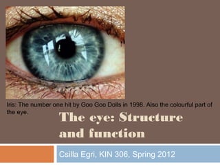

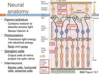
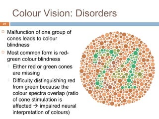
![Phototransduction: Dark current
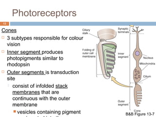
14
Partially active guanylyl cyclase
keeps cytoplasmic [cGMP] high in the
dark
Outer segment contains cGMP-gated
cation channels
Influx of Na+ and Ca2+
Inner segment contains non-gated K+
selective channels
K+ efflux
Resting, or dark Vm is -40 mV
concentration gradients maintained
by Na+/K+ pump and NCX
Kandel Figure 26-5A](https://image.slidesharecdn.com/0gv50marmsyzuocn7ng4-signature-877c3c8e10e04f4ec6f52a04e46b4c13371f8b21670841002d959bd97f2e7e4a-poli-141108120751-conversion-gate02/85/Visual-System-Structure-and-Function-14-320.jpg)
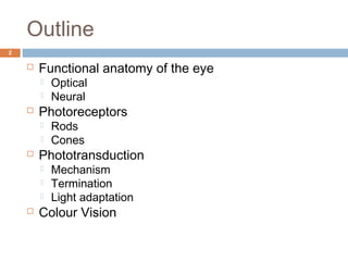
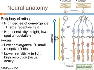
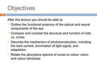
![Phototransduction
15
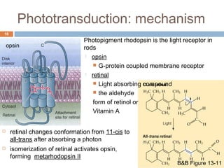
Photoreceptors hyperpolarize in response to light and release less
neurotransmitter
In darkness, the Vm of -40 mV keeps CaV channels in the
synaptic terminal open
photoreceptors continuously release neurotransmitter
glutamate
absorption of light by photopigment ’s [cGMP]
cation channels close
K+ efflux predominates, hyperpolarizes cell (-70mV)
CaV channels close, decreased release of glutamate](https://image.slidesharecdn.com/0gv50marmsyzuocn7ng4-signature-877c3c8e10e04f4ec6f52a04e46b4c13371f8b21670841002d959bd97f2e7e4a-poli-141108120751-conversion-gate02/85/Visual-System-Structure-and-Function-15-320.jpg)
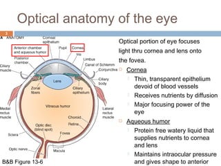
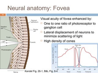
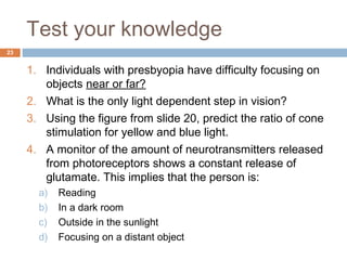
![Phototransduction: mechanism
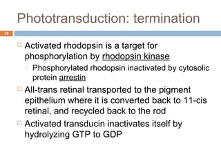
17
light
1. Absorption of a photon isomerizes retinal
a) Converts opsin to metarhodopsin II
2. Metarodophsin II activates the G-protein transducin
a) Activates cGMP phosphodiesterase (PDE)
3. PDE hydrolyzes cGMP to GMP
a) Decreased [cGMP] closes cGMP gated cation channels
b) Photoreceptor hyperpolarizes, less glutamate released Kandel Figure 26-4](https://image.slidesharecdn.com/0gv50marmsyzuocn7ng4-signature-877c3c8e10e04f4ec6f52a04e46b4c13371f8b21670841002d959bd97f2e7e4a-poli-141108120751-conversion-gate02/85/Visual-System-Structure-and-Function-17-320.jpg)
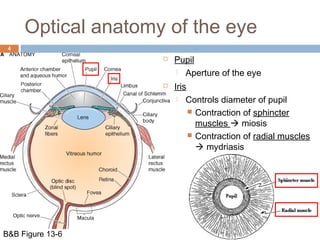
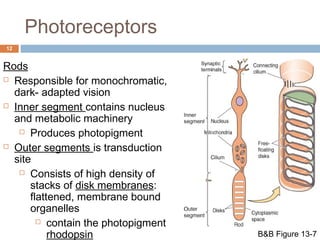
![Phototransduction: light
adaptation 19
Eyes adapt to increased light intensity and
remain sensitive to further changes in light
Optic adaptation:
Constriction of pupils to allow in less light
Photoreceptor adaptation:
The closure of cGMP gated channel reduces
inward flux of Ca2+ decreased [Ca2+]i
Ca2+ induced inhibition of guanylyl cyclase
removed
More cGMP made reopening of some cGMP gated
channels influx of cations slight depolarization
Photoreceptor can once again be stimulated
(hyperpolarized) by photons](https://image.slidesharecdn.com/0gv50marmsyzuocn7ng4-signature-877c3c8e10e04f4ec6f52a04e46b4c13371f8b21670841002d959bd97f2e7e4a-poli-141108120751-conversion-gate02/85/Visual-System-Structure-and-Function-19-320.jpg)

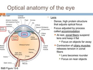
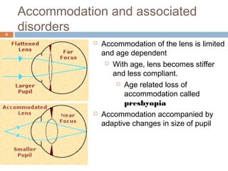
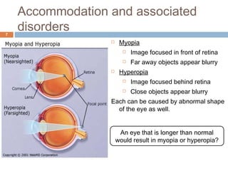
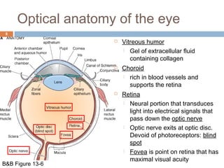
This document provides an overview of the structure and function of the eye. It discusses the optical and neural anatomy of the eye, including the cornea, iris, lens, retina, and photoreceptors. It explains the mechanisms of phototransduction, color vision, and how the eye adapts to light levels. Disorders like presbyopia, myopia, and color blindness are also summarized. The document is presented as a lecture outline with definitions and diagrams to aid understanding of the key components and processes of vision.


![PERI-PROSTHETIC FRACTURE NAIL-PLATE CONSTRUCT [NPC].pptx](https://cdn.slidesharecdn.com/ss_thumbnails/drarunkumardrmohamedashrafperiprostheticfrasturenail-plateconstructnpc-260209164459-7e9d15a1-thumbnail.jpg?width=640&height=640&fit=bounds)


