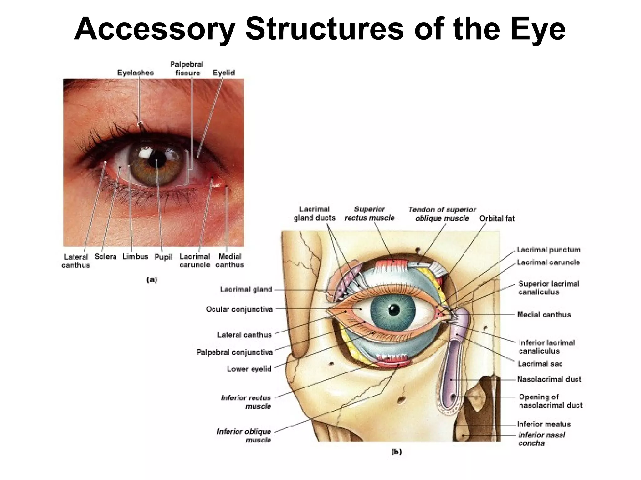
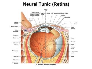
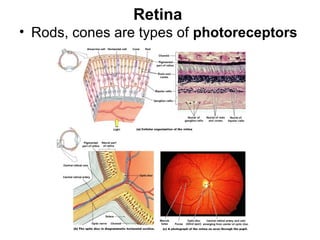
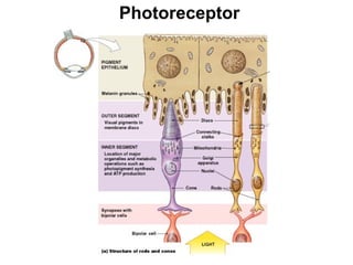
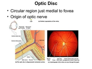
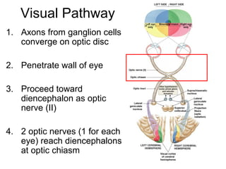
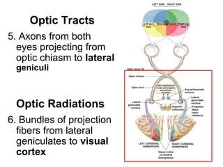
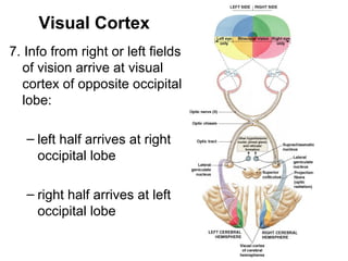
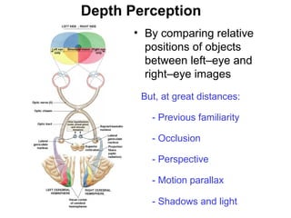
Accessory Structures of the Eye

Figure 17–3
Neural Tunic (Retina)

Figure 17–4c
Retina
• Rods, cones are types of photoreceptors

Figure 17–6
Rods
• Highly sensitive to light, do not discriminate colors
• Rods dominate peripheral areas of retina
• Provide low-resolution black–and–white vision in
dimly lit environments

Cones
• Sensitive to colored light
• Densely clustered in fovea
• Provide high–resolution color vision in brightly lit
environments
Photoreceptor

Figure 17–13a
• Rods and cones synapse with neurons
called bipolar cells
• Bipolar cells then synapse with neurons
called ganglion cells

Figure 17–6a
Horizontal Cells
• Where receptors synapse with bipolar cells

Amacrine Cells
• Where bipolar cells synapse with ganglion
cells
• Both facilitate or inhibit communication
between photoreceptors and ganglion cells
• Alter sensitivity of retina
Optic Disc
• Circular region just medial to fovea
• Origin of optic nerve

Figure 17–6b, c
Blind Spot

Figure 17–7
Visual Pigments
• Where light absorption
occurs
• Derivatives of rhodopsin
(opsin plus retinal)
• Retinal:
synthesized from vitamin A

Figure 17–13b
Phototransduction
1) Photon strikes retinal portion of rhodopsin
2) Opsin is activated:
– Goes from 11-cis form to 11-trans form
3) Opsin activates transducin (G protein), then
activates phosphodiesterase (PDE)
4) Cyclic-GMP (cGMP) levels decline; gated sodium
channels close
5) Dark current is reduced; neurotransmitter release
declines
In dark,
photoreceptors are
activated (stimulated)

Dark Current
In light,
photoreceptors are
inactivated (inhibited)

Figure 17–14
Bleaching
• Rhodopsin molecule breaks down into
retinal and opsin

Figure 17–15
Dark-Adapted State
• Pupil dilates
• Most pigments can be activated
• A single photon can be detected

Light-Adapted State
• Pupil constricts
• Bleaching of visual pigments occurs
• “Blinding” when go from dark to light room
Pupillary Muscle Reflexes

Figure 17–5
Color Sensitivity
• Integration of
information from
red, green, and
blue cones
• All wavelengths
reflected off an
object looks white

Figure 17–16
Color Blindness
• Inability to detect certain colors

Figure 17–17
Visual Pathway
• Begins at photoreceptors
• Ends at visual cortex of cerebral
hemispheres
• Message crosses 2 synapses before goes
toward brain:
–photoreceptor --> bipolar cell
–bipolar cell --> ganglion cell (sensory neuron)
“processing in retina”
Convergence
• Each ganglion cell
receives input from
many photoreceptors
• Therefore,
Receptive field of
ganglion cell is
monitored by many
photoreceptors
M Cells
• ganglion cells that monitor rods
• Provide information about:
–general form of object
–motion
–shadows in dim lighting

P Cells
• ganglion cells that monitor cones
• Provide information about:
–edges
–fine detail
–color
Visual Pathway
1. Axons from ganglion cells
converge on optic disc
2. Penetrate wall of eye
3. Proceed toward
diencephalon as optic
nerve (II)
4. 2 optic nerves (1 for each
eye) reach diencephalons
at optic chiasm
Figure 17–19
Optic Tracts
5. Axons from both
eyes projecting from
optic chiasm to lateral
geniculi

Optic Radiations
6. Bundles of projection
fibers from lateral
geniculates to visual
cortex
Visual Cortex
7. Info from right or left fields
of vision arrive at visual
cortex of opposite occipital
lobe:
– left half arrives at right
occipital lobe
– right half arrives at left
occipital lobe
Depth Perception
• By comparing relative
positions of objects
between left–eye and
right–eye images
But, at great distances:
- Previous familiarity
- Occlusion
- Perspective
- Motion parallax
- Shadows and light

Figure 17–19

Lecture12 6 13

  • 1.
    Accessory Structures ofthe Eye Figure 17–3
  • 2.
  • 3.
    Retina • Rods, conesare types of photoreceptors Figure 17–6
  • 4.
    Rods • Highly sensitiveto light, do not discriminate colors • Rods dominate peripheral areas of retina • Provide low-resolution black–and–white vision in dimly lit environments Cones • Sensitive to colored light • Densely clustered in fovea • Provide high–resolution color vision in brightly lit environments
  • 5.
  • 6.
    • Rods andcones synapse with neurons called bipolar cells • Bipolar cells then synapse with neurons called ganglion cells Figure 17–6a
  • 7.
    Horizontal Cells • Wherereceptors synapse with bipolar cells Amacrine Cells • Where bipolar cells synapse with ganglion cells • Both facilitate or inhibit communication between photoreceptors and ganglion cells • Alter sensitivity of retina
  • 8.
    Optic Disc • Circularregion just medial to fovea • Origin of optic nerve Figure 17–6b, c
  • 9.
  • 10.
    Visual Pigments • Wherelight absorption occurs • Derivatives of rhodopsin (opsin plus retinal) • Retinal: synthesized from vitamin A Figure 17–13b
  • 11.
    Phototransduction 1) Photon strikesretinal portion of rhodopsin 2) Opsin is activated: – Goes from 11-cis form to 11-trans form 3) Opsin activates transducin (G protein), then activates phosphodiesterase (PDE) 4) Cyclic-GMP (cGMP) levels decline; gated sodium channels close 5) Dark current is reduced; neurotransmitter release declines
  • 12.
    In dark, photoreceptors are activated(stimulated) Dark Current In light, photoreceptors are inactivated (inhibited) Figure 17–14
  • 13.
    Bleaching • Rhodopsin moleculebreaks down into retinal and opsin Figure 17–15
  • 14.
    Dark-Adapted State • Pupildilates • Most pigments can be activated • A single photon can be detected Light-Adapted State • Pupil constricts • Bleaching of visual pigments occurs • “Blinding” when go from dark to light room
  • 15.
  • 16.
    Color Sensitivity • Integrationof information from red, green, and blue cones • All wavelengths reflected off an object looks white Figure 17–16
  • 17.
    Color Blindness • Inabilityto detect certain colors Figure 17–17
  • 18.
    Visual Pathway • Beginsat photoreceptors • Ends at visual cortex of cerebral hemispheres • Message crosses 2 synapses before goes toward brain: –photoreceptor --> bipolar cell –bipolar cell --> ganglion cell (sensory neuron) “processing in retina”
  • 19.
    Convergence • Each ganglioncell receives input from many photoreceptors • Therefore, Receptive field of ganglion cell is monitored by many photoreceptors
  • 20.
    M Cells • ganglioncells that monitor rods • Provide information about: –general form of object –motion –shadows in dim lighting P Cells • ganglion cells that monitor cones • Provide information about: –edges –fine detail –color
  • 21.
    Visual Pathway 1. Axonsfrom ganglion cells converge on optic disc 2. Penetrate wall of eye 3. Proceed toward diencephalon as optic nerve (II) 4. 2 optic nerves (1 for each eye) reach diencephalons at optic chiasm Figure 17–19
  • 22.
    Optic Tracts 5. Axonsfrom both eyes projecting from optic chiasm to lateral geniculi Optic Radiations 6. Bundles of projection fibers from lateral geniculates to visual cortex
  • 23.
    Visual Cortex 7. Infofrom right or left fields of vision arrive at visual cortex of opposite occipital lobe: – left half arrives at right occipital lobe – right half arrives at left occipital lobe
  • 24.
    Depth Perception • Bycomparing relative positions of objects between left–eye and right–eye images But, at great distances: - Previous familiarity - Occlusion - Perspective - Motion parallax - Shadows and light Figure 17–19