This document discusses the human sense organs and the structure and function of the eye. It contains the following key points:
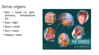
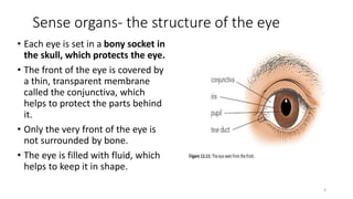
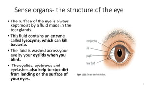
1. The five main human sense organs are the eyes, ears, nose, tongue, and skin, each of which contains receptor cells that respond to stimuli like light, sound, smell, taste, and touch.
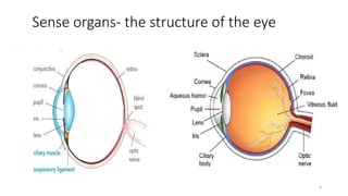
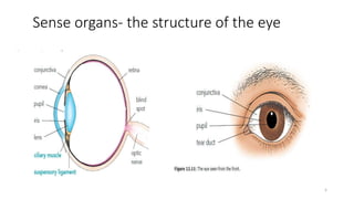
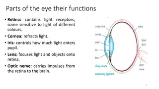
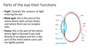
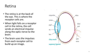
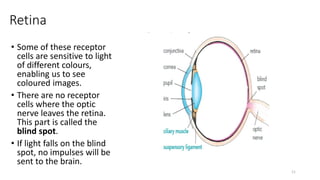
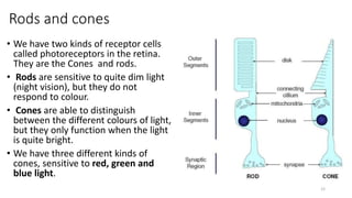
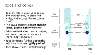
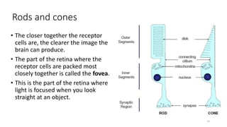
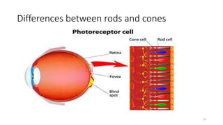
2. The eye contains specialized structures like the retina, cornea, iris, lens, and optic nerve that work together to allow vision. Rod and cone cells in the retina detect light and send signals to the brain.
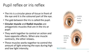
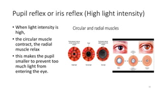
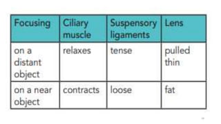
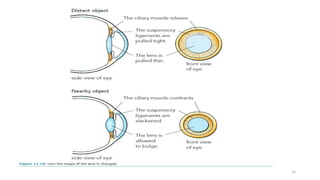
3. The iris controls the size of the pupil to regulate the amount of light entering the eye. The lens and ciliary muscles work together to focus


![Coronary heart diseṁḥṁṣhyxase[CHD] (1).pptx](https://cdn.slidesharecdn.com/ss_thumbnails/coronaryheartdiseasechd1-240415172747-bea8086f-thumbnail.jpg?width=640&height=640&fit=bounds)


