Download to read offline
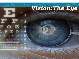
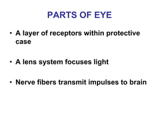
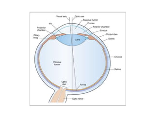
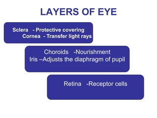
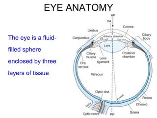
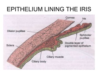
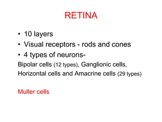
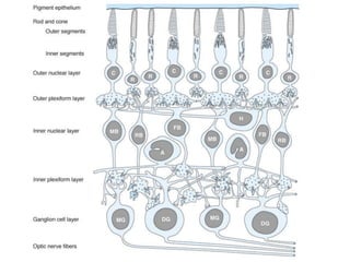
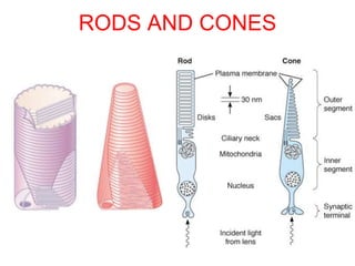
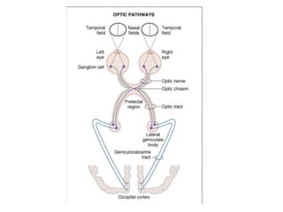
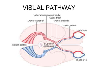
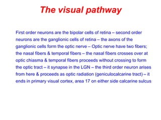
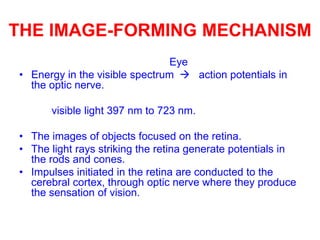
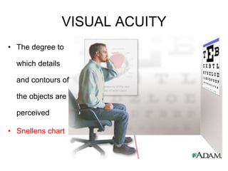
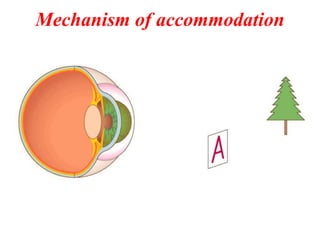
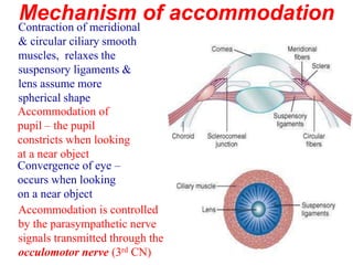
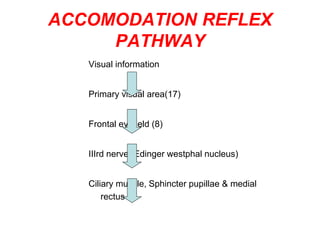
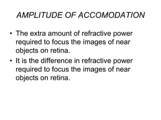
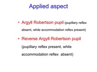
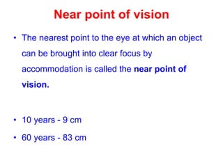
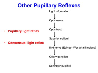
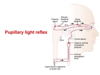
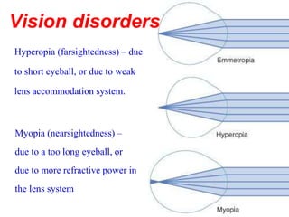
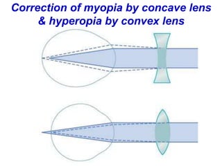
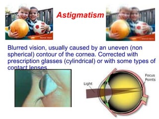
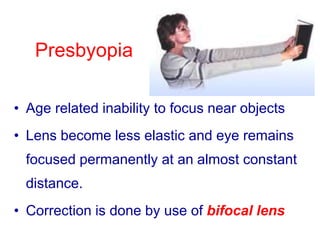
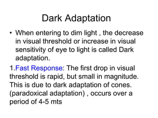
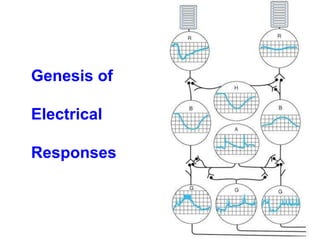
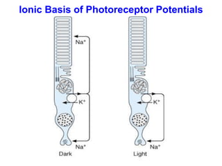
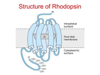
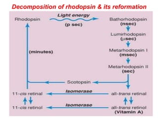

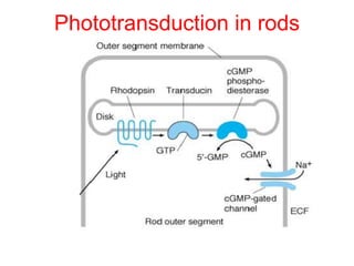
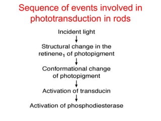
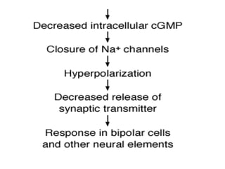
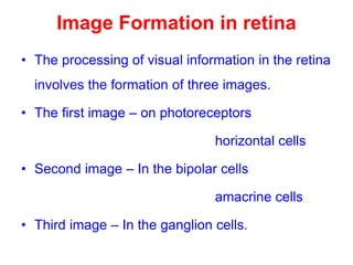
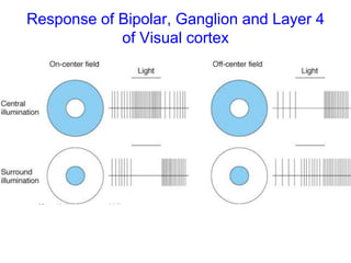
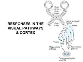

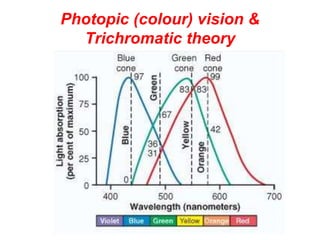
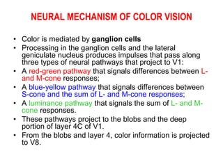
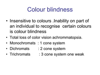
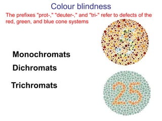


The document provides an overview of the anatomy and physiology of the visual system. It discusses the major parts of the eye including the sclera, cornea, iris, retina, rods and cones. It describes how light is focused on the retina through the lens system and how visual signals are transmitted via the optic nerve and pathways to the visual cortex. It also covers topics like color vision, accommodation, dark adaptation and various eye movements.


