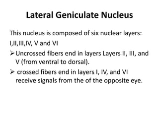
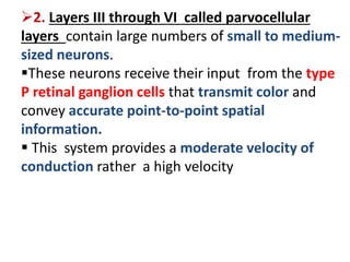
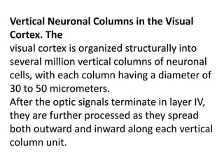
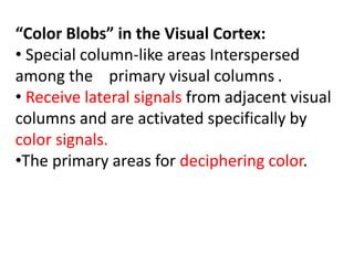
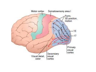
Visual Cortex
Dr. Nusrat Tariq
Ganglion Cells Of Retina
 Axons of ganglion cells form the optic nerve.
Three types of ganglion cell; W,X and Y cells.
• W cells: important for crude rod vision under dark conditions.
• X cells:
• Responsible for color vision as receives input from at least one cone.
• The X cells have small fields thus X cells transmit fine details of visual images.
• Y cells:
• Are the largest of all
• Respond to rapid changes in visual images. Give clues that make the eyes
move toward the exciting vision.
 Another classification :
• M cells.(magnocellular).
• are sensitive to low-contrast, black and white stimuli.
• P cells.(parvocellular) The P cells are sensitive to the color of a stimulus.
Lateral Geniculate Nucleus
This nucleus is composed of six nuclear layers:
I,II,III,IV, V and VI
Uncrossed fibers end in layers Layers II, III, and
V (from ventral to dorsal).
 crossed fibers end in layers I, IV, and VI
receive signals from the of the opposite eye.
The dorsal lateral geniculate nucleus is divided in
to:
1. Layers I and II called magnocellular layers
because they contain large neurons. These
neurons
receive input from the large type M retinal
ganglion cells.
This magnocellular system provides a rapidly
conducting pathway to the visual cortex.
This system is color blind, transmitting only black
and white information.
Its point-to-point transmission is poor.
2. Layers III through VI called parvocellular
layers contain large numbers of small to medium-
sized neurons.
These neurons receive their input from the type
P retinal ganglion cells that transmit color and
convey accurate point-to-point spatial
information.
 This system provides a moderate velocity of
conduction rather a high velocity
THE PRIMARY VISUAL CORTEX
the primary visual cortex has six major layers:
I
II
III
IV: a, b, cα, cβ
V
VI
Vertical Neuronal Columns in the Visual
Cortex. The
visual cortex is organized structurally into
several million vertical columns of neuronal
cells, with each column having a diameter of
30 to 50 micrometers.
After the optic signals terminate in layer IV,
they are further processed as they spread
both outward and inward along each vertical
column unit.
“Color Blobs” in the Visual Cortex:
• Special column-like areas Interspersed
among the primary visual columns .
• Receive lateral signals from adjacent visual
columns and are activated specifically by
color signals.
•The primary areas for deciphering color.
VISUAL CORTEX :
Divided into a primary visual cortex and secondary visual areas
1- Primary Visual Cortex.
 The primary visual cortex lies in the calcarine fissure area
(occipital pole).
This area is the terminus of direct visual signals from the eyes.
The peripheral retinal representation occupies the anterior part of
visual cortex.
Macular representation occupies the posterior part of visual
cortex near occipital pole.
The upper portion of the retina is represented superiorly and the
lower portion is represented inferiorly.
The primary visual cortex is also called visual area I or the striate
cortex because this area has a grossly striated appearance.
2-Secondary Visual Cortex.( visual association
areas).
Brodmann’s area 18 : called visual area II or
simply V-2.
The other, more distant secondary visual areas
have specific designations—V-3, V-4, and so forth—
up to more than a dozen areas.
lie lateral, anterior, superior, and inferior to the
primary visual cortex.
 Concerned with the interpretation of visual
impulses
various aspects of the visual image are
progressively dissected and analyzed.
TWO MAJOR PATHWAYS FOR ANALYSIS OF VISUAL
INFORMATION pathways in the secondary visual
areas :
1. THE FAST“POSITION” AND “MOTION” PATHWAY:
Analysis of Third-Dimensional Position, Gross Form, and Motion
of Objects .
2. THE ACCURATE COLOR PATHWAY:
Analysis of Visual Detail and Color.
Functions of visual cortex
Functions of primary visual cortex.(Brodmann’s
area 17).
1-Analysis of visual information by
(1) the fast“position” and “motion” pathway
(2) the accurate color pathway
2-Analysis of Contrasts in the Visual Image
3-Detects Orientation of Lines and Borders
by“Simple” Cells.
4-Detection of Line Orientation When a Line Is Displaced Laterally
or Vertically in the Visual Field by “Complex” Cells.
5-Detection of Lines of Specific Lengths, Angles, or Other Shapes
6-Detection Of Color.
Functions of secondary visual cortex/ visual association
areas/Brodmann’s area 18 and 19/ peristriate areas.
1-various aspects of the visual image are progressively dissected and
analyzed.
2-Relates visual information received by primary visual cortex to past
experience.
3- person recognize and appreciates what he is seeing.
 Removal of the primary visual cortex in the human being causes:
• loss of conscious vision—that is, blindness.
• such “blind” people can still, at times, react subconsciously to
changes in light intensity, to movement in the visual scene, or,
rarely, even to some gross patterns of vision. These reactions
include turning the eyes, turning the head, and avoidance.
• This vision is believed to be subserved by neuronal pathways that
pass from the optic tracts mainly into the superior colliculi and
other portions of the older visual system.
 Removal of secondary visual cortex causes:
 DYSLEXIA or word blindness. Person does not understand meaning
of seen words.
 Person is unable to perceive shape size and meaning of objects.

Visual cortex

  • 1.
  • 3.
    Ganglion Cells OfRetina  Axons of ganglion cells form the optic nerve. Three types of ganglion cell; W,X and Y cells. • W cells: important for crude rod vision under dark conditions. • X cells: • Responsible for color vision as receives input from at least one cone. • The X cells have small fields thus X cells transmit fine details of visual images. • Y cells: • Are the largest of all • Respond to rapid changes in visual images. Give clues that make the eyes move toward the exciting vision.  Another classification : • M cells.(magnocellular). • are sensitive to low-contrast, black and white stimuli. • P cells.(parvocellular) The P cells are sensitive to the color of a stimulus.
  • 4.
    Lateral Geniculate Nucleus Thisnucleus is composed of six nuclear layers: I,II,III,IV, V and VI Uncrossed fibers end in layers Layers II, III, and V (from ventral to dorsal).  crossed fibers end in layers I, IV, and VI receive signals from the of the opposite eye.
  • 5.
    The dorsal lateralgeniculate nucleus is divided in to: 1. Layers I and II called magnocellular layers because they contain large neurons. These neurons receive input from the large type M retinal ganglion cells. This magnocellular system provides a rapidly conducting pathway to the visual cortex. This system is color blind, transmitting only black and white information. Its point-to-point transmission is poor.
  • 6.
    2. Layers IIIthrough VI called parvocellular layers contain large numbers of small to medium- sized neurons. These neurons receive their input from the type P retinal ganglion cells that transmit color and convey accurate point-to-point spatial information.  This system provides a moderate velocity of conduction rather a high velocity
  • 7.
    THE PRIMARY VISUALCORTEX the primary visual cortex has six major layers: I II III IV: a, b, cα, cβ V VI
  • 9.
    Vertical Neuronal Columnsin the Visual Cortex. The visual cortex is organized structurally into several million vertical columns of neuronal cells, with each column having a diameter of 30 to 50 micrometers. After the optic signals terminate in layer IV, they are further processed as they spread both outward and inward along each vertical column unit.
  • 10.
    “Color Blobs” inthe Visual Cortex: • Special column-like areas Interspersed among the primary visual columns . • Receive lateral signals from adjacent visual columns and are activated specifically by color signals. •The primary areas for deciphering color.
  • 11.
    VISUAL CORTEX : Dividedinto a primary visual cortex and secondary visual areas 1- Primary Visual Cortex.  The primary visual cortex lies in the calcarine fissure area (occipital pole). This area is the terminus of direct visual signals from the eyes. The peripheral retinal representation occupies the anterior part of visual cortex. Macular representation occupies the posterior part of visual cortex near occipital pole. The upper portion of the retina is represented superiorly and the lower portion is represented inferiorly. The primary visual cortex is also called visual area I or the striate cortex because this area has a grossly striated appearance.
  • 12.
    2-Secondary Visual Cortex.(visual association areas). Brodmann’s area 18 : called visual area II or simply V-2. The other, more distant secondary visual areas have specific designations—V-3, V-4, and so forth— up to more than a dozen areas. lie lateral, anterior, superior, and inferior to the primary visual cortex.  Concerned with the interpretation of visual impulses various aspects of the visual image are progressively dissected and analyzed.
  • 15.
    TWO MAJOR PATHWAYSFOR ANALYSIS OF VISUAL INFORMATION pathways in the secondary visual areas : 1. THE FAST“POSITION” AND “MOTION” PATHWAY: Analysis of Third-Dimensional Position, Gross Form, and Motion of Objects . 2. THE ACCURATE COLOR PATHWAY: Analysis of Visual Detail and Color.
  • 17.
    Functions of visualcortex Functions of primary visual cortex.(Brodmann’s area 17). 1-Analysis of visual information by (1) the fast“position” and “motion” pathway (2) the accurate color pathway 2-Analysis of Contrasts in the Visual Image 3-Detects Orientation of Lines and Borders by“Simple” Cells.
  • 18.
    4-Detection of LineOrientation When a Line Is Displaced Laterally or Vertically in the Visual Field by “Complex” Cells. 5-Detection of Lines of Specific Lengths, Angles, or Other Shapes 6-Detection Of Color. Functions of secondary visual cortex/ visual association areas/Brodmann’s area 18 and 19/ peristriate areas. 1-various aspects of the visual image are progressively dissected and analyzed. 2-Relates visual information received by primary visual cortex to past experience. 3- person recognize and appreciates what he is seeing.
  • 19.
     Removal ofthe primary visual cortex in the human being causes: • loss of conscious vision—that is, blindness. • such “blind” people can still, at times, react subconsciously to changes in light intensity, to movement in the visual scene, or, rarely, even to some gross patterns of vision. These reactions include turning the eyes, turning the head, and avoidance. • This vision is believed to be subserved by neuronal pathways that pass from the optic tracts mainly into the superior colliculi and other portions of the older visual system.  Removal of secondary visual cortex causes:  DYSLEXIA or word blindness. Person does not understand meaning of seen words.  Person is unable to perceive shape size and meaning of objects.

Editor's Notes

  • #6 Its point-to-point transmission is poor because there are not many M ganglion cells, and their dendrites spread widely in the retina.