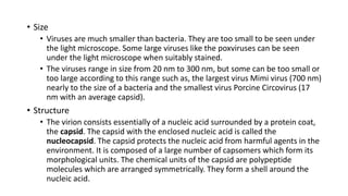
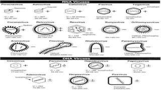
Virology is the study of viruses – submicroscopic, parasitic particles of genetic material contained in a protein coat and virus-like agents. It focuses on the following aspects of viruses: their structure, classification and evolution, their ways to infect and exploit host cells for reproduction, their interaction with host organism physiology and immunity, the diseases they cause, the techniques to isolate and culture them, and their use in research and therapy. Virology is considered to be a subfield of microbiology or of medicine.
Viruses are infectious intracellular obligate parasites with subcellular level of organisation and without protoplasm, cell, cell organells and the molecular machineries for energy metabolism and protein synthesis,Grouped on the basis of size and shape, chemical composition and structure of the genome and mode of replication
most viruses have a specific shape that is determined by the capsomeres or the envelope.
Capsid symmetry - Three types
(1) HELICAL CAPSID
(2) ICOSAHEDRALCAPSID
(3) COMPLEX CAPSID
Capsid - large macromolecular structures.
Made up of proteins called capsomers.
Chemical unit of capsomers are polypeptide chain.
Capsid - surrounded by lipoprotein layer called envelop
Envelop is made up of proteins and glycoproteins
Presence of lipid -envelope seems flexible and loose.
Envelope is composed of both the host viral components.
projections on the envelope known as spikes/peplomers which are arranged into distinct units.
VIRUSES CLASSIFICATION , LIFE CYCLE OF VIRUSES. CHARACTERISTICS OF VIRUSES Shylesh M
VIRUSES
LIFE CYCLE OF BACTERIOPHAGES
The word virus is derived from Latin word venom which means poisonous fluid that causes infection.
The branch of science that deals with the study of viruses is called Virology. It is the branch of Microbiology.
They show living characters inside the host and non living characters outside the host.
They contain either DNA or RNA as genetic material.
They have different size and shape. They cause diseases in plants, animals and micro-organisms .
Not cellular
Cannot carry on metabolic activities independently.
Contain either DNA or RNA, not both ( true cells contain both ).
Lack ribosomes and enzymes necessary for protein synthesis.
Reproduce only within cells they infect.
CLASSIFICATION OF VIRUSES
Holmes, in 1948, proposed a simple system of classifying viruses based on the type of cell (host) they infect:
Phytophagineae: They infect plants and they RNA as their genetic material. Eg: TMV,CaMV.
Zoophagineae: They infect animals and they have mostly DNA as their genetic material. Eg: Polio virus.
Pagineae: They infect bacterial cells, called bacteriophages they usually have DNA as genetic material.
Based on the viral envelope
Named after David Baltimore, a noble prize winning biologist n 1971.
1. dsDNA viruses Eg: Adenoviruses, Herpiviruses.
2. ssDNA viruses Eg: Paravoviruses.
3. dsRNA viruses Eg: Reoviruses.
4. (+)ssRNA viruses Eg: Picornaviruses.
5. (-)ssRNA viruses Eg: Orthomyxoviruses.
6. ssRNA-RT viruses Eg: Retroviruses.
7. dsDNA-RT viruses Eg: Hepadnaviruses.
Tobacco mosaic:
Causative agent: Tobacco mosaic virus (TMV)
Symptoms: The leaves of infected plants develop mosaic patches ,it is due to destruction of chlorophyll or due to production of abnormal chlorophyll .blisters appear in the region of dark green spots these may be regular or irregular in advanced stages leaves curl and get distorted.
Adsorption of the virion to the bacterial cell.
Penetration and decoating of the nucleic acid .
Protein synthesis.
Breakdown of bacterial DNA.
Arrest of host cell development.
Replication of phage DNA.
Maturation of infective progeny.
Lysis and release of newly formed phages.
Holmes, in 1948, proposed a simple system of classifying viruses based on the type of cell (host) they infect:
Phytophagineae: They infect plants and they RNA as their genetic material. Eg: TMV,CaMV.
Zoophagineae: They infect animals and they have mostly DNA as their genetic material. Eg: Polio virus.
Pagineae: They infect bacterial cells, called bacteriophages they usually have DNA as genetic material.
This presentation gives a detail overview on Viruses - Morphology and Classification. The presentation is helpful for students of B. Pharm Second Year and those who wants to gain basic knowledge about Viruses.
Subject - Microbiology
Viruses are microscopic organisms that exist almost everywhere on earth. They can infect animals, plants, fungi, and even bacteria.Viruses vary in complexity. They consist of genetic material, RNA or DNA, surrounded by a coat of protein, lipid (fat), or glycoprotein. Viruses cannot replicate without a host, so they are classified as parasitic.They are considered the most abundant biological entity on the planet.
Here we discuss the general properties of viruses in detail.
Concept of virology
Viruses
Types of viruses
Viral characteristics
Virion
Size and Shape
Structure
Replication
Viral Variation
Classification
Presentation
BEST OF LUCK
General Characters and Classification of Viruses. Includes ICTV classification and Baltimore classification of viruses. A brief explanation of the Viral structure and Lifecycle.
Bacteriophage is the most common and extensively studied virus. The life cycle of bacteriophages. The transfer of their genetic system via the process of transduction (Generalised and Specialised) and studying the gene mapping in phages. This theoretical explanation about viruses and their genetic system will help the learner in the fields of biotechnology, microbiology, basic science, life science, and various other fields of biology.
Lecture 1 - Introduction To Virology.pptxJonesChipinga
This lecture outlines why we study viruses, the virosphere,bacteriophage plaque assay, the nature of viruses, properties of viruese, viral genomics and comparison of viruses to other cells.
Slide 1: Title Slide
Extrachromosomal Inheritance
Slide 2: Introduction to Extrachromosomal Inheritance
Definition: Extrachromosomal inheritance refers to the transmission of genetic material that is not found within the nucleus.
Key Components: Involves genes located in mitochondria, chloroplasts, and plasmids.
Slide 3: Mitochondrial Inheritance
Mitochondria: Organelles responsible for energy production.
Mitochondrial DNA (mtDNA): Circular DNA molecule found in mitochondria.
Inheritance Pattern: Maternally inherited, meaning it is passed from mothers to all their offspring.
Diseases: Examples include Leber’s hereditary optic neuropathy (LHON) and mitochondrial myopathy.
Slide 4: Chloroplast Inheritance
Chloroplasts: Organelles responsible for photosynthesis in plants.
Chloroplast DNA (cpDNA): Circular DNA molecule found in chloroplasts.
Inheritance Pattern: Often maternally inherited in most plants, but can vary in some species.
Examples: Variegation in plants, where leaf color patterns are determined by chloroplast DNA.
Slide 5: Plasmid Inheritance
Plasmids: Small, circular DNA molecules found in bacteria and some eukaryotes.
Features: Can carry antibiotic resistance genes and can be transferred between cells through processes like conjugation.
Significance: Important in biotechnology for gene cloning and genetic engineering.
Slide 6: Mechanisms of Extrachromosomal Inheritance
Non-Mendelian Patterns: Do not follow Mendel’s laws of inheritance.
Cytoplasmic Segregation: During cell division, organelles like mitochondria and chloroplasts are randomly distributed to daughter cells.
Heteroplasmy: Presence of more than one type of organellar genome within a cell, leading to variation in expression.
Slide 7: Examples of Extrachromosomal Inheritance
Four O’clock Plant (Mirabilis jalapa): Shows variegated leaves due to different cpDNA in leaf cells.
Petite Mutants in Yeast: Result from mutations in mitochondrial DNA affecting respiration.
Slide 8: Importance of Extrachromosomal Inheritance
Evolution: Provides insight into the evolution of eukaryotic cells.
Medicine: Understanding mitochondrial inheritance helps in diagnosing and treating mitochondrial diseases.
Agriculture: Chloroplast inheritance can be used in plant breeding and genetic modification.
Slide 9: Recent Research and Advances
Gene Editing: Techniques like CRISPR-Cas9 are being used to edit mitochondrial and chloroplast DNA.
Therapies: Development of mitochondrial replacement therapy (MRT) for preventing mitochondrial diseases.
Slide 10: Conclusion
Summary: Extrachromosomal inheritance involves the transmission of genetic material outside the nucleus and plays a crucial role in genetics, medicine, and biotechnology.
Future Directions: Continued research and technological advancements hold promise for new treatments and applications.
Slide 11: Questions and Discussion
Invite Audience: Open the floor for any questions or further discussion on the topic.
Multi-source connectivity as the driver of solar wind variability in the heli...Sérgio Sacani
The ambient solar wind that flls the heliosphere originates from multiple
sources in the solar corona and is highly structured. It is often described
as high-speed, relatively homogeneous, plasma streams from coronal
holes and slow-speed, highly variable, streams whose source regions are
under debate. A key goal of ESA/NASA’s Solar Orbiter mission is to identify
solar wind sources and understand what drives the complexity seen in the
heliosphere. By combining magnetic feld modelling and spectroscopic
techniques with high-resolution observations and measurements, we show
that the solar wind variability detected in situ by Solar Orbiter in March
2022 is driven by spatio-temporal changes in the magnetic connectivity to
multiple sources in the solar atmosphere. The magnetic feld footpoints
connected to the spacecraft moved from the boundaries of a coronal hole
to one active region (12961) and then across to another region (12957). This
is refected in the in situ measurements, which show the transition from fast
to highly Alfvénic then to slow solar wind that is disrupted by the arrival of
a coronal mass ejection. Our results describe solar wind variability at 0.5 au
but are applicable to near-Earth observatories.
Richard's entangled aventures in wonderlandRichard Gill
Since the loophole-free Bell experiments of 2020 and the Nobel prizes in physics of 2022, critics of Bell's work have retreated to the fortress of super-determinism. Now, super-determinism is a derogatory word - it just means "determinism". Palmer, Hance and Hossenfelder argue that quantum mechanics and determinism are not incompatible, using a sophisticated mathematical construction based on a subtle thinning of allowed states and measurements in quantum mechanics, such that what is left appears to make Bell's argument fail, without altering the empirical predictions of quantum mechanics. I think however that it is a smoke screen, and the slogan "lost in math" comes to my mind. I will discuss some other recent disproofs of Bell's theorem using the language of causality based on causal graphs. Causal thinking is also central to law and justice. I will mention surprising connections to my work on serial killer nurse cases, in particular the Dutch case of Lucia de Berk and the current UK case of Lucy Letby.
Observation of Io’s Resurfacing via Plume Deposition Using Ground-based Adapt...Sérgio Sacani
Since volcanic activity was first discovered on Io from Voyager images in 1979, changes
on Io’s surface have been monitored from both spacecraft and ground-based telescopes.
Here, we present the highest spatial resolution images of Io ever obtained from a groundbased telescope. These images, acquired by the SHARK-VIS instrument on the Large
Binocular Telescope, show evidence of a major resurfacing event on Io’s trailing hemisphere. When compared to the most recent spacecraft images, the SHARK-VIS images
show that a plume deposit from a powerful eruption at Pillan Patera has covered part
of the long-lived Pele plume deposit. Although this type of resurfacing event may be common on Io, few have been detected due to the rarity of spacecraft visits and the previously low spatial resolution available from Earth-based telescopes. The SHARK-VIS instrument ushers in a new era of high resolution imaging of Io’s surface using adaptive
optics at visible wavelengths.
3. • The word “Virus” means Poison.
• Viruses infect all types of organisms, including animals and plants, as well as
bacteria and archaea.
• They stays at the borderline of living and non-living. When it enters to the cells
then only it becomes living organism otherwise it stays as non-living particle.
• They can only be seen under electron microscope. Most viruses are so tiny they
are only observable with at least a conventional optical microscope.
• They don’t have any cellular organisation like ribosome, cytoplasm, mitochondria
etc.
• They are resistant to antibiotic.
• They can replicate only inside the cells of a host organism so that they are called
obligate intracellular parasite.
4. Like all other biological entities viruses also have nucleic acid. But they have
only one type of nucleic acid, either DNA or RNA.
Viruses are classified on the basis of their characteristic feature. This
classification is called ICTV Classification. Classification on the basis of their
genome i.e. single stranded or double stranded is called as Baltimore
classification.
Example:- Double Stranded Genome viruses- Herpesvirus, Smallpox.
Single Stranded Genome Viruses- Influenza Virus, Polio Virus.
Viruses are found in almost every ecosystem on Earth and are the most
abundant type of biological entity.
The study of viruses is known as virology, a sub-speciality of microbiology.
8. • Despite his other successes, Louis Pasteur (1822–1895) was unable to find
a causative agent for rabies and speculated about a pathogen too small to
be detected using a microscope.
• In 1876, Adolf Mayer, who directed the Agricultural Experimental Station in
Wageningen was the first to show that what he called "Tobacco Mosaic
Disease" was infectious, he thought that “soluble, enzyme-like infectious
principle was involved”.
• Later, in 1892, the Russian biologist Dmitry Ivanovsky (1864–1920) used a
Chamberland filter(a filter which can filter bacterias from a sample, which
was founded by Charles Chamberland) to study the thing, what is now
known as the tobacco mosaic virus. His experiments showed that crushed
leaf extracts from infected tobacco plants remain infectious after filtration.
Ivanovsky suggested the infection might be caused by a toxin produced by
bacteria, but did not pursue the idea.
• In 1898, the Dutch microbiologist Martinus Beijerinck (1851–1931), a
microbiology teacher at the Agricultural School in Wageningen repeated
experiments by Adolf Mayer and became convinced that filtrate contained
a new form of infectious agent.
9. • He observed that the agent multiplied only in cells that were dividing and
he called it a contagium vivum fluidum (soluble living germ) and re-
introduced the word Virus and he gave the name of virus as Tobacco
Mosaic Virus.
• Beijerinck maintained that viruses were liquid in nature, a theory later
discredited by the American biochemist and virologist Wendell Meredith
Stanley (1904–1971), who proved that they were in fact, particles(non
living thing).
• In the same year Friedrich Loeffler (1852–1915) and Paul Frosch (1860–
1928) passed the first discovered animal virus named Picornavirus,
through a similar filter and discovered the cause of foot-and-mouth
disease.
• Afterwards about 5,000 virus species have been described in detail,
although there are millions of types.
11. • There is much debate among virologists about this question. Three main
hypotheses have been articulated:-
1. The progressive Hypothesis(Escape)
This hypothesis states that viruses arose from genetic elements that gained the ability to move
between cells
2. The regressive Hypothesis(Reduction)
This hypothesis asserts that viruses are remnants of cellular organisms. This hypothesis follows
that existing viruses may have evolved from more complex, possibly free-living organisms that lost
genetic information over time, as they adopted a parasitic approach to replication.
3. The Virus first hypothesis(Existence)
This hypothesis states that viruses are existed before cells.
No single hypothesis can not describe the actual origin of virus. There are
too much debate about this question.
Microbiologists generally agree that certain bacteria
that are obligate intracellular parasites, like
Chlamydia and Rickettsia species, evolved from free-
living ancestors. Indeed, genomic studies, according
to Andersson et al. , in 1998, indicate that the
mitochondria of eukaryotic cells and Rickettsia
prowazekii may share a common, free-living
ancestor.
Recently, several investigators proposed that viruses
may have been the first replicating entities. Koonin
and Martin (2005) postulated that viruses existed in
a precellular world as self-replicating units. Over
time these units, they argue, became more
organized and more complex. Eventually, enzymes
for the synthesis of membranes and cell walls
evolved, resulting in the formation of cells. Viruses,
then, may have existed before bacteria, archaea, or
eukaryotes.
13. • The International Committee on Taxonomy of Viruses (ICTV) authorizes and organizes the
taxonomic classification of and the nomenclatures for viruses.
• The ICTV have developed a universal taxonomic-scheme for viruses, and means to
describe, name, and classify every virus that affects living organisms.
• The members of the International Committee on Taxonomy of Viruses are considered
expert virologists.
• The ICTV was formed from and is governed by the Virology Division of the International
Union of Microbiological Societies.
• The name of a taxon has no official status until it has been approved by ICTV, and names
will only be accepted if they are linked to approved hierarchical taxa.
Introduction to ICTV
14. Objectives
The objectives of the International Committee on Taxonomy of Viruses are:
1. To develop an internationally agreed taxonomy for viruses.
2. To establish internationally agreed names for virus taxa.
3. To communicate the decisions reached concerning the classification and nomenclature
of viruses to virologists by holding meetings and publishing reports.
4. To maintain an Official Index of agreed names of virus taxa.
5. To study the virus effects in modern society and their behaviour. To avoid or reject the
use of names which might cause error or confusion.
6. To avoid the unnecessary creation of names.
15. • Orders
• An order is a group of families sharing certain common characters. An order name
must be a single word ending in the suffix -virales.
• Families
• A family is a group of genera, whether or not these are organized into subfamilies,
sharing certain common characters. A family name must be a single word ending in
the suffix -viridae.
• Subfamilies
• A subfamily is a group of genera sharing certain common characters. The taxon shall
be used only when it is needed to solve a complex hierarchical problem. A subfamily
name must be a single word ending in the suffix -virinae.
Rules for Taxa
16. • Genera
• A virus genus is a group of related species that share some significant properties and
often only differ in host range and virulence. A genus name must be a single word
ending in the suffix -virus.
• Species
• A species name shall consist of as few words as practicable but must not consist only
of a host name and the word virus. A species name must provide an appropriately
unambiguous identification of the species. Numbers, letters, or combinations thereof
may be used as species epithets.
Continued…
17. • Some Viruses and their Classification
Species Name Genus Subfamily Family Order
Human alphaherpesvirus Simplexvirus Alphaherpesvirinae Herpesviridae Herpesvirals
Escherichia virus Lambda Lambdavirus Tunavirinae Siphoviridae Caudovirales
Orthopoxvirus Orthopoxvirus Chordopoxvirinae Poxviradae Poxvirales
Flanders hapavirus Hapavirus Novirhabdovirinae Rhabdoviridae Mononegavirales
Avian avulavirus 1 Avulavirus Paramyoxovirinae Paramyoxoviridae Mononegavirales
19. • Size
• Viruses are much smaller than bacteria. They are too small to be seen under
the light microscope. Some large viruses like the poxviruses can be seen
under the light microscope when suitably stained.
• The viruses range in size from 20 nm to 300 nm, but some can be too small or
too large according to this range such as, the largest virus Mimi virus (700 nm)
nearly to the size of a bacteria and the smallest virus Porcine Circovirus (17
nm with an average capsid).
• Structure
• The virion consists essentially of a nucleic acid surrounded by a protein coat,
the capsid. The capsid with the enclosed nucleic acid is called the
nucleocapsid. The capsid protects the nucleic acid from harmful agents in the
environment. It is composed of a large number of capsomers which form its
morphological units. The chemical units of the capsid are polypeptide
molecules which are arranged symmetrically. They form a shell around the
nucleic acid.
20. Continued…
• Virions may be enveloped or nonenveloped. The envelope of viruses is
derived from the host cell membrane. This occurs when the virus is
released from the host cell by budding. Protein subunits may be present as
projecting spikes on the surface of the envelope. They are called
peplomers.
• The influenza virus carries two kinds of peplomers:
a) Haemagglutinin, is a triangular spike
b) Neuraminidase is mushroom-shaped.
• Envelope is sensitive to the action of lipid solvents. Envelopes confer
chemical, antigenic and biological properties on viruses.
Shape
• The overall shape of the virus particle varies in different groups of viruses.
Most animal viruses are roughly spherical. The rabies virus is bullet shaped.
Poxviruses are brick-shaped.
21.
22. Symmetry
• The capsid shows two kinds of symmetry –
i. Icosahedral (cubical)- An icosahedron is a polygon with 12 vertices and 20
facets or sides. Each facet is in the shape of an equilateral triangle. Two
types of capsomers are present in the icosahedral capsid. They are-
a. The pentagonal capsomers at the vertices (pentons)
b. The hexagonal capsomers making up the facets (hexons).
There are always 12 pentons but the number of hexons varies with
the virus group. Examples of viruses with icosahedral symmetry of the
capsid are Adenovirus and Herpes Simplex Virus.
ii. Helical- In the nucleocapsids with helical symmetry, the capsomers
and nucleic acid are wound together to form a helical or spiral tube,
for example tobacco mosaic virus.
iii. All viruses do not show the typical icosahedral or helical symmetry.
Some, like the poxviruses, show a complex symmetry.
23.
24. Figure 1 An array of viruses. (a) The helical virus of rabies. (b) The segmented helical virus of influenza. (c) A
bacteriophage with an icosahedral head and helical tail. (d) An enveloped icosahedral herpes simplex virus. (e) The
unenveloped polio virus. (f) The icosahedral HIV with spikes on its envelope.
25. • Chemical Properties:
• Viruses contain only one type of nucleic acid, either DNA or RNA. Viruses are unique
because they can carry genetic information on RNA. This property is not seen in any
other organism in nature. Viruses also contain protein which makes up the capsid.
Enveloped viruses contain lipids derived from the host cell membrane. Most viruses
do not have enzymes for the synthesis of viral components or for energy production.
Some viruses have enzymes, for example the influenza virus has neuraminidase.
• Resistance:
• Viruses are destroyed by heat except a few. They are stable at low temperatures. For
long term storage, they are kept at -70°C. A better method for prolonged storage is
lyophilisation or freeze-drying. Viruses are inactivated by sunlight, UV rays and
ionising radiation. They are, in general, more resistant than bacteria to chemical
disinfectants. Phenolic disinfectants have a weak action on viruses.
26. Multiplication of Virus
• Multiplication of virus is called Viral Replication. Viruses are
intracellular obligate parasites which means that they cannot
replicate or express their genes without the help of a living cell.
A single virus particle (Virion) is in and of itself essentially inert. It
lacks needed components that cells have to reproduce. When a
virus infects a cell, it marshals the cell's ribosomes, enzymes and
much of the cellular machinery to replicate.
• Process of Viral Replication- When a virus infects a cell, nucleic
acid will be uncoated and gain access to metabolic machinery of
cell. Then new virus formed.
27. • Steps involved in viral
replication-
1. Attachment & adsorption
2. Penetration
3. Uncoating
4. Synthesis(Replication and
Protein Production)
5. Assembly
6. Release
29. 1. Viruses contain either DNA or RNA as their genetic material, but not both. This
nucleic acid usually has unique chemical and/or physical features which makes
it distinguishable from human nucleic acid.
2. Viral nucleic acid is enclosed in a capsid made up of protein subunits called
protomeres.
3. Some species of viruses have a membrane, the envelope, surrounding the
capsid; other species do not have an envelope, i.e., they are naked.
30. Continued…
4. The morphology of a virus is determined by the arrangement of the protomeres.
When protomeres aggregate into units of five or six it forms capsomeres and then
condense to form a geometric figure having 20 equal triangular faces and 12 apices,
the virus is said to have icosahedral (cubic) morphology. When protomeres aggregate
to form a capped tube, they are said to have helical morphology. Any other
arrangement of the protomeres results in a complex morphology.
5. All viruses undergo a replication cycle in their host cell consisting of adsorption,
penetration, uncoating, nucleic acid replication, maturation and release stages.
6. In general, all DNA-containing viruses replicate in the host cell nucleus. The
exceptions to the rule are the poxviruses.
7. In general, all RNA-containing viruses replicate in the host cell cytoplasm. The
exceptions to the rule are the retroviruses and the orthomyxoviruses.
31. Continued…
8. A virally-infected cell generally presents three signals that it is infected.
The first is the production of double-stranded RNA, which induces
interferon; the second is the expression of viral protein on the surface of
the plasma membrane, thus causing activation of cytotoxic T-cells, natural
killer cells and sometimes induction of antibody synthesis. The third is the
formation of an inclusion body either within the cytoplasm or the nucleus
or very rarely within both the cytoplasm and nucleus.
32. Reference
• Mayer, Adolf (1886). "Über die Mosaikkrankheit des Tabaks.". Die
Landwirtschaftliche Versuchs-stationen (in German). 32: 451–467.
Translated into English in Johnson, J., Ed. (1942) Phytopathological
classics (St. Paul, Minnesota: American Phytopathological Society) No.
7, pp. 11–24.
• www.talk.ictvonline.org
• www.biologydiscussion.com
• Zimmer, Carl (5 September 2013). "A Catalog for All the World’s
Viruses?". New York Times. Retrieved 6 September 2013.