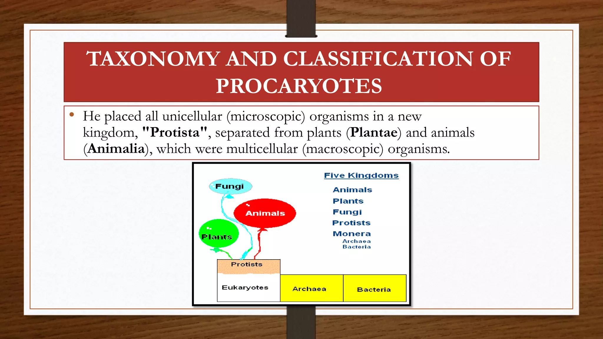
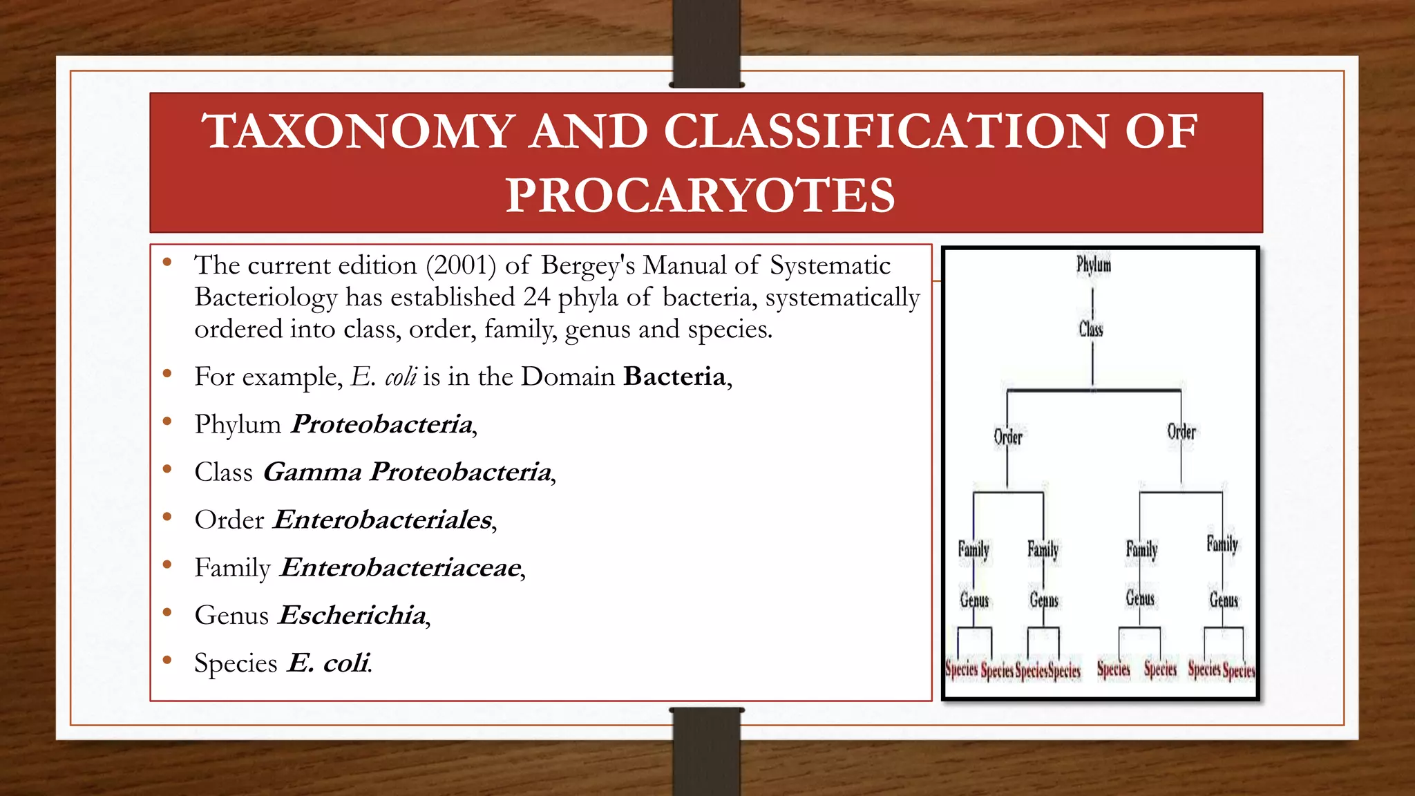
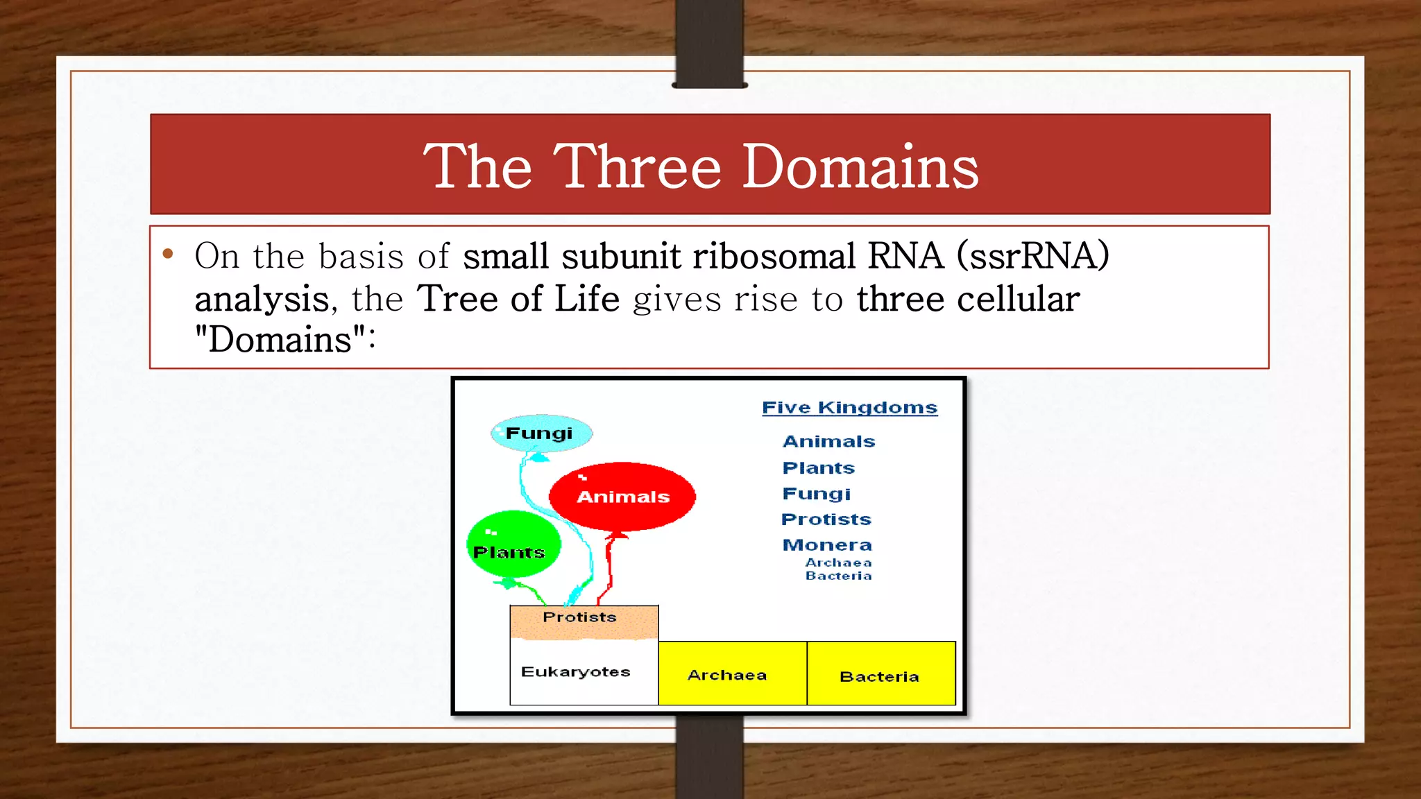
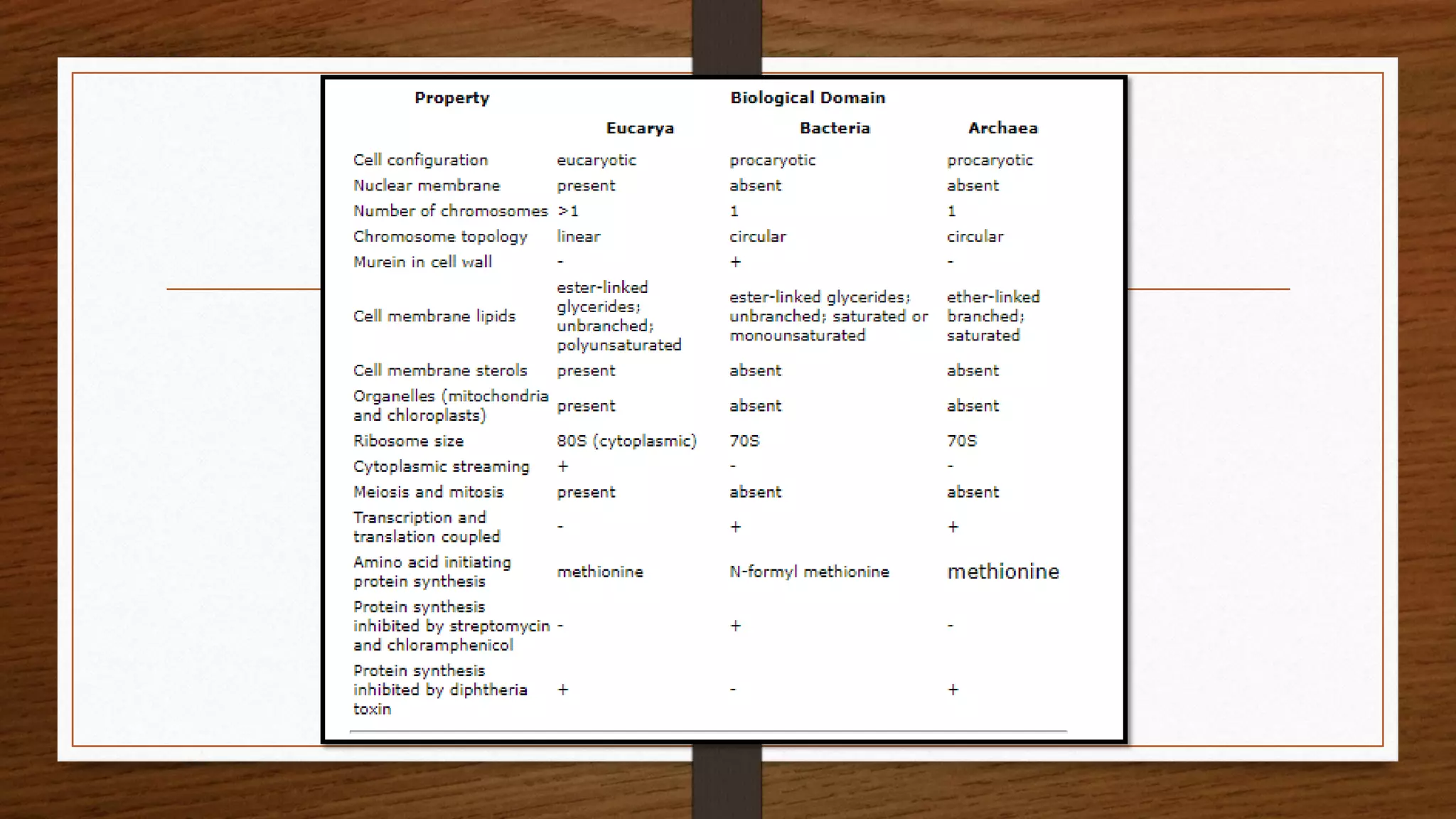
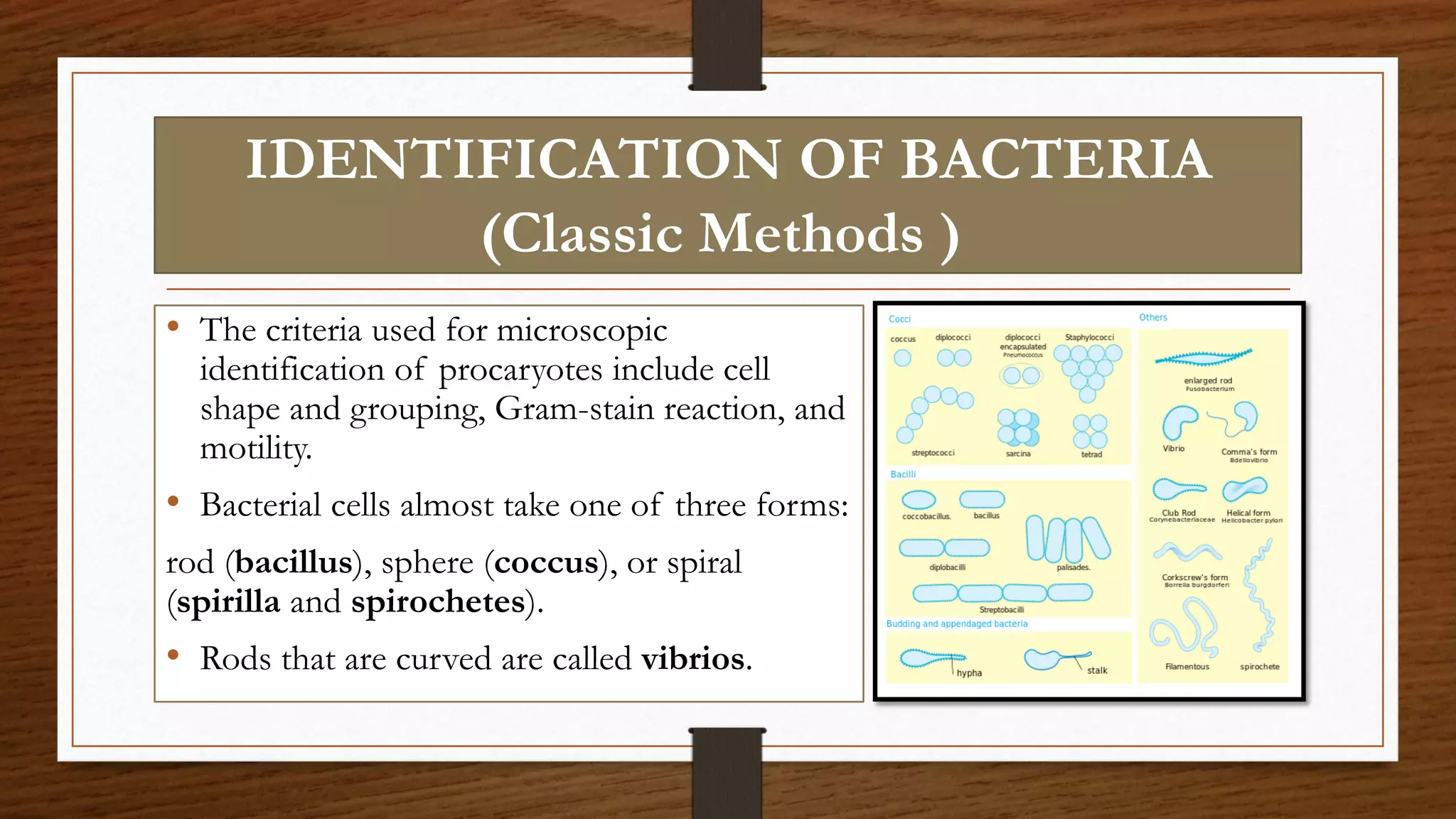
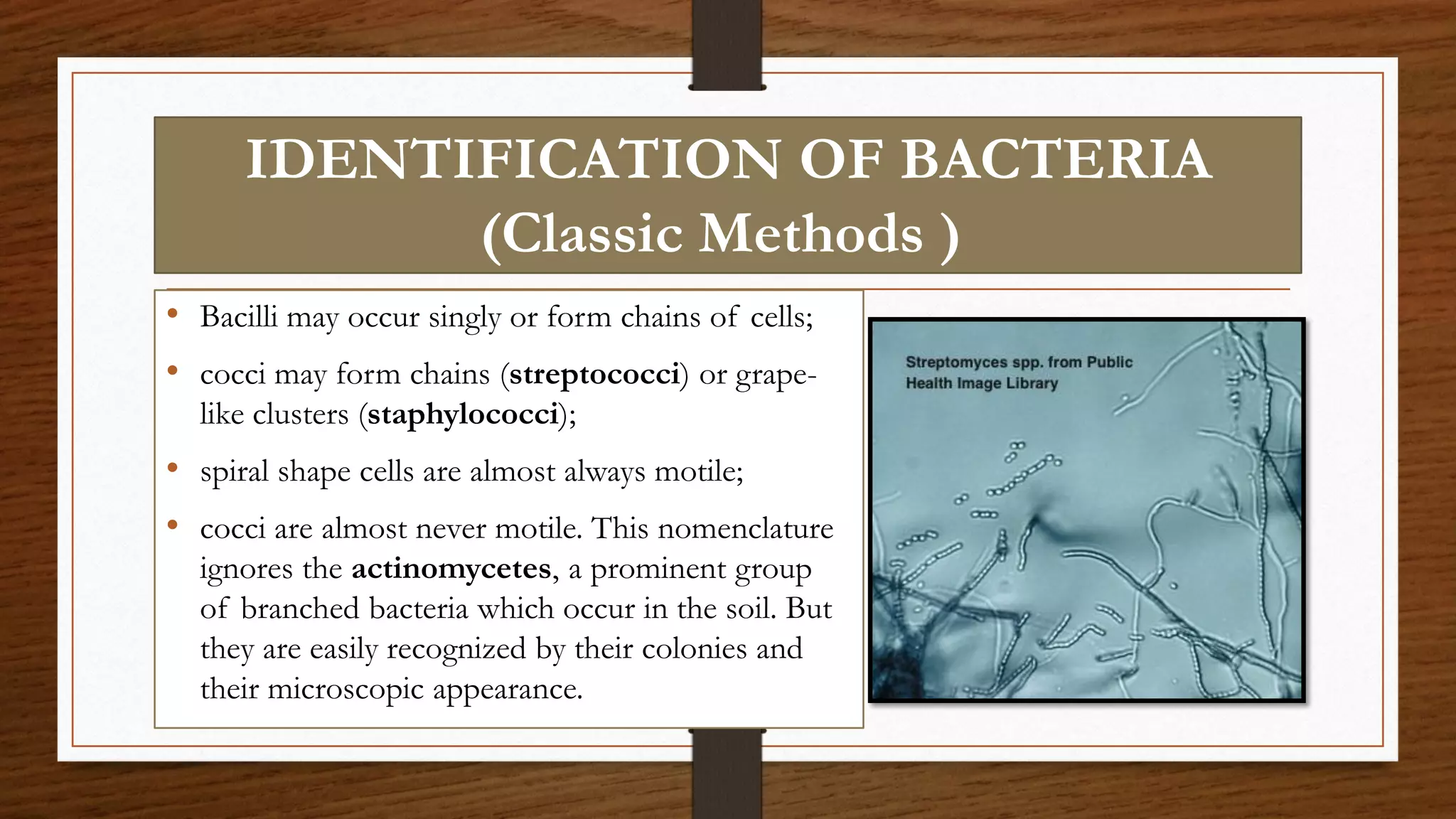
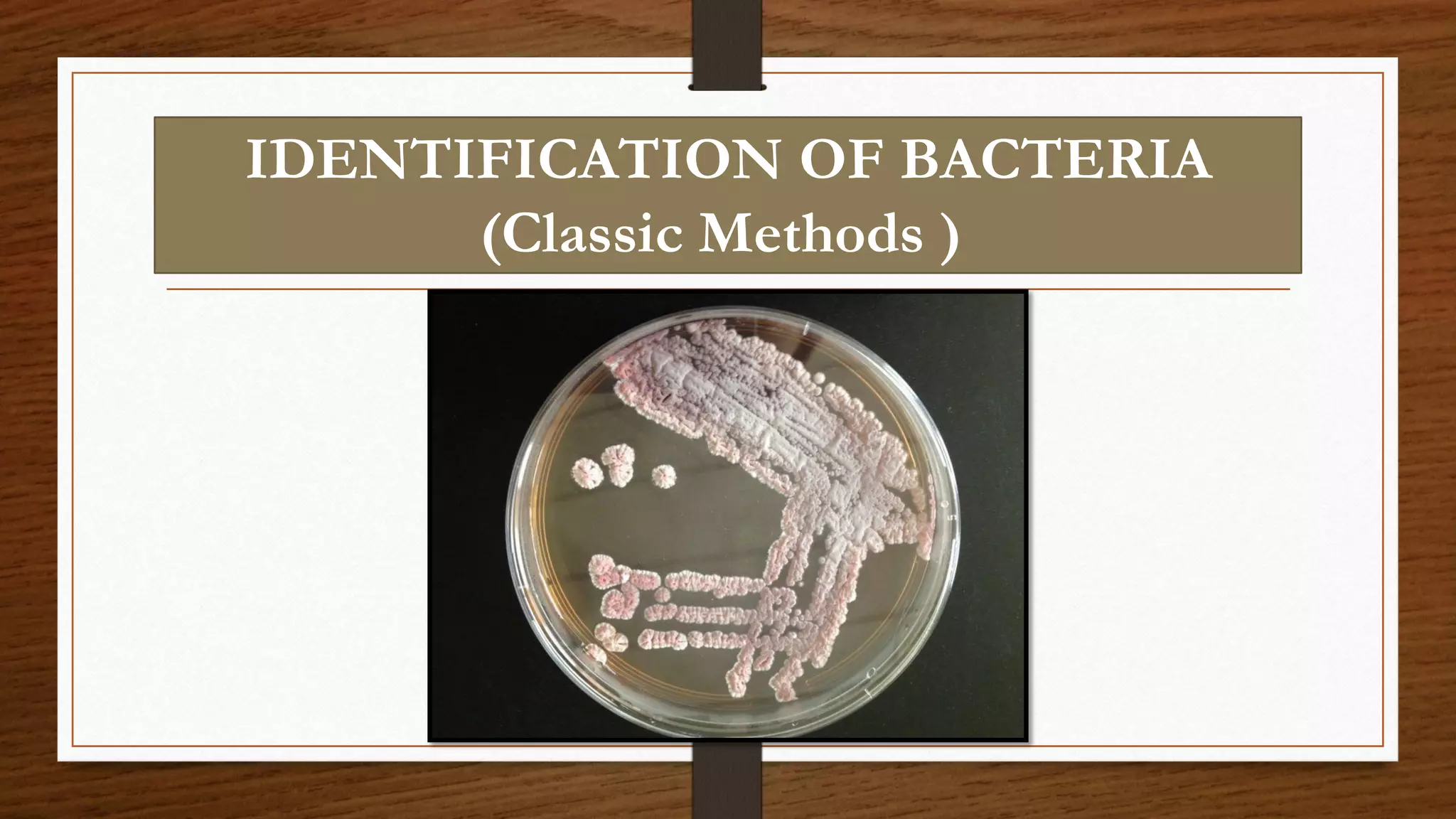
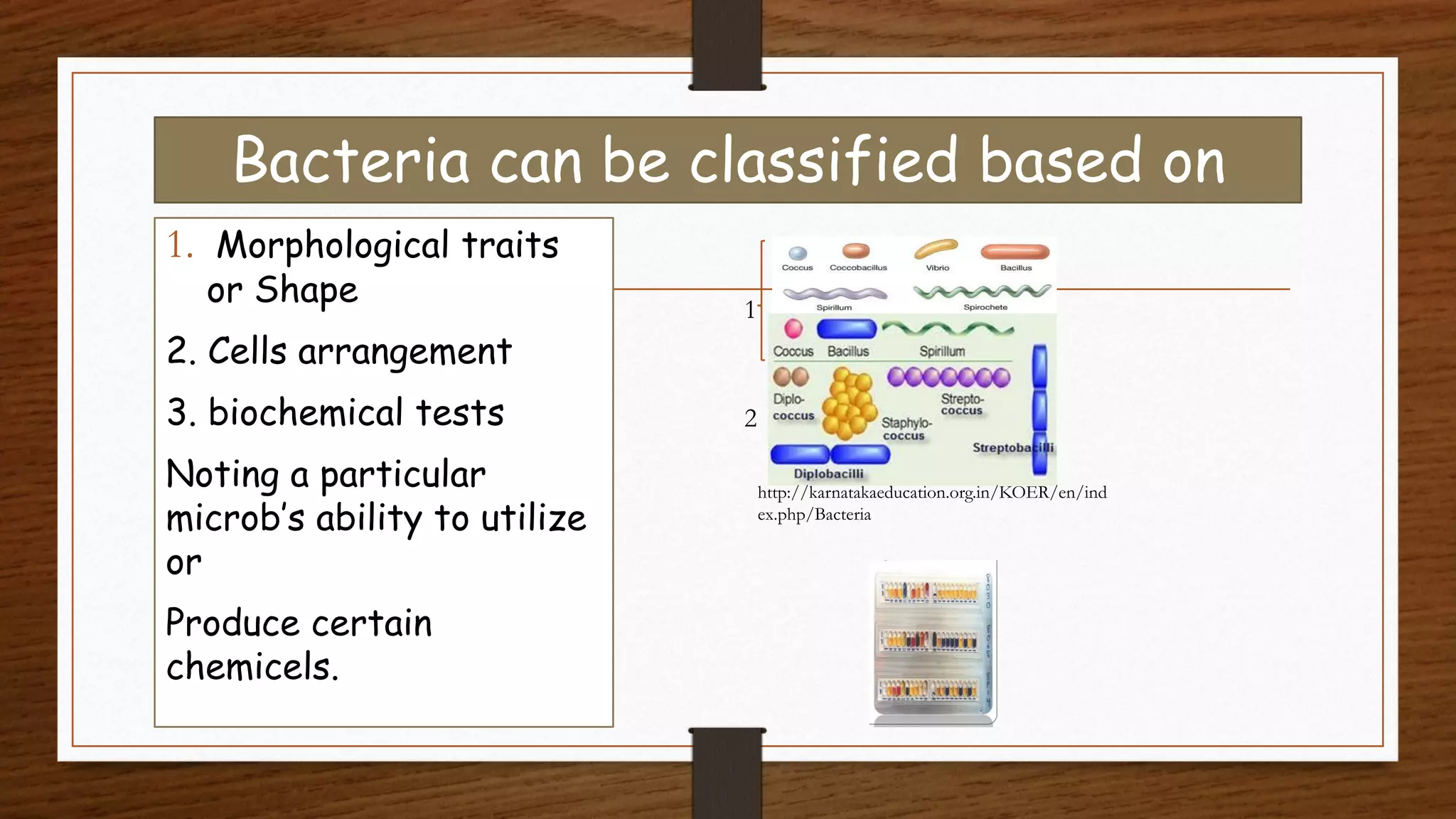
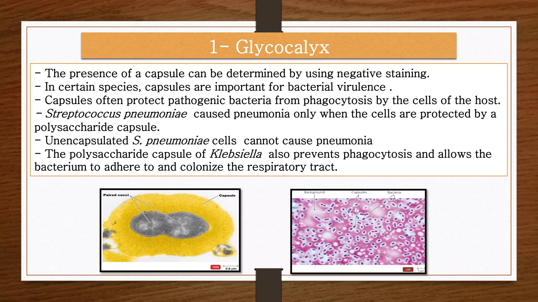
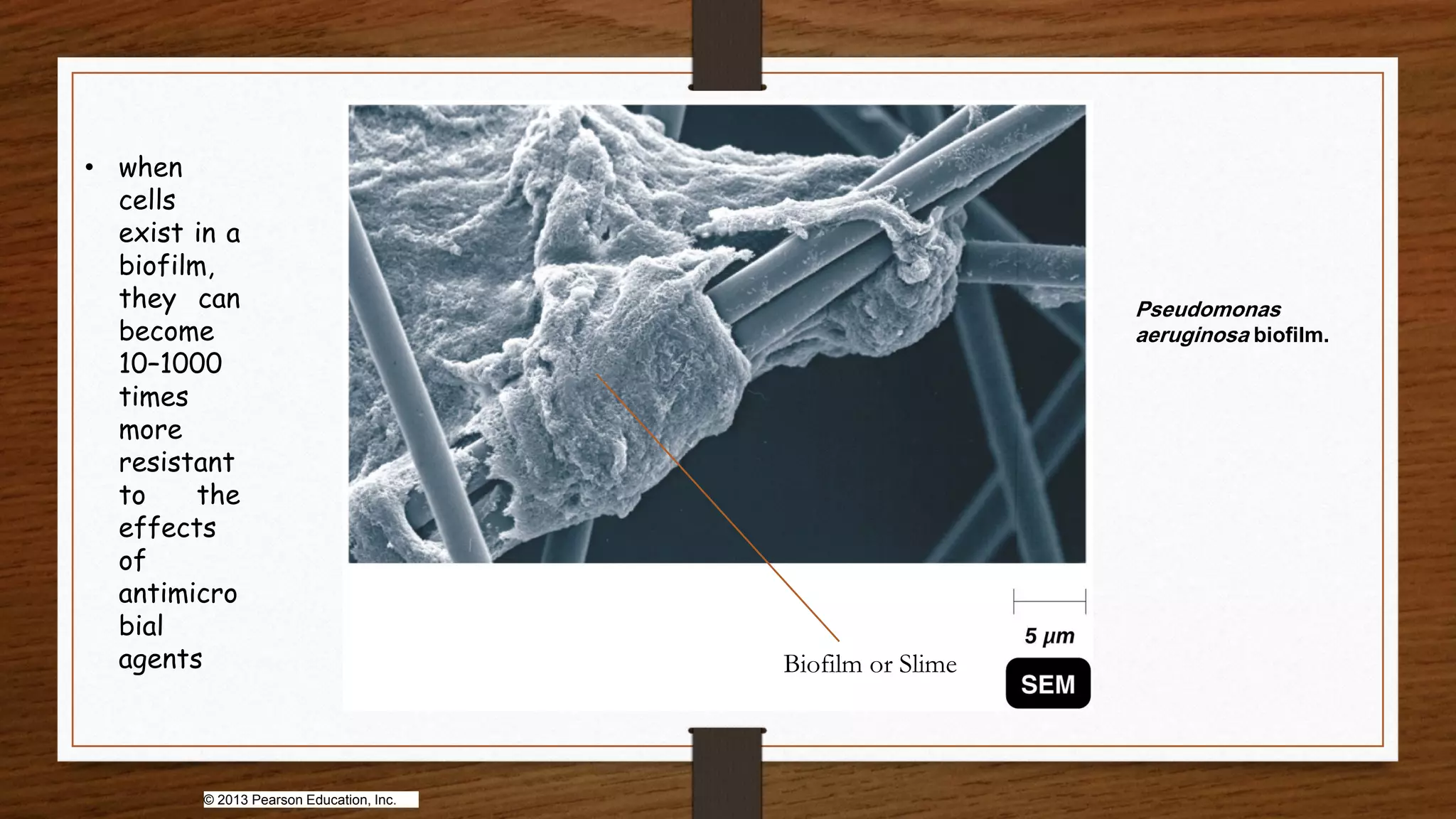
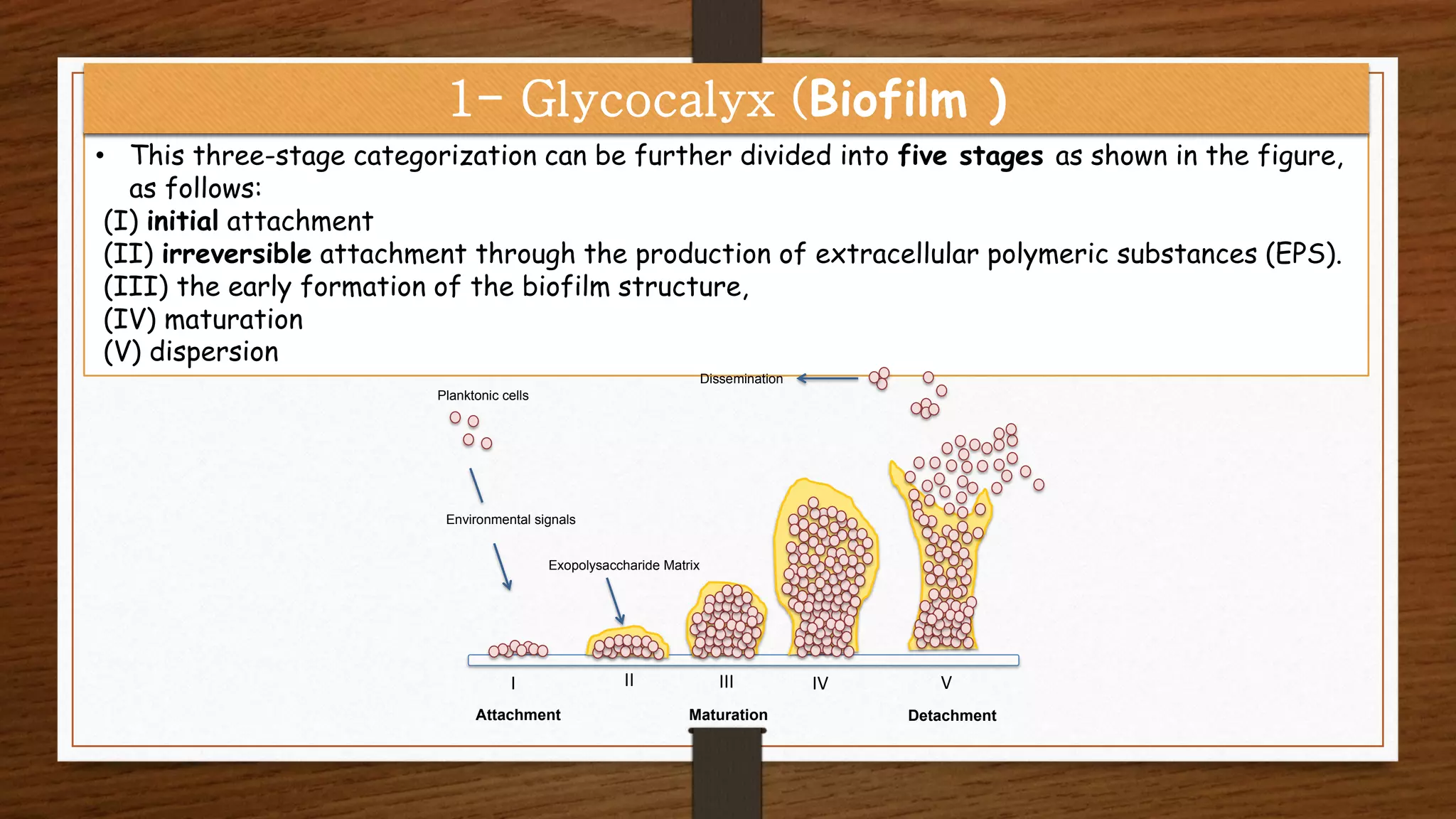
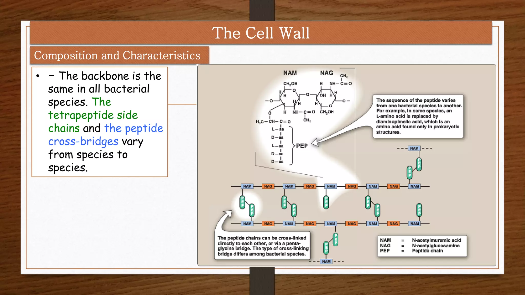
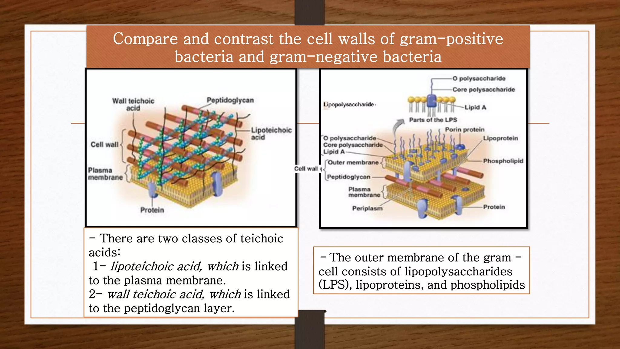
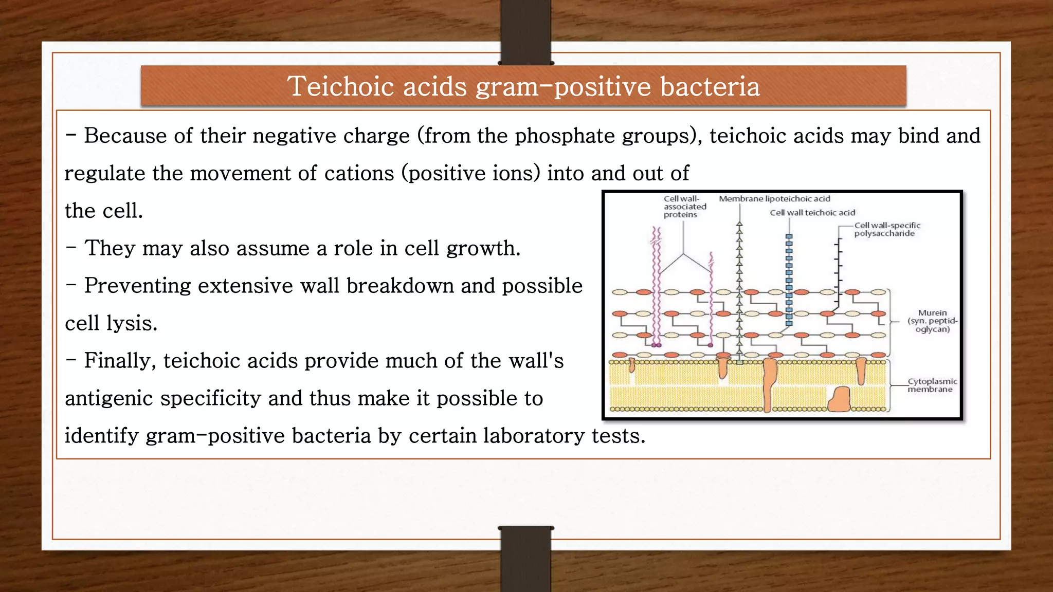
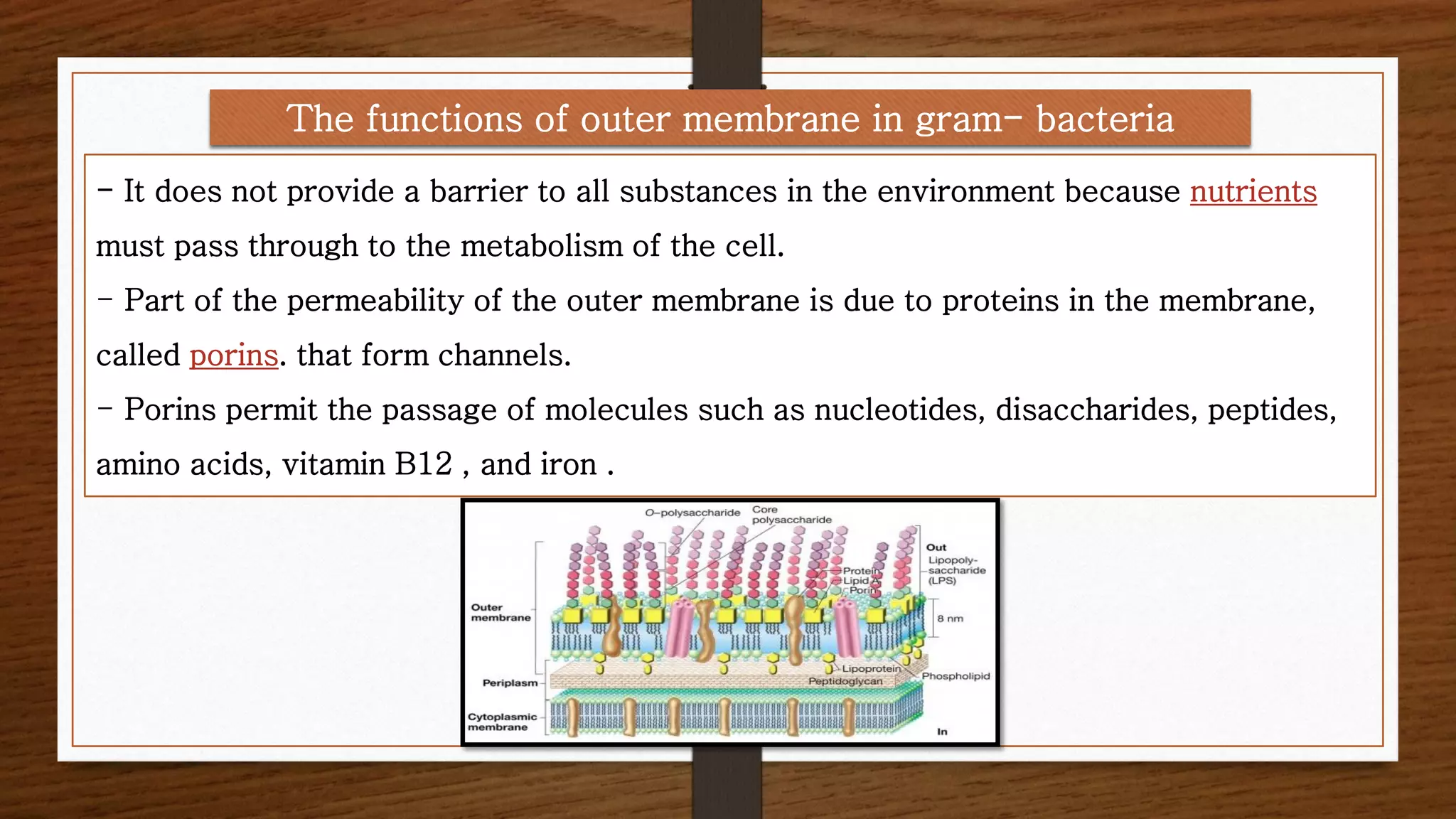
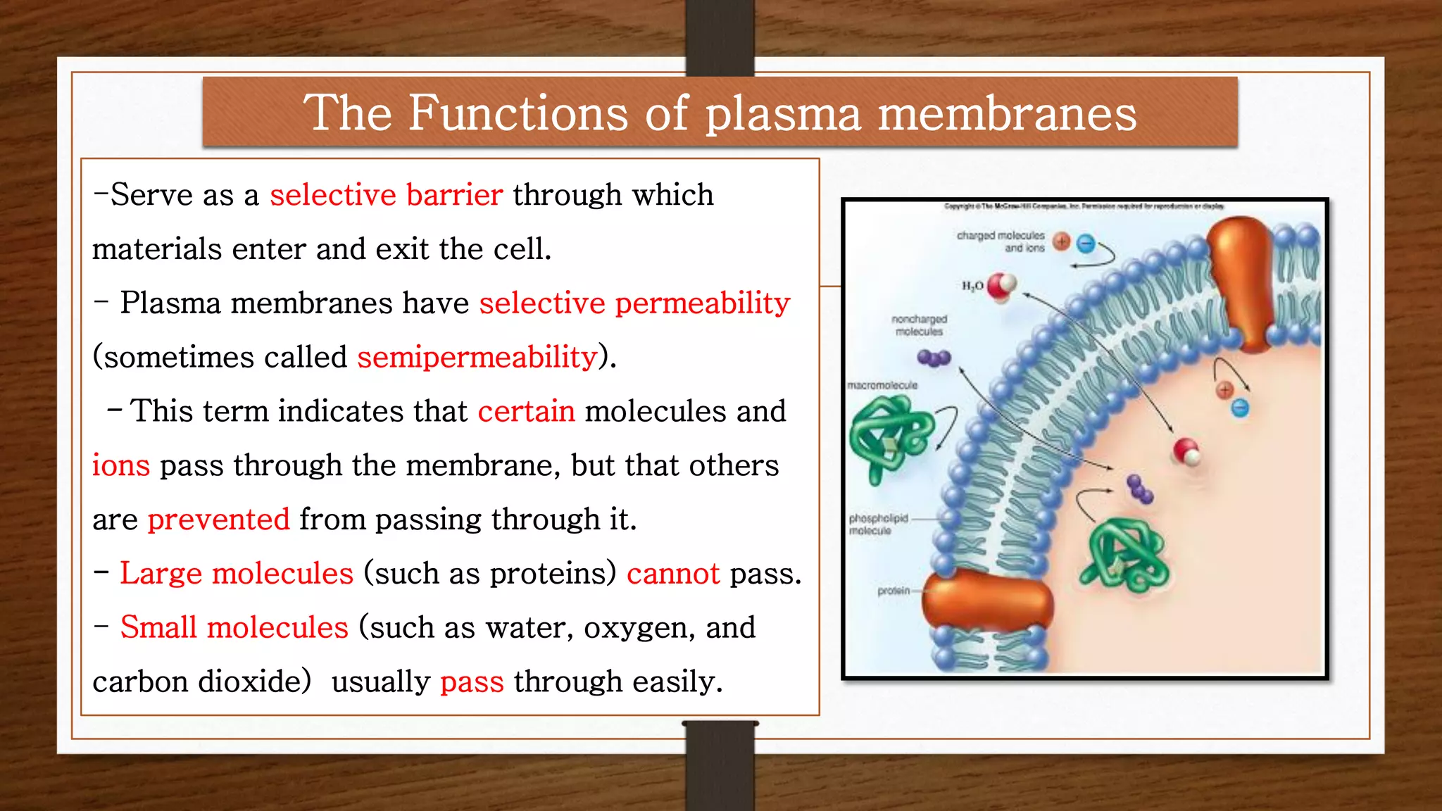
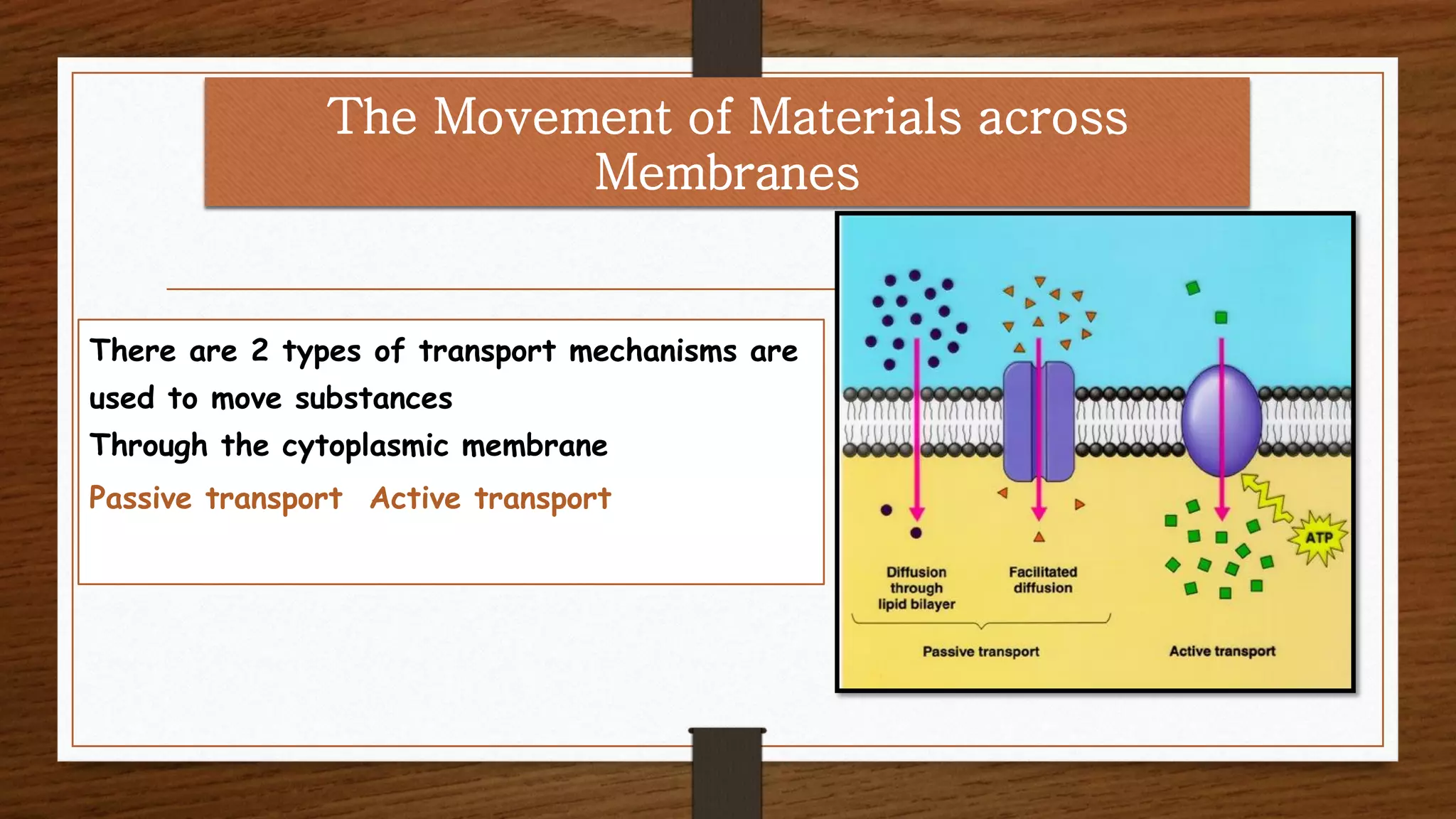
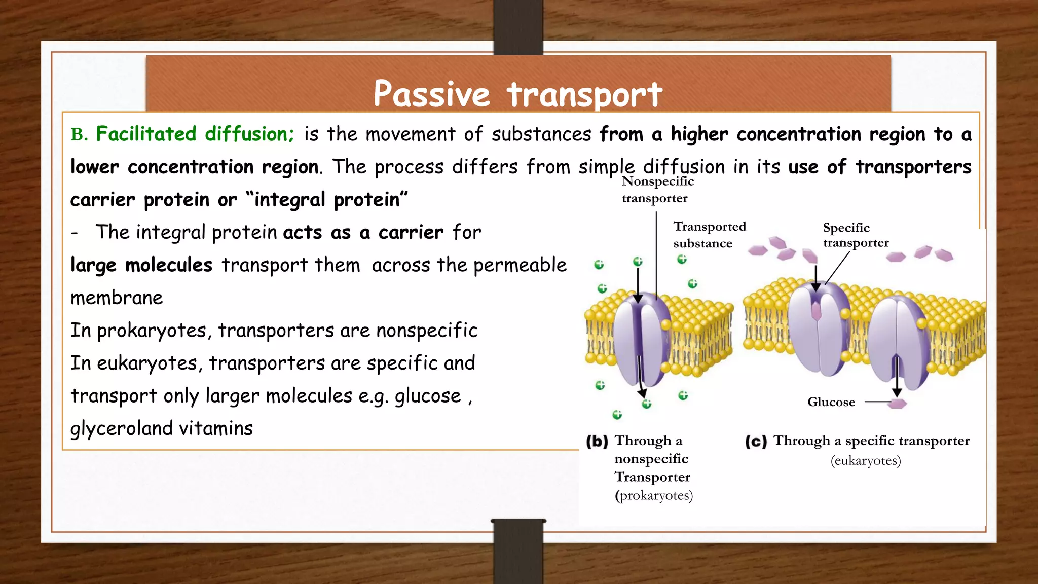
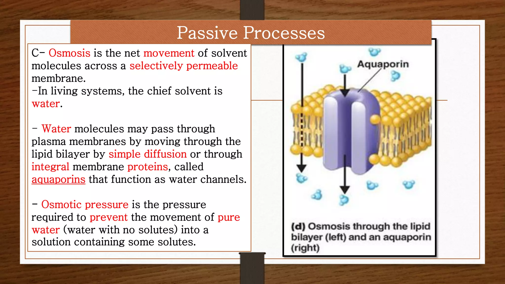
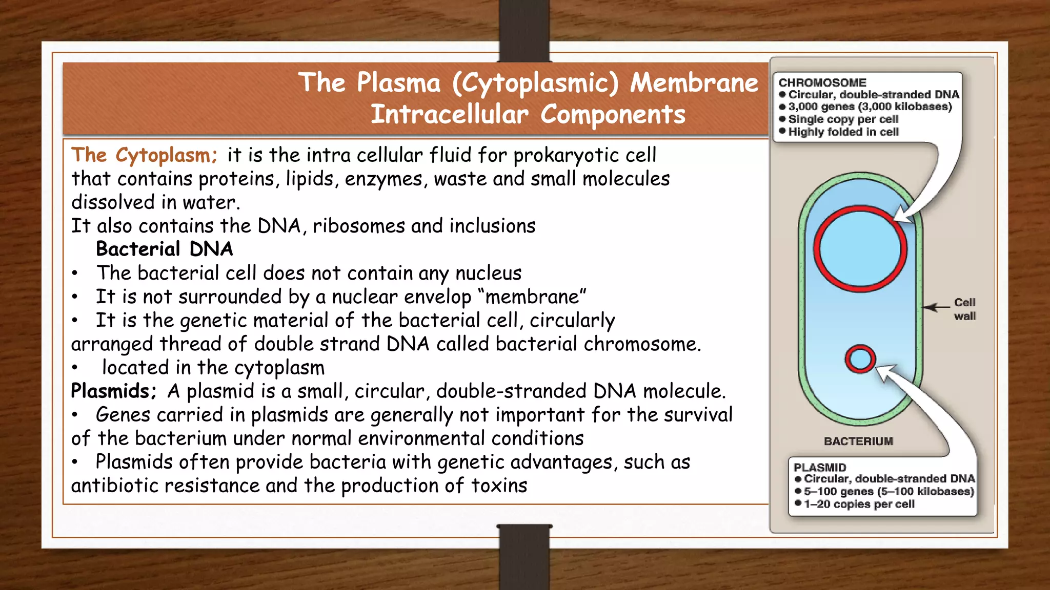
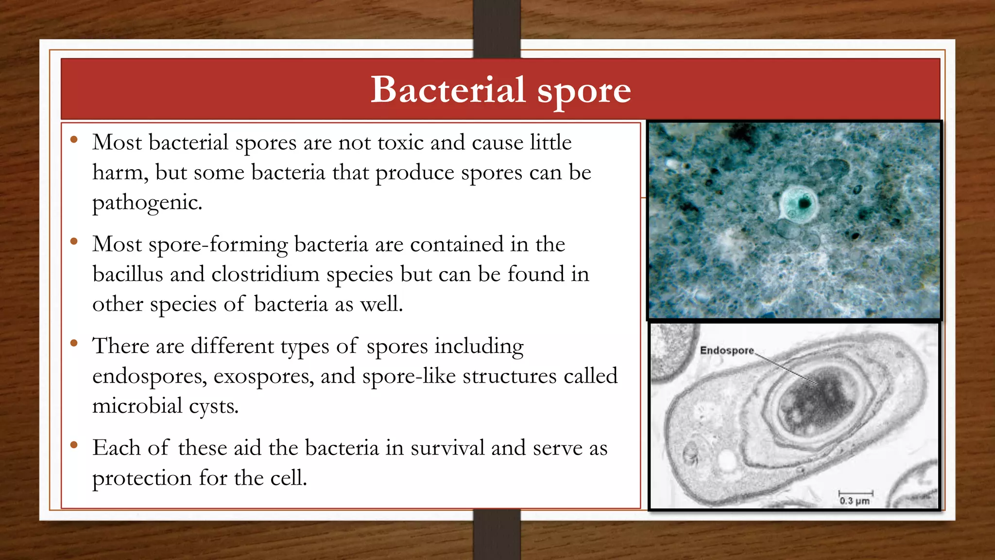
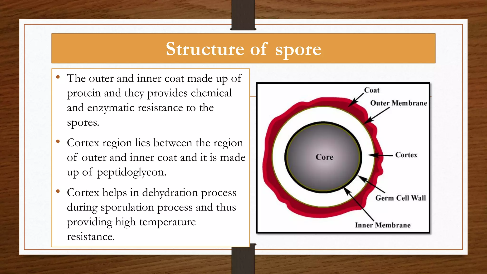
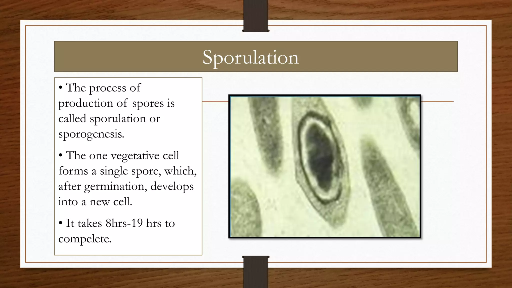
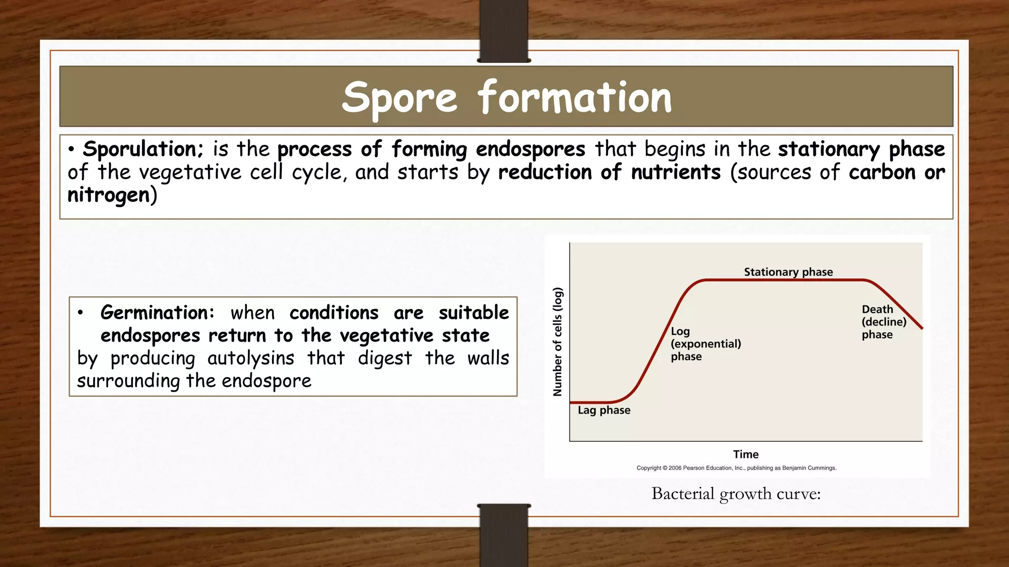
This document provides an overview of the topics to be covered in a bacteriology course. The course will last 5 weeks and cover cell structure and functions, gram reaction, spore formation, nutrition and respiration, growth curves and factors affecting growth, bacterial relationships, bacterial division, and classification. Students will be evaluated through exams, labs, activities, and a final exam. Learning resources include medical microbiology textbooks and online sources. The document then provides background information on bacteria and their classification, including an overview of prokaryotic life, the universal tree of life consisting of three domains, and methods for identifying bacteria.
2. Topics to be Covered
Topic No of
Weeks
Contac
thours
Cell structure and functions 2 4
Gram reaction, cell wall 1 2
Spore formatiom 1 2
Nutrition of bacteria and respiration 3 6
Growth curve, Growth, Factors affecting growth, Antibiotics 3 6
Bacterial relation ships 1 2
Division of bacteria 1 2
Classsificaion 2 4
White, David. "The physiology and biochemistry of
prokaryotes." (2007). Third edition. Indiana University.
Course Description
3. Methods of evaluation % of grade:
First exam (sixth week ) 15%
Midterm exam ( tenth week ) 20%
Lab exam 25%
Activity 10%
Final exam 30%
-------------------------------------------------------------------------------
Total 100
4. Learning Resources
1. Geo. F et al. 2007. Medical Microbiology. 24th ed. McGraw-Hill Publishing.
2. Baron S et al. 1996. Medical Microbiology. 4th ed. Galveston (TX).
Available at:
http://www.ncbi.nlm.nih.gov/books/NBK7627/?term=Structure%20and%20Function%
20of%20Bacterial%20Cells
3. Any microbiology textbook that explains the discussed topics.
5. MICROORGANISMS
• A microorganism can be one cell or a cluster of cells that
can be seen only by using a microscope.
• Microorganisms are organized into six fields of study:
bacteriology, virology, mycology, phycology, protozoology,
and parasitology.
• Bacteriology is the study of bacteria.
6. What are bacteria?
• Bacteria are prokaryotic organisms.
• Bacteria are microscopic single-celled organisms “unicellular”
that reproduce by binary fission.
• Most bacteria are capable of independent survival and growth, but
species
Chlamydia and Rickettsia can only survive as intracellular organisms.
• Some bacteria can move freely in their environment while others are
stationary.
• Bacteria can be found almost every where on the earth. They can live
in variety of environments including a wide range of temperature, PH
and salt concentration.
• They can be beneficial or harmful by causing diseases.
7. Overview Of Bacteriology (PROCARYOTIC LIFE)
• The Bacteria are a group of single-cell microorganisms
with procaryotic cellular configuration.
• Bacteria are the oldest and simplest organisms, but they
are metabolically much more diverse than all other life-
forms combined.
• Procaryotes are found in all of the habitats where
eucaryotes live.
8. Classifying and naming microorganisms
Taxonomy: it is the science concerned with classification and
put organisms into categories or taxa to show degrees of
similarities among organisms.
Taxa: is a group of organisms seen by taxonomists to form a
unit as this group show degrees of similarities among
organisms. These similarities are due to relatedness
Why Taxonomy is important?
- Provides a reference for identifying organisms
- Provides universal names for organisms
9. TAXONOMY AND CLASSIFICATION OF
PROCARYOTES
• Anton van Leeuwenhoek, using a simple microscope, was the
first to observe microorganisms (1673).
10. TAXONOMY AND CLASSIFICATION OF
PROCARYOTES
• Haeckel (1866) was the first to create a natural Kingdom for the
microorganisms, which had been discovered nearly two centuries before by
van Leeuwenhoek.
11. TAXONOMY AND CLASSIFICATION OF
PROCARYOTES
• He placed all unicellular (microscopic) organisms in a new
kingdom, "Protista", separated from plants (Plantae) and animals
(Animalia), which were multicellular (macroscopic) organisms.
12. TAXONOMY AND CLASSIFICATION OF
PROCARYOTES
• The development of the electron
microscope in the 1950's revealed a
fundamental dichotomy among
Haeckel's "Protista": some cells
contained a membrane-enclosed nucleus,
and some cells lacked this intracellular
structure.
• The latter were temporarily shifted to a
fourth kingdom, Monera (or Moneres),
the procaryotes (also
called Procaryotae).
13. TAXONOMY AND CLASSIFICATION OF
PROCARYOTES
• Protista remained as a kingdom of
unicellular eucaryotic
microorganisms.
• Whittaker refined the system into
five kingdoms in 1967, by
identifying the Fungi as a separate
multicellular eucaryotic kingdom of
organisms, distinguished by their
absorptive mode of heterotrophic
nutrition.
14. • The current edition (2001) of Bergey's Manual of Systematic
Bacteriology has established 24 phyla of bacteria, systematically
ordered into class, order, family, genus and species.
• For example, E. coli is in the Domain Bacteria,
• Phylum Proteobacteria,
• Class Gamma Proteobacteria,
• Order Enterobacteriales,
• Family Enterobacteriaceae,
• Genus Escherichia,
• Species E. coli.
TAXONOMY AND CLASSIFICATION OF
PROCARYOTES
15. Placing Bacteria
1735 Kingdoms Plantae and Animalia
1857 Bacteria and fungi put in the Kingdom Plantae—“Flora”
1866 Kingdom Protista proposed for bacteria, protozoa, algae, and fungi
1937 Prokaryote introduced for cells “without a nucleus”
1961 Prokaryote defined as cell in which nucleoplasm is not surrounded by a
nuclear membrane
1959 Kingdom Fungi
1968 Kingdom Prokaryotae proposed
1978 Two types of prokaryotic cells found
16. The Universal Tree of Life
• In, 1978 Carl R. Woese developed a new
classification system with three classification
groups called domains.
• A domain; is larger than a kingdom
Organisms is classified in 3 domine system based
on:
1. Cell type
2. Difference in ribosomal RNA(rRNA)
3. Membrane lipid structure
4. Transfer RNA molecules
5. Sensitivity to antibiotics
17. The Three Domains
• On the basis of small subunit ribosomal RNA (ssrRNA)
analysis, the Tree of Life gives rise to three cellular
"Domains":
18. • The difference between
Archaea and Bacteria is based on fundamental
differences in the nucleotide base sequence in
the 16S ribosomal RNA.
• There are many biochemical and phenotypic
differences between the two groups of
procaryotes.
• The phylogenetic tree indicates that Archaea are
more closely related to Eucarya than
are Bacteria.
The Archaea and Bacteria
19. • This relatedness seems most evident in the
similarities between transcription and translation in
the Archaea and the Eucarya.
• However, it is also evident that the Bacteria have
evolved into chloroplasts and mitochondria, so that
these eucaryotic organelles derive their lineage from
this group of procaryotes.
• Perhaps the biological success of eucaryotic cells
springs from the evolutionary merger of the two
procaryotic life forms.
The Archaea and Bacteria
20. The Archaea and Bacteria
• The archaea and bacteria differ
fundamentally in their structure from
eucaryotic cells, which always contain
a membrane-enclosed nucleus,
multiple chromosomes, and various
other membranous organelles such as
mitochondria, chloroplasts, the golgi
apparatus, vacuoles, etc.
21. • Unlike plants and animals, archaea and bacteria are unicellular organisms
that do not develop or differentiate into multicellular forms.
• Some bacteria grow in filaments or masses of cells, but each cell in the
colony is identical and capable of independent existence.
• The cells may be adjacent to one another because they did not separate
after cell division or because they remained enclosed in a common sheath
or slime secreted by the cells, but typically there is no continuity or
communication between the cells.
The Archaea and Bacteria
22.
23. Archaea
• Archaea consist of prokaryotic cells; they lack
peptidoglycan in
• their cell walls.
• Archaea Live in extreme environments
• Include:
• 1) Methanogens produce methane as a
waste product from respiration
• 2) Extreme halophiles (halo = salt; philic
=loving) live in extremely salty environments
such as the Dead Sea
• 3) Extreme thermophiles (therm = heat)
live in hot sul furous water, such as hot
springs at Yellowstone National Park.
• Archaea are not known to cause disease in
humans.
24. The Origin of Life
• When life arose on Earth about 4 billion years ago, the first
types of cells to evolve were procaryotic cells.
• For approximately 2 billion years, procaryotic-type cells
were the only form of life on Earth.
• The oldest known sedimentary rocks, from Greenland, are
about 3.8 billion years old.
• The oldest known fossils are procaryotic cells, 3.5 billion
years in age, found in Western Australia and South Africa.
25. • The nature of these fossils, and the chemical composition of
the rocks in which they are found, indicates
that lithotrophic and fermentative modes of metabolism were
the first to evolve in early procaryotes.
• Photosynthesis developed in bacteria a bit later, at least 3
billion years ago.
• Anoxygenic photosynthesis (bacterial photosynthesis, which is
anaerobic and does not produce O2) preceded oxygenic
photosynthesis (plant-type photosynthesis, which yields O2).
The Origin of Life
26. • Oxygenic photosynthesis also
arose in procaryotes,
specifically in the cyanobacteria,
which existed millions of years
before the evolution of green
algae and plants.
• Larger, more complicated
eucaryotic cells did not appear
until much later, between 1.5
and 2 billion years ago.
The Origin of Life
27. IDENTIFICATION OF BACTERIA
(Classic Methods )
• The criteria used for microscopic
identification of procaryotes include cell
shape and grouping, Gram-stain reaction, and
motility.
• Bacterial cells almost take one of three forms:
rod (bacillus), sphere (coccus), or spiral
(spirilla and spirochetes).
• Rods that are curved are called vibrios.
28. IDENTIFICATION OF BACTERIA
(Classic Methods )
• Fixed bacterial cells
stain either Gram-
positive (purple) or
Gram-negative (pink);
motility is easily
determined by
observing living
specimens.
29. IDENTIFICATION OF BACTERIA
(Classic Methods )
• Bacilli may occur singly or form chains of cells;
• cocci may form chains (streptococci) or grape-
like clusters (staphylococci);
• spiral shape cells are almost always motile;
• cocci are almost never motile. This nomenclature
ignores the actinomycetes, a prominent group
of branched bacteria which occur in the soil. But
they are easily recognized by their colonies and
their microscopic appearance.
31. • Bergey's Manual describes affiliated groups
of Bacteria and Archaea based on a few easily
observed microscopic and physiologic
characteristics.
• Further identification requires biochemical tests
which will distinguish genera among families and
species among genera.
• Strains within a single species are usually
distinguished by genetic or immunological
criteria.
IDENTIFICATION OF BACTERIA
(Classic Methods )
32. IDENTIFICATION OF BACTERIA
(Molecular Techniques)
• The sciences of genomics and bioinformatics have led to a radical
reclassification of procaryotes based on comparative analysis of organismal
DNA.
• Genomics involves the study of all of the nucleotide sequences, including
structural genes, regulatory sequences, and noncoding DNA segments, in the
chromosomes of an organism.
• To date over 200 bacterial genomes have been sequenced.
33. IDENTIFICATION OF BACTERIA
(Molecular Techniques)
• We have seen how highly conserved genetic sequences, such as those that
encode for the small subunit ribosomal RNAs (16S rRNA) of an organism,
can be analyzed to specifically relate two organisms.
• So can the identification of certain genes provide information about specific
properties of an organism, and analysis of specific nucleotide sequences may
be used to indicate identity and degrees of genetic relatedness among
organisms.
34. IDENTIFICATION OF BACTERIA
(Molecular Techniques)
• The newest editions of Bergey's Manual are adapted to the new phylogenetic
classification.
• This has resulted in the formation of several new taxa of bacteria and
archaea at every hierarchical level.
• Occasionally, organisms thought to be more or less distantly related become
unified; but more likely, organisms thought to be closely-related due to
similar phenotypic properties are found to be genetically distinct and warrant
separation into a new taxa.
35. Binominal Nomenclature
• Carl Linnaeus developed this system for naming organisms in 1735.
• Each organism is given two latinized names
• The first name is called the genus
• The second name is the specific epithet, and is not capitalized.
• The genus and the species name appear underlined or italicized. The
name itself describes the organism.
• Sometimes an organism is named for a researcher, e.g.
Escherichia coli
Genus
Theodor Escherich
bacterium lives in the colon
specific epithet
36. Binominal Nomenclature
• different scientists can classify the same organisms
differently. For example, clinical microbiologists are interested
in virulence factors and pathogenicity focusing on antimicrobial
resistance pattern, and toxins in Escherichia coli, whereas
geneticists are concerned with specific mutations and plasmids.
37. Bacteria can be classified based on
1. Morphological traits
or Shape
2. Cells arrangement
3. biochemical tests
Noting a particular
microb’s ability to utilize
or
Produce certain
chemicels.
1
2
http://karnatakaeducation.org.in/KOER/en/ind
ex.php/Bacteria
38. Bacteria can be classified based on
4. Ability to accept gram
stain
5. By serology test
6. O2 requirement
7. Habitat (PH level, salt
concentration and
temperature)
8. Classification based on
bacterial genetic sequences
using molecular techniques
39. The Size, Shape, and Arrangement of Bacterial Cells
• Bacterial cells are extremely small and measured in microns (µm) (0.001 mm).
• Bacteria range in size from large cells such as Bacillus anthracis (1.0 to 1.3µm )
to very small cells such as Pasteurella tularensis (0.2 to 0.7µm) Mycoplasmas (atypical
pneumonia group) are even smaller, measuring (0.1 to 0.2 µm) in diameter.
40. The Size, Shape, and Arrangement of Bacterial Cells
- Most bacteria are monomorphic; they maintain a single shape.
- Some bacteria are genetically pleomorphic., such as Rhizobium sp. and Corynebacterium
sp. which means they can have many shapes.
42. - Cocci are usually round but can be oval.
- When cocci divide to reproduce, the cells can remain attached to one another.
- Cocci that remain in pairs after dividing are called diplococci.
- Those that divide and remain attached in chain like patterns are called streptococci.
Plane of
division
Diplococci
Streptococci
Coccus (spherical); Arrangements
43. - Those that divide in two planes and remain in groups of four are
known as tetrads.
Tetrad
Coccus (spherical); Arrangements
44. - Those that divide in three planes and remain attached in cube like groups of
eight are called sarcinae.
Sarcinae
Coccus (spherical); Arrangements
45. - Those that divide in multiple planes and form grape like broad sheets are called staphylococci .
- These group characteristics are helpful in identifying cocci.
Staphylococci
Coccus (spherical); Arrangements
46. - Bacilli divide only across their short axis, so there are fewer groupings of bacilli than of cocci.
- Most bacilli appear as single rods .
- Scientific name: Bacillus or shape: bacillus
Bacillus (rod-shaped) ; Arrangements
Single bacillus
47. -Diplobacilli appear in pairs after division
Bacillus (rod-shaped) ; Arrangements
Diplobacilli
49. - Others are oval and look so much like cocci that they are called
coccobacilli .
Bacillus (rod-shaped) ; Arrangements
Coccobacillus
50. - Spiral bacteria have one or more twists; they are never straight.
- Bacteria that look like curved rods are called vibrios.
Spiral bacteria ; Arrangements
Vibrio
51. -Spirillum, have a helical shape, like a corkscrew, and fairly rigid
bodies .
- The spirilla are used propeller-like external appendages called
flagella to move.
Spiral bacteria ; Arrangements
Spirillum
52. Spiral bacteria ; Arrangements
- Spirals they are helical and flexible are called spirochetes.
- Spirochetes move by means of axial filaments
Spirochete
53. - There are star-shaped cells (genus Stella).
- Rectangular, flat cells (halophilic archaea) of the genus Haloarcula .
In addition to the three basic shapes
Star-shaped bacteria Rectangular bacteria
54. -There are prokaryotes that look very much like triangles Haloarcula
japonica
- Or squares Holoquadratum walsbyi .
In addition to the three basic shapes
55. 1) Because we are Microbiologists!
• Form about half of the biosphere-
Both in terms of biomass:
~5 x 1030 microbial cells on Earth
5,000,000,000,000,000,000,000,000,000,000 cells
and biodiversity:
4,000 “species” in a gram of soil. (http://whyfiles.org/shorties/count_bact.html)
• Play essential roles in recycling carbon, nitrogen, sulfur, and other
chemical elements used by living things
• Many bacteria help decompose (break down) dead organisms and
animal wastes into chemical elements
• Interactions with eukaryotes (either beneficial or harmful)
But why care about bacteria?
56. 2. Because they kill us!
Tuberculosis (Mycobacterium tuberculosis) kills about 2 million people a year,
mostly in sub-Saharan Africa.
Water, and food-bourne diseases such as Cholera (Vibrio cholerae) shigellosis
(Shigella spp.) and Salmonella.
Air-bourne (e.g. Streptococcus pneumoniae)
• The spread of antibiotic resistance and the emergence of hospital “superbugs”
(e.g. Staphylococcus aureus (MRSA), Escherichia coli and Clostridium difficile).
• The development of effective vaccines (e.g. Neisseria meningitidis)
• Epidemiological surveillance and the management of outbreaks.
But why care about bacteria?
57. 3. Bacteria are useful!
Fermentation: beer, wine, baking, cheese, butter, pickles,
soya sauce, vinegar, and yogurt.
Chemical manufacturing: ethanol, enzymes, perfumes.
Pharmaceuticals: antibiotics and vaccines
Bio-remediation: sewage treatment, oil spills.
But why care about bacteria?
58. Structures External to the Cell Wall
The external structures in the prokaryotic cell wall are;
- Glycocalyx.
- Flagella.
- Axial filaments.
- Fimbriae.
- Pili.
59. - Many prokaryotes secrete it on their surface.
- Glycocalyx (meaning sugar coat) that surround cells.
- It is a sticky, gelatinous polymer that is external to the cell wall.
- It composed of polysaccharide, polypeptide, or both.
- It is made inside the cell and secreted to the cell surface.
- If the substance is organized and attached to the cell wall , the glycocalyx is described as a
capsule.
-If the substance is unorganized and only loosely attached to the cell wall, the glycocalyx is
described as a slime layer.
- A glycocalyx that helps cells in a biofilm attach to their target environment and to each other is
called an extracellular polymeric substance (EPS). The EPS protects the cells. It can provide
nutrients.
1- Glycocalyx
60. - The presence of a capsule can be determined by using negative staining.
- In certain species, capsules are important for bacterial virulence .
- Capsules often protect pathogenic bacteria from phagocytosis by the cells of the host.
- Streptococcus pneumoniae caused pneumonia only when the cells are protected by a
polysaccharide capsule.
- Unencapsulated S. pneumoniae cells cannot cause pneumonia
- The polysaccharide capsule of Klebsiella also prevents phagocytosis and allows the
bacterium to adhere to and colonize the respiratory tract.
1- Glycocalyx
61. - Many prokaryotes secrete it on their surface.
- Glycocalyx (meaning sugar coat) that surround cells.
- It is a sticky, gelatinous polymer that is external to the cell wall.
- It composed of polysaccharide, polypeptide, or both.
- It is made inside the cell and secreted to the cell surface.
- If the substance is organized and attached to the cell wall , the glycocalyx is described as a
capsule.
-If the substance is unorganized and only loosely attached to the cell wall, the glycocalyx is
described as a slime layer.
- A glycocalyx that helps cells in a biofilm attach to their target environment and to each other is
called an extracellular polymeric substance (EPS). The EPS protects the cells. It can provide
nutrients.
1- Glycocalyx
62. - The presence of a capsule can be determined by using negative staining.
- In certain species, capsules are important for bacterial virulence .
- Capsules often protect pathogenic bacteria from phagocytosis by the cells of the host.
- Streptococcus pneumoniae caused pneumonia only when the cells are protected by a
polysaccharide capsule.
- Unencapsulated S. pneumoniae cells cannot cause pneumonia
- The polysaccharide capsule of Klebsiella also prevents phagocytosis and allows the
bacterium to adhere to and colonize the respiratory tract.
1- Glycocalyx
63. - A glycocalyx that helps cells in a biofilm attach to their target environment and to each other is
called an extracellular polymeric substance (EPS). The EPS protects the cells. It can provide
nutrients.
• Biofilm can be defined as a group of microorganisms surrounded by an extracellular polymeric
substances (EPS) also known as an exopolysaccharide matrix or slime and attached to various
surfaces.
• The main biofilm matrix components are “microbial cells, polysaccharides and water, together
with excreted cellular products”
• Essential nutrients can transport within the biofilm and reach the deepest regions through
complex channel networks
• Biofilms provide resistance to antibiotics as well as protection against the host immune system
• Bbacterial cells in the biofilm are up to 1000 times more resistant to antibiotics than in their
planktonic form
1- Glycocalyx (Biofilm )
65. Dental plaque and gum disease
Medical issues caused by Biofilms
Catheters
1. 2.
3.
www.medicalnewstoday.comwww.ijmm.orgwww.mdpi.com www.geistlich-pharma.com
• S. aureus, and S. epidermidis are a major cause of implant related infections
which results in the infections being chronic and irreducible
Dental
hip
knee implants
• S. aureus can adhere to plastic surfaces such as catheters as well as form a specific
attachment either to human tissue or to an abiotic surface covered by the host matrix proteins.
67. • There are three main stages in the development of biofilm:
- Attachment; is a critical step in establishing an infection
- Growth (or maturation); biofilm develops into a 3D structure
- Detachment; enables dissemination to infect other sites
1- Glycocalyx (Biofilm )
Dissemination
Planktonic cells
Environmental signals
Exopolysaccharide Matrix
Attachment DetachmentMaturation
I II III IV V
The figure is adapted from
Michael O., 2009. The figure
illustrates the biofilm life cycle. (I)
Initial attachment stage: In
response to environmental
signals, planktonic bacteria
adhere to abiotic or biotic
surfaces; (II) Biofilm
development stage with
production of the
exopolysaccharide matrix and
irreversible adherence; (III and
IV) Biofilm maturation stages;
(V) detachment and
dissemination of single cells or
clumps of the biofilm body.
68. • This three-stage categorization can be further divided into five stages as shown in the figure,
as follows:
(I) initial attachment
(II) irreversible attachment through the production of extracellular polymeric substances (EPS).
(III) the early formation of the biofilm structure,
(IV) maturation
(V) dispersion
1- Glycocalyx (Biofilm )
Dissemination
Planktonic cells
Environmental signals
Exopolysaccharide Matrix
Attachment DetachmentMaturation
I II III IV V
69. - Some prokaryotic cells have flagella (singular:
flagellum ).
-Flagella are relatively long filamentous
appendages consisting of a filament that contains
of the globular (roughly spherical) protein
flagellin arranged in several chains that
intertwine and form a helix around attached to a
hook ( consisting of a different protein) and basal
body , which anchors the flagellum to the cell
wall and plasma membrane.
2- Flagella
71. 2- Flagella
• In Gram negative bacteria, the basal body consists of 4 rings. 2 attached to the plasma
membrane, one to the peptidoglycan layer and other to the outer lipopolysaccharide (LPS) layer.
• In Gram positive bacteria, outer LPS layer is absent and basal body has 2 rings one attached to
the plasma membrane and one attached to the peptidoglycan wall
Plasma
membrane
Cell wall
Basal body
Gram-
nega ve
Pep doglycan
Outer
membrane
Hook
Basal body
Pep doglycan
Hook
Filament
Cytoplasm
Flagellum
Plasma
membrane
Cell wall
Gram-
posi ve
Filament
Cytoplasm
Flagellum
Parts and a achment of a flagellum of a gram-
nega ve bacterium
Parts and a achment of a flagellum of a gram-
posi ve bacterium
Inner pair of ringsouter pair of
rings
72. - Prokaryotic flagella rotate to push the cell.
- When a bacterium moves in one direction for a
length of time, the movement is called a "run" or
"swim.“
- Random changes in direction called "tumbles.
- "Tumbles" are caused by a reversal of flagellar
rotation
- One advantage of motility is that it enables a
bacterium to move toward a favorable environment
or away from a particular stimulus (taxis).
- Such stimuli include chemicals (chemotaxis) and
light (phototaxis).
-Bacterial cells can alter the speed and direction of
rotation of flagella and thus are capable of various
patterns of motility.
-Motility the ability of an organism to move by itself.
2- Flagella
Run
Run
Tumble
Tumble
Tumble
A bacterium running and tumbling. Notice
that the direction of flagellar rotation (blue
arrows) determines which of these
movements occurs. Gray arrows indicate
direction of movement of the microbe.
73. Peritrichous Monotrichous and polar
Lophotrichous and polar Amphitrichous and polar
- Bacteria that lack flagella are referred to as atrichous .
- Flagella may be peritrichous (distributed over the entire cell)
- Polar (at one or both poles or ends of the cell).
- If polar, flagella may be monotrichous (a single flagellum at one pole)
- lophotrichous (a tuft of flagella coming from one pole).
- Amphitrichous (flagella at both poles of the cell).
2- Flagella
74. - Flagellar protein is an antigen.
- The flagellar protein called H antigen is useful for distinguishing a variations species
of gram negative bacteria .
- For example, there are at least 50 different H antigens for E. coli. Those variations
species identified as E. coli 0157:H7 are associated with food borne epidemics.
2- Flagella
75. - They are structures similar to that of flagella
(endoflagellum).
- Spiral cells that move by an endoflagellum are
called spirochetes.
- One of the best-known spirochetes is
Treponema pallidum
- Axial filaments are anchored at one end of
the spirochete.
- The rotation of the filaments produces a
movement of the outer sheath that propels the
spirochetes in a spiral motion.
- This type of movement is similar to the way a
corkscrew moves through a cork.
- This corkscrew motion probably enables a
bacterium such as T. pallidum to move
effectively through body fluids.
3- Axial Filaments
Outer sheath
Axial filament
Cell wall
A photomicrograph of the spirochete
Leptospira, showing an axial filament
76. A axial filaments wrapping around part of a
spirochete for a cross section of axial filaments)
3- Axial Filaments
77. 4- Fimbriae and Pili
- Many gram-negative bacteria contain hairlike appendages that are shorter, straighter,
and thinner than flagella .
- They are used for attachment and transfer of DNA rather than for motility.
- These structures, which consist of a protein called pilin arranged helically around a
central core, are divided into two types, fimbriae
and pili, having very different functions.
78. - Fimbriae (singular: fimbria)help cells adhere to surfaces.
- They can number anywhere from a few to several hundred per cell
- They are involved in forming biofilms
- Fimbriae can also help bacteria adhere to epithelial surfaces in the
body.
4- Fimbriae and Pili
79. The Cell Wall
- The cell wall of the bacterial cell is a complex.
-The cell wall of the bacterial cell is semi rigid structure.
- It is responsible for the shape of the cell.
- It protects the interior of the cell from adverse changes in the outside environment.
-Serves as a point of anchorage for flagella.
- It contributes to the ability of some species to cause disease.
- It is the site of action of some antibiotics.
- The chemical composition of the cell wall is used to differentiate major types of bacteria.
80. Composition and Characteristics
- The bacterial cell wall is composed of
a macromolecular network called
peplidoglycan (also known as murein).
- It is present either alone or in
combination with other substances.
- Peptidoglycan consists of a repeating
disaccharide attached by polypeptides to
form a lattice that surrounds and protects
the entire cell.
The Cell Wall
81. - The disaccharide portion is
made up of monosaccharides
called N-acetylglucosamine
(NAG) and N-acetylmuramic
acid (NAM) , (from murus,
meaning wall), which are
related to glucose.
Composition and Characteristics
The Cell Wall
82. - Alternating NAM and NAG molecules
are linked in rows of 10 to 65 sugars to
form a carbohydrate "backbone" ( the
glycan portion of peptidoglycan).
- Adjacent rows are linked by
polypeptides (the peptide portion of
peptidoglycan), it always includes
tetrapeptide side chains, which consist
of four amino acids attached to NAMs in
the backbone.
Composition and Characteristics
The Cell Wall
83. - The amino acids occur in an
alternating pattern of D and L forms.
This is unique because the amino acids
found in other proteins are L forms.
-Parallel tetrapeptide side chains may
be directly bonded to each other or
linked by a peptide cross-bridge,
consisting of a short chain of amino
acids.
Composition and Characteristics
The Cell Wall
84. • - The backbone is the
same in all bacterial
species. The
tetrapeptide side
chains and the peptide
cross-bridges vary
from species to
species.
Composition and Characteristics
The Cell Wall
85. Between:
D-alanine and Diaminopimelic acid
Between:
D-alanine and L-lysine
L-alanine
D-glutamic acid
Diaminopimelic acid
D-alanine
L-alanine
D-glutamine
L- lysine
D-alanine
Peptide chains can be cross linked directly (a) or
via penta glycine bridge (b)
86. - Gram + cell wall consists of many layers of
peptidoglycan, forming a thick, rigid structure.
- The cell walls contain teichoic acids, which
consist of an alcohol (such as glycerol or
ribitol) and phosphate.
-Gram - cell wall contain only one or a very few
layers of peptidoglycan.
- The peptidoglycan is bonded to lipoproteins in
the outer membrane, and is in the periplasm, a
gel-like fluid between the outer mem. and the
plasma mem.. The periplasm contains a high
concentration of degradative enzymes and
transport proteins.
Compare and contrast the cell walls of gram-positive bacteria and
gram-negative bacteria
87. Compare and contrast the cell walls of gram-positive
bacteria and gram-negative bacteria
- There are two classes of teichoic
acids:
1- lipoteichoic acid, which is linked
to the plasma membrane.
2- wall teichoic acid, which is linked
to the peptidoglycan layer.
- The outer membrane of the gram -
cell consists of lipopolysaccharides
(LPS), lipoproteins, and phospholipids
88. - Because of their negative charge (from the phosphate groups), teichoic acids may bind and
regulate the movement of cations (positive ions) into and out of
the cell.
- They may also assume a role in cell growth.
- Preventing extensive wall breakdown and possible
cell lysis.
- Finally, teichoic acids provide much of the wall's
antigenic specificity and thus make it possible to
identify gram-positive bacteria by certain laboratory tests.
Teichoic acids gram-positive bacteria
89. - lts strong negative charge is an important factor in evading phagocytosis and the actions of
complement (lyses cells and promotes phagocytosis), two components of the defenses of the
host.
- The outer membrane also provides a barrier to certain antibiotics (for example, penicillin),
digestive enzymes such as lysozyme, detergents, heavy metals, and certain dyes.
The functions of outer membrane in gram- bacteria
90. - It does not provide a barrier to all substances in the environment because nutrients
must pass through to the metabolism of the cell.
- Part of the permeability of the outer membrane is due to proteins in the membrane,
called porins. that form channels.
- Porins permit the passage of molecules such as nucleotides, disaccharides, peptides,
amino acids, vitamin B12 , and iron .
The functions of outer membrane in gram- bacteria
91. - It is consists of three components:
1- lipid A, Lipid A is the lipid portion of the LPS and is embedded in the top
layer of the outer membrane.
- When gram- bacteria die, they release lipid A, which functions as an
endotoxin.
- Lipid A is responsible for the symptoms associated with infections by gram-
bacteria such as fever, shock, and blood clotting.
2- A core polysaccharide, The core polysaccharide is attached to lipid A and
contains unusual sugars.
- Its role is structural-to provide stability.
3- An O polysaccharide. The O polysaccharide extends outward from the core
polysaccharide and is composed of sugar molecules.
-The O polysaccharide functions as an antigen.
- For example, the food E. coli 0157:H7.
The lipopolysaccharide (LPS) of the outer membrane
92. 1- Mycoplasma is a bacterial genus
that naturally lacks cell walls.
Atypical Cell Walls.
2- Archaea have pseudomurein; they
lack peptidoglycan.
3- Acid-fast cell walls have a layer of
mycolic acid outside a thin
peptidoglycan layer.
93. Damage to the Cell Wall
- Chemicals can damage bacterial cell walls, or interfere with their synthesis.
-Thus, cell wall synthesis is the target for some antimicrobial drugs.
- One way the cell wall can be damaged is by exposure to the digestive
enzyme lysozyme. (occurs naturally in some eukaryotic cells, tears and saliva.
94.
95. - In the presenee of lysozyme, gram-positive cell walls are destroyed, and the
remaining cellular contents (wall-less cell) are referred to as a protoplast.
- In the presence of lysozyme, gram-negative cell walls are not completely
destroyed, and the remaining cellular contents are referred to as a spheroplast.
For lysozyme, to exert its effect on gram- cells, the cells are first treated with
EDTA (ethylenediaminetetraacetic acid). EDTA weakens ionic bonds in the
outer membrane and thereby damages it, giving the lysozyme access to the
peptidoglycan layer.
Damage to the Cell Wall
96. - Some members of the genus Proteus can lose their cell walls and swell into
irregularly shaped cells called L forms. named for the Lister Institute.
- They form spontaneously or develop in response to penicillin (which inhibits
cell wall formation ) or lysozyme (which removes the cell wall ).
- L forms can live and divide or return to the walled state.
- L forms are gram-positive or gram-negative bacteria that do not make a cell
wall.
- Antibiotics such as penicillin interfering with the formation of the peptide
cross-bridges of peptidoglycan with cell wall synthesis.
Damage to the Cell Wall
97. The Structure of a Prokaryotic Cell
Capsule
Cell wall
Plasma
membrane
Fimbriae
Cytoplasm
Pilus
70S Ribosomes
Plasma membrane
Inclusions
Nucleoid containing DNA
Plasmid
Flagella
98. The Plasma (Cytoplasmic) Membrane
- The plasma (cytoplasmic) membrane (or inner
membrane) is a thin structure lying inside the cell wall.
- It enclosing the cytoplasm of the cell.
- The plasma membrane of prokaryotes consists of
phospholipids and proteins.
- Eukaryotic plasma membranes also contain
carbohydrates and sterols, such as cholesterol.
- Prokaryotic plasma membranes are less rigid
because they lack sterols than eukaryotic membranes.
- Mycoplasma, contains membrane sterols.
99. - Prokaryotic and eukaryotic plasma membranes look like two-layered structures.
-The phospholipid molecules are arranged in two parallel rows, called a lipid bilayer .
Structure 1- The phospholipid
100. - Each phospholipid molecule contains a
polar head, composed of a phosphate group
and glycerol that is hydrophilic (water-
loving) and soluble in water, and nonpolar
tails, composed of fatty acids that are
hydrophobic (water-fearing) and insoluble in
water .
- The polar heads are on the surfaces of the
lipid bilayer, and the nonpolar tails are in the
interior of the bilayer.
Structure 1- The phospholipid
101. Structure 2- The protein
There are 2 kinds of proteins within the cytoplasmic membrane
1. Integral membrane proteins (IMP); extends into the lipid layers e.g.
- Transmembrane protein; regulates the movement of molecules through
the cytoplasmic membrane
- Channel proteins; forms pores or channels in
the cytoplasmic membrane that permit the flow
of molecules through the cytoplasmic membrane
2. Peripheral proteins;They hang out on either
side of cell membranes. They are loosely attached
to other proteins or the membrane itself through hydrogen bonds and
allow many molecules to be carried around the cell.
102. The Functions of plasma membranes
-Serve as a selective barrier through which
materials enter and exit the cell.
- Plasma membranes have selective permeability
(sometimes called semipermeability).
- This term indicates that certain molecules and
ions pass through the membrane, but that others
are prevented from passing through it.
- Large molecules (such as proteins) cannot pass.
- Small molecules (such as water, oxygen, and
carbon dioxide) usually pass through easily.
103. The Movement of Materials across
Membranes
There are 2 types of transport mechanisms are
used to move substances
Through the cytoplasmic membrane
Passive transport Active transport
104. Passive transport
1. It is a movement of molecular substances across cell membranes
without need of energy input
• There are 3 types of passive transport;
a. Simple diffusion; is the movement of substances from a high
concentration
Region to a lower concentration region through the cytoplasmic
membrane
-The movement continues until the molecules or ions are evenly distributed
(equilibrium)
- Only small chemicals e.g. O2, CO2 and H2O
- Large molecules e.g. glucose and monosacharide are too large to enter
Simple diffusion
through
the lipid bilayer
Outside
Inside
Plasma
membrane
105. Passive transport
B. Facilitated diffusion; is the movement of substances from a higher concentration region to a
lower concentration region. The process differs from simple diffusion in its use of transporters
carrier protein or “integral protein”
- The integral protein acts as a carrier for
large molecules transport them across the permeable
membrane
In prokaryotes, transporters are nonspecific
In eukaryotes, transporters are specific and
transport only larger molecules e.g. glucose ,
glyceroland vitamins
Through a
nonspecific
Transporter
(prokaryotes)
Through a specific transporter
Nonspecific
transporter
Transported
substance
Specific
transporter
Glucose
(eukaryotes)
106. Passive Processes
C- Osmosis is the net movement of solvent
molecules across a selectively permeable
membrane.
-In living systems, the chief solvent is
water.
- Water molecules may pass through
plasma membranes by moving through the
lipid bilayer by simple diffusion or through
integral membrane proteins, called
aquaporins that function as water channels.
- Osmotic pressure is the pressure
required to prevent the movement of pure
water (water with no solutes) into a
solution containing some solutes.
107. - Isotonic (iso means equal).
- Hypotonic, (hypo means under or less)
- Hypertonic, (hyper means above or more).
A bacterial cell may be subjected to any of three kinds
of osmotic solutions:
108. Passive transport
c. Osmosis; How Osmosis works?
i. Isotonic solution;
Means that there is the same concentration of (solute and solvent) inside and
outside of the cell and there is an equal movement of substances into and out of the cell
ii. Hypotonic solution;
The cell is placed in a an environment where there is more solute concentrations
inside of the cell. The water outside will move into the cell by osmosis causing the cell
to swell and break apart (Lysis)
iii. Hypertonic solution;
The cell is placed in an environment where there is a higher concentration of solute.
What happens is that the water inside the cell membrane move out of the cell by
Osmosis causing it to shrink (Crenation)
109. Active transport
• 2. Active transport; The movement of a substance against its the concentration
(from low to high) allowing a cell to accumulate needed materials
even when they are in low concentration outside the cell
- The cell uses energy in the form of ATP to move substances
across the plasma membrane e.g. Na+ and K+ pump
- These substances can be moved into cells by passive processes from high
to low Concentration
- It depends on transporter protein in the plasma membrane
- By moving a phosphate (P) from (ATP) energy will be released
- Energy then is used to change the shape of the
integral membrane protein
Inside
High concentra0on
Out ide
Low concentra0on
s
110. Active transport
• 2. Active transport by group translocation;
• The movement of a substance against its the
concentration
(from low to high)
• Requires energy supplied by high -energy phosphate
compounds, phosphoenolpyruvic acid (PEP)
• the substance is chemically altered during transport
across the membrane e.g. Glucose is modified to
phosphorylated glucose as it enters to the cell membrane
Inside
High concentra0on
Out ide
Low concentra0on
s
111. The Plasma (Cytoplasmic) Membrane
Intracellular Components
The Cytoplasm; it is the intra cellular fluid for prokaryotic cell that contains proteins, lipids,
enzymes, waste and small molecules dissolved in water.
It also contains the DNA, ribosomes and inclusions
Bacterial DNA
• The bacterial cell does not contain any nucleus
• It is not surrounded by a nuclear envelop “membrane”
• It is the genetic material of the bacterial cell, circularly
arranged thread of double strand DNA called bacterial chromosome.
• located in the cytoplasm
Plasmids; A plasmid is a small, circular, double-stranded DNA molecule.
• Genes carried in plasmids are generally not important for the survival of the bacterium under
normal environmental conditions
• Plasmids often provide bacteria with genetic advantages, such as antibiotic resistance and the
production of toxins
112. The Plasma (Cytoplasmic) Membrane
Intracellular Components
The Cytoplasm; it is the intra cellular fluid for prokaryotic cell
that contains proteins, lipids, enzymes, waste and small molecules
dissolved in water.
It also contains the DNA, ribosomes and inclusions
Bacterial DNA
• The bacterial cell does not contain any nucleus
• It is not surrounded by a nuclear envelop “membrane”
• It is the genetic material of the bacterial cell, circularly
arranged thread of double strand DNA called bacterial chromosome.
• located in the cytoplasm
Plasmids; A plasmid is a small, circular, double-stranded DNA molecule.
• Genes carried in plasmids are generally not important for the survival
of the bacterium under normal environmental conditions
• Plasmids often provide bacteria with genetic advantages, such as
antibiotic resistance and the production of toxins
113. The Plasma (Cytoplasmic) Membrane
Intracellular Components
Inclusions; is a storage area for lipids, nitrogen, phosphate, starch and sulfur within
cytoplasm
- They can be used to identify bacteria
• Inclusions are classified as
- Granule inclusion: this type has many granules each containing specific substances
e.g. Polyphosphate granules inclusion have granules of Polyphosphate that are used to
synthesized ATP
- Vesicle inclusions; found in aquatic photosynthesis bacteria and Cyanobacteria. And used
to store gas that help cells to float at depth where light, co2 and nutrients are available
for photosynthesis
114. The Plasma (Cytoplasmic) Membrane
Intracellular Components
A ribosome; is an organelle within the cell that synthesizes
protein
• There are thousands of ribosomes in the cytoplasm of the
bacterial cell
• Ribosomes are composed of two units, each of which consist
of protein and type of RNA called ribosomal RNA (rRNA)
• Prokaryotic ribosomes are called 70S (50S + 30S) subunits
compared to the 80S sedimentation rat of Eukaryotic
ribosome
• Ribosomes and their subunits are identified by their
sedimentation rat
• sedimentation rat is the rate which ribosomes are drown to
the bottom of a test tube during high speed centrifugation
and it is expressed in Svederg (S) which reflects the
weight and shape of a particle.
Small subunit
30S
Large subunit
50S
Complete 70S
ribosome
50S
30S
115. Intracellular Components (Bacterial spore)
• A bacterial spore is a structure produced by
bacteria that is resistant to many
environmental or induced factors that the
bacteria may be subjected to.
• Bacterial spores are highly resistant,
dormant structures (i.e. no metabolic
activity) formed in response to adverse
environmental conditions.
• Spore formation (sporulation) occurs when
nutrients, such as sources of carbon and
nitrogen are depleted.
116. Bacterial spore
• Spores help bacteria survive by being
resistant to extreme changes in the
bacteria's habitat including extreme
temperatures, lack of moisture
(Dehydration) , or being exposed to
chemicals and radiation, but it is still not
clear where they get nutrition from in
these conditions.
• Bacterial spores can also survive at low
nutrient levels, as well as being resistant to
antibiotics and disinfectants.
117. Bacterial spore
• These factors make it nearly
impossible to eliminate bacterial
spores, as they are found in many
places, especially in food products.
• The ability to form a spore just
before they die off, that will allow
them to resume life or become a
vegetative cell again if conditions
improve.
• It is a survival mechanism not a
reproductive method.
118. • Most bacterial spores are not toxic and cause little
harm, but some bacteria that produce spores can be
pathogenic.
• Most spore-forming bacteria are contained in the
bacillus and clostridium species but can be found in
other species of bacteria as well.
• There are different types of spores including
endospores, exospores, and spore-like structures called
microbial cysts.
• Each of these aid the bacteria in survival and serve as
protection for the cell.
Bacterial spore
119. Types of spores
• There are two types of spore
1) endospores.
2) exospores.
• Endospores- are formed within
the vegetative cell. (inside the
cell).
• Exospores- are formed in either
one of the ends of the vegetative
cell. (on the surface of the cell).
120. Structure of spore
• The outer and inner coat made up of
protein and they provides chemical
and enzymatic resistance to the
spores.
• Cortex region lies between the region
of outer and inner coat and it is made
up of peptidoglycon.
• Cortex helps in dehydration process
during sporulation process and thus
providing high temperature
resistance.
121. Structure of spore
• Germ cell wall protects from
potentially damaging chemicals and it
protects the central core.
• The central core portion consists of
DNA, small amounts of RNA,
ribosomes, enzymes and nearly 40%
of dipiclonic acid. (DPA)
• This DPA helps in preventing the
damage against DNA by chemicals
present in the environment.
122. The factors that plays major role for the
resistance of Bacterial Spore:
• Calcium dipicolinate in core
• Keratin spore coat
• New enzymes (i.e., dipicolinic
acid synthetase, heat-resistant
catalase)
• Increases or decreases in other
enzymes.
123. Sporulation
• The process of
production of spores is
called sporulation or
sporogenesis.
• The one vegetative cell
forms a single spore, which,
after germination, develops
into a new cell.
• It takes 8hrs-19 hrs to
compelete.
125. Spore formation
• Sporulation; is the process of forming endospores that begins in the stationary phase
of the vegetative cell cycle, and starts by reduction of nutrients (sources of carbon or
nitrogen)
Bacterial growth curve:
• Germination: when conditions are suitable
endospores return to the vegetative state
by producing autolysins that digest the walls
surrounding the endospore
126.
127. Exospores formation
• In exospores formation spores developed outside the body.
• They developed attached with a outer surface of the cell wall.
• During the unfavorable conditions Primarily, the mother cell and the daughter cells are
divided by means of the septum and later it forms a bud like structure at the outer
covering.
128. Exospores formation
• The cytoplasmic division results in the bud and it is covered by a double layered
membrane.
• Later it is followed by outer and inner coat development and thus resulting exospores.
• During favorable conditions the bud get detached from the body and it is followed by
germination.
129. Germination
• Sporulation is followed by germination.
• An endospores and exospores returns
to its vegetative state by a process called
germination.
• Germination is triggered by physical or
chemical damage to the endospores coat.
130. Germination
• When the environmental conditions
become favor of the bacteria, the spores
are reactivated and thus giving rise to a
new bacterial cell.
• This is not a process of reproduction.
• Vegetative cell produces a single spore
which in turn forms a new bacterial cell.