Download as PDF, PPTX

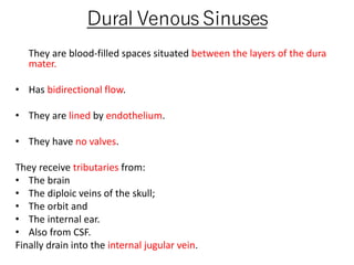
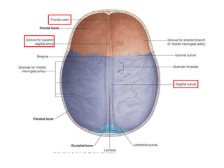
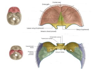
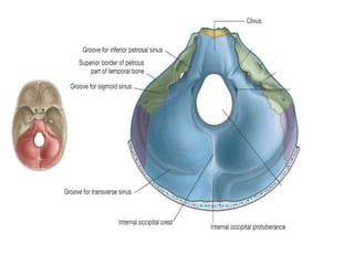
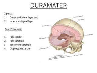
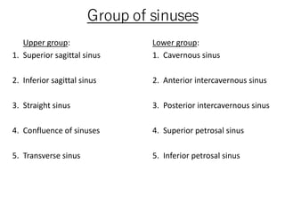
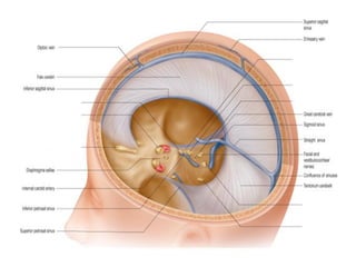
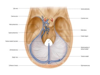
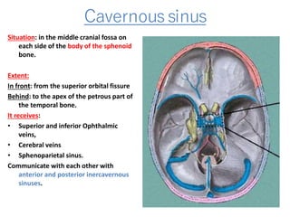
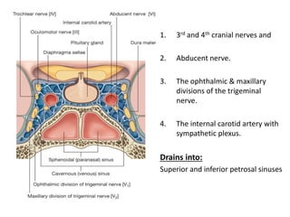
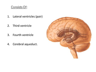
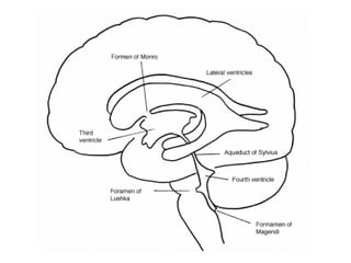
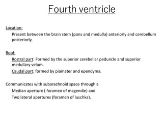
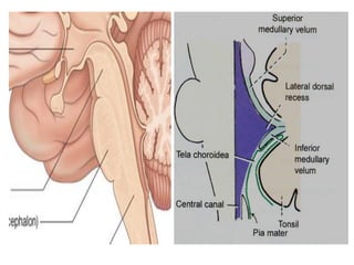
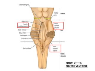

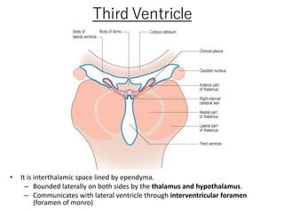
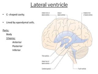
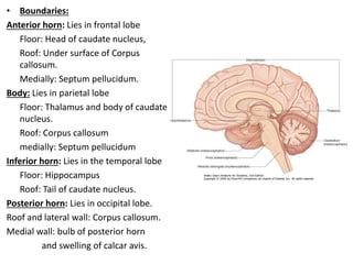
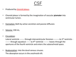
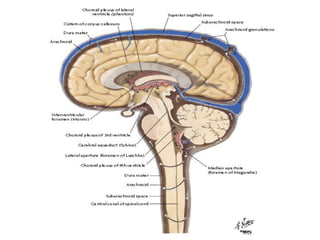
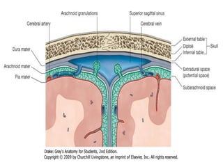
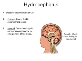


This document summarizes the dural sinuses, ventricles, and cerebrospinal fluid (CSF) systems. It describes the anatomy and flow of blood within the dural sinuses, located between the layers of the dura mater, and lists the various sinuses. It also outlines the anatomy and structures of the four ventricles - lateral, third, fourth, and their interconnections via the cerebral aqueduct. Finally, it discusses CSF production, circulation from the ventricles to subarachnoid space, reabsorption into dural sinuses, and conditions like hydrocephalus.


![CTEV [ clubfoot] DR ARUN LAL ,DR MOHAMED ASHRAF travancore medical college k...](https://cdn.slidesharecdn.com/ss_thumbnails/ctevclubfootdrarunlaldrmohamedashraftravancoremedicalcollegekollamkeralaindia-260208063247-18fc466c-thumbnail.jpg?width=640&height=640&fit=bounds)


![PERI-PROSTHETIC FRACTURE NAIL-PLATE CONSTRUCT [NPC].pptx](https://cdn.slidesharecdn.com/ss_thumbnails/drarunkumardrmohamedashrafperiprostheticfrasturenail-plateconstructnpc-260209164459-7e9d15a1-thumbnail.jpg?width=640&height=640&fit=bounds)


