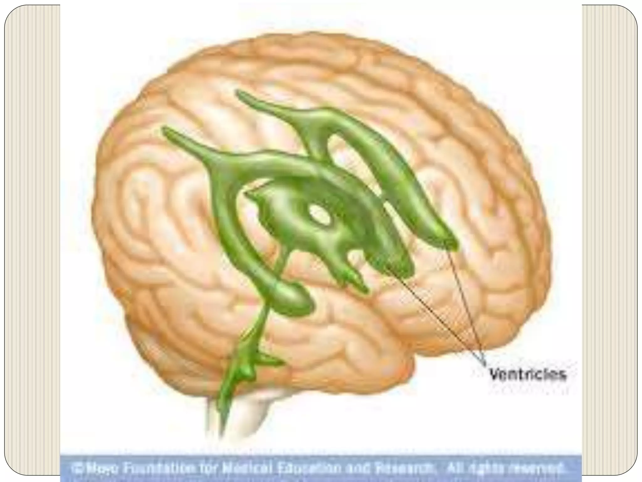
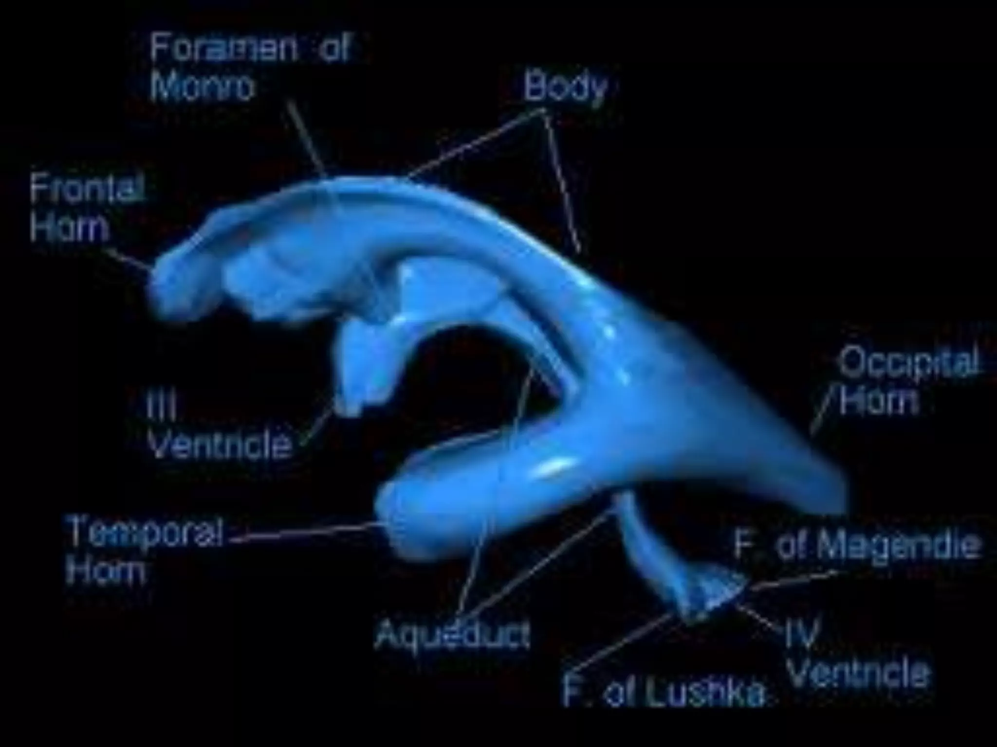
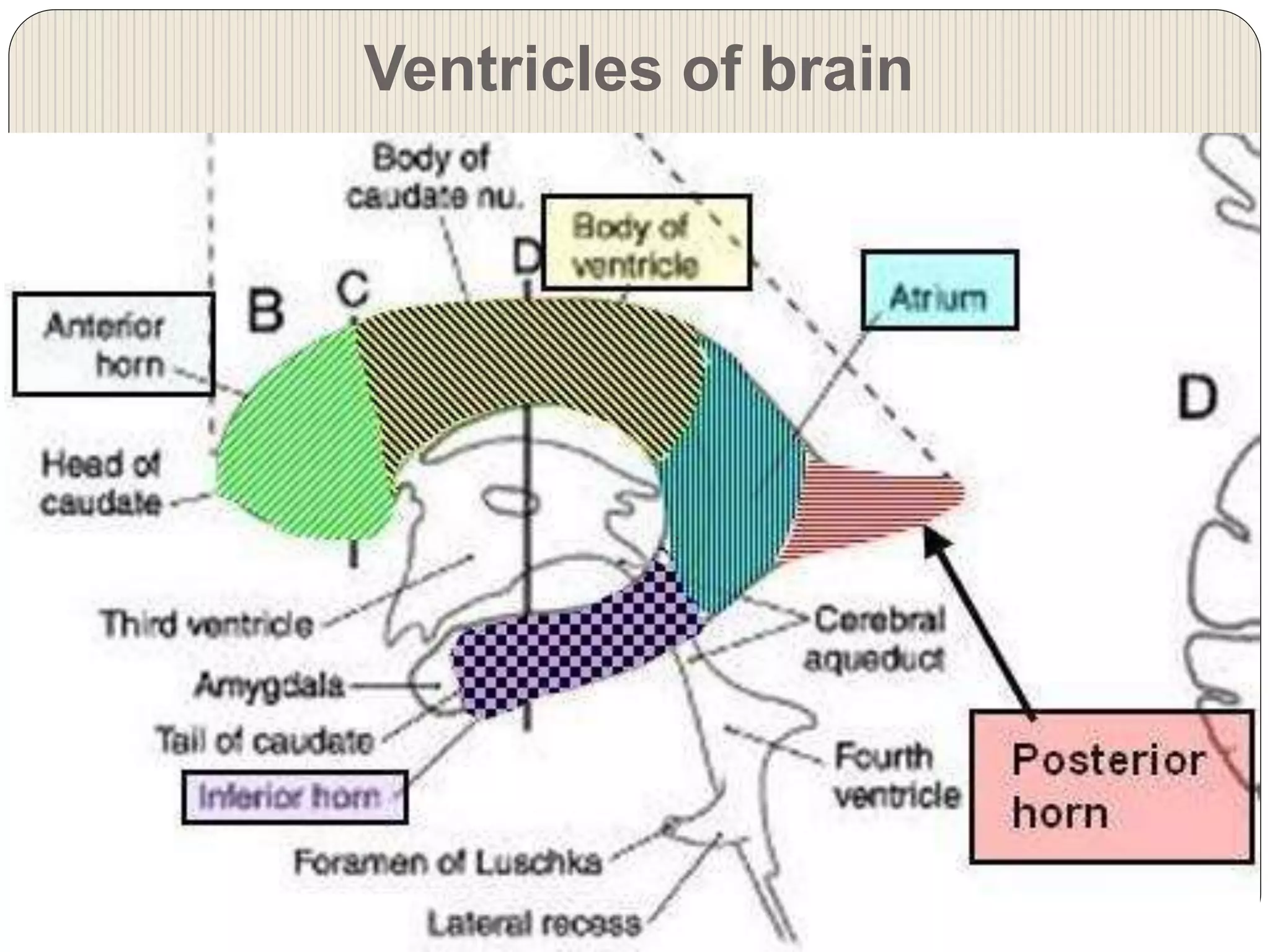
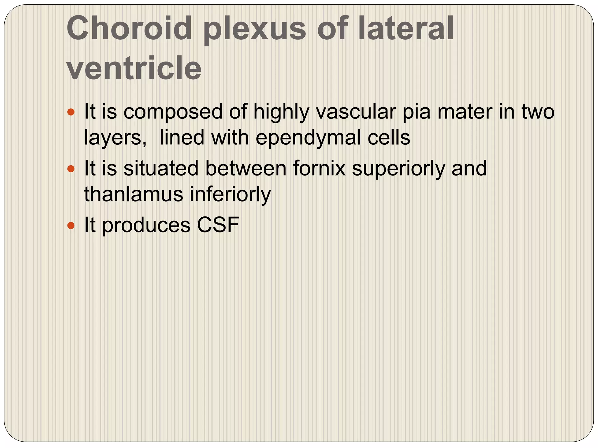
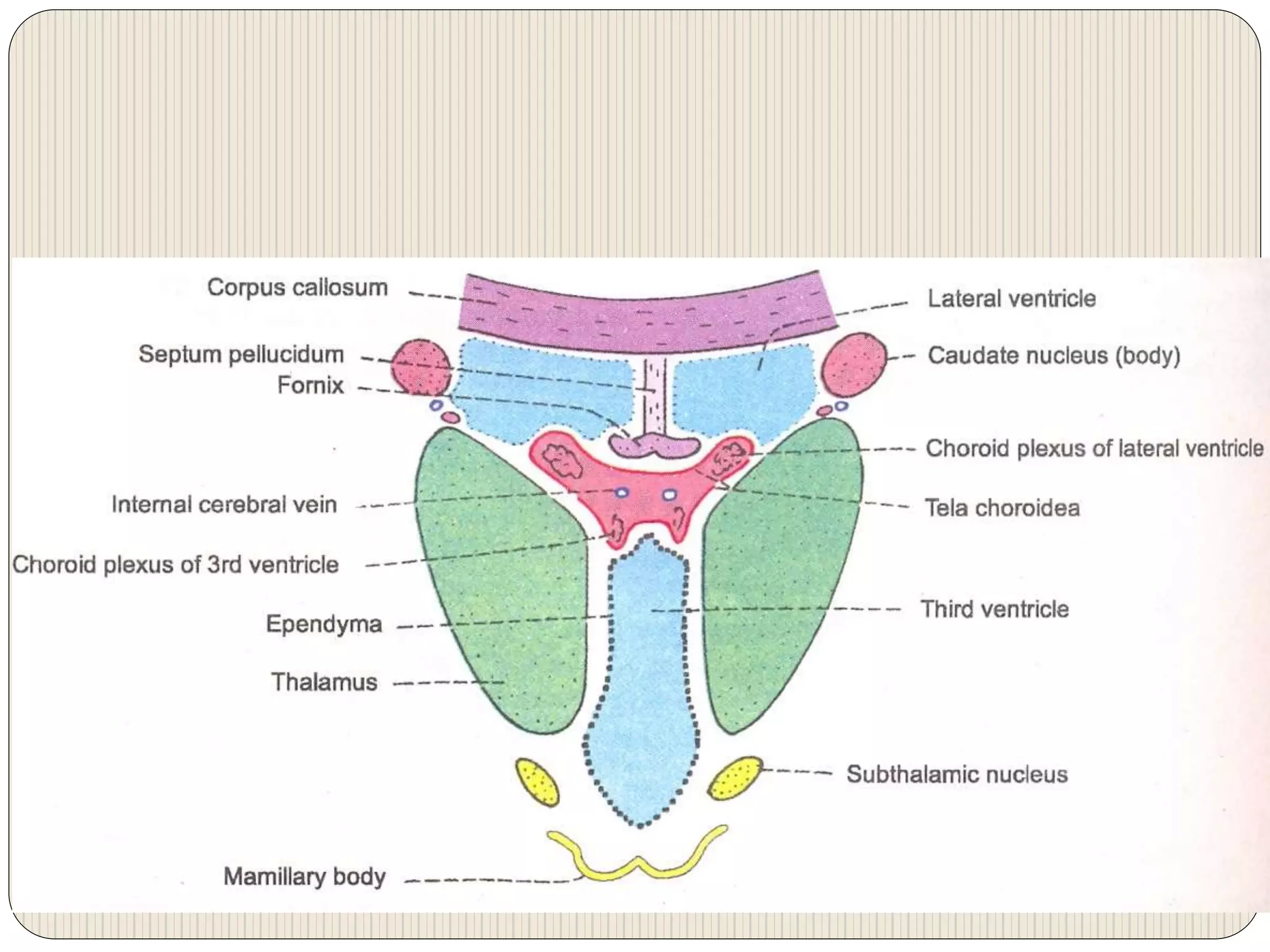
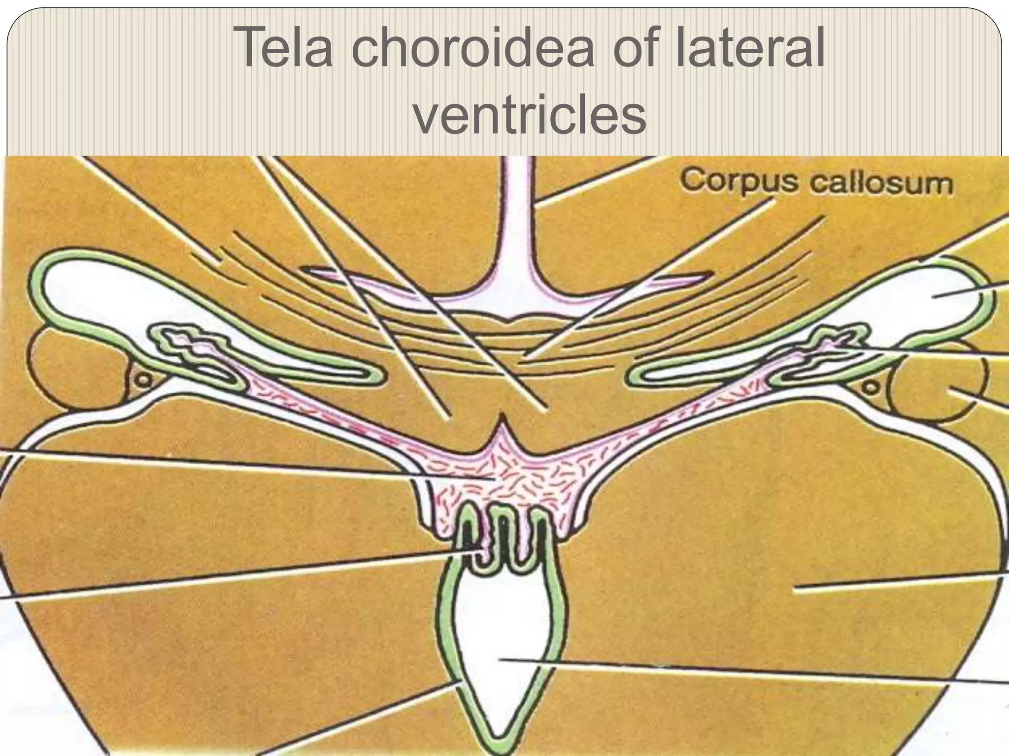
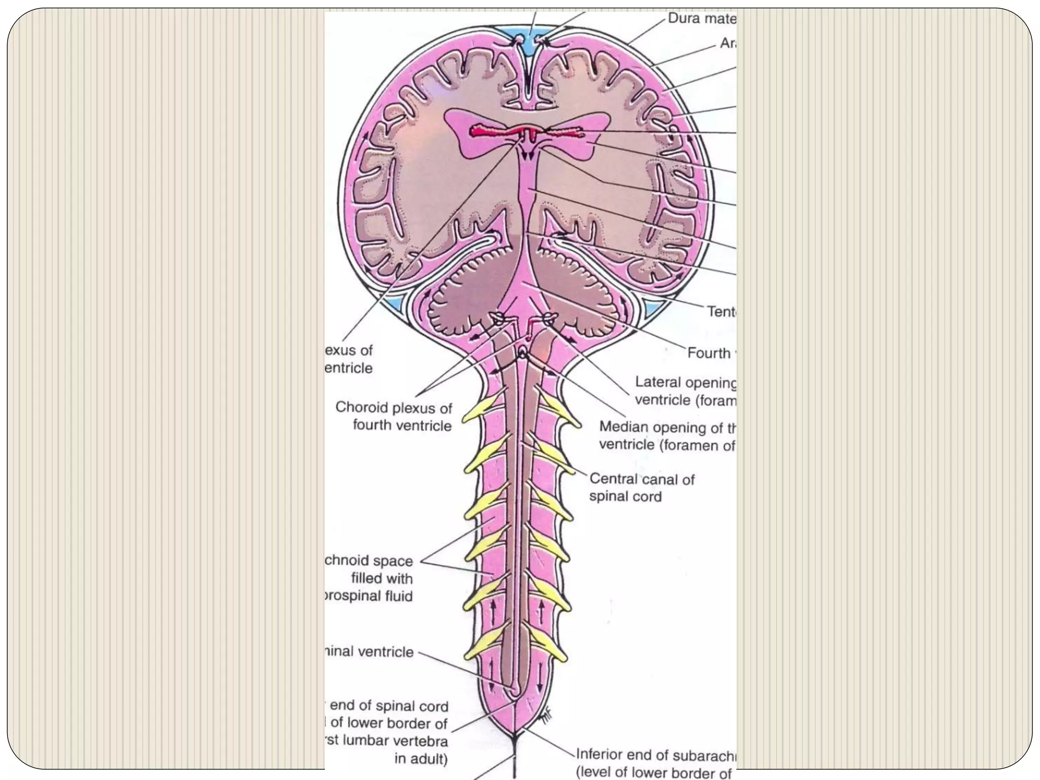
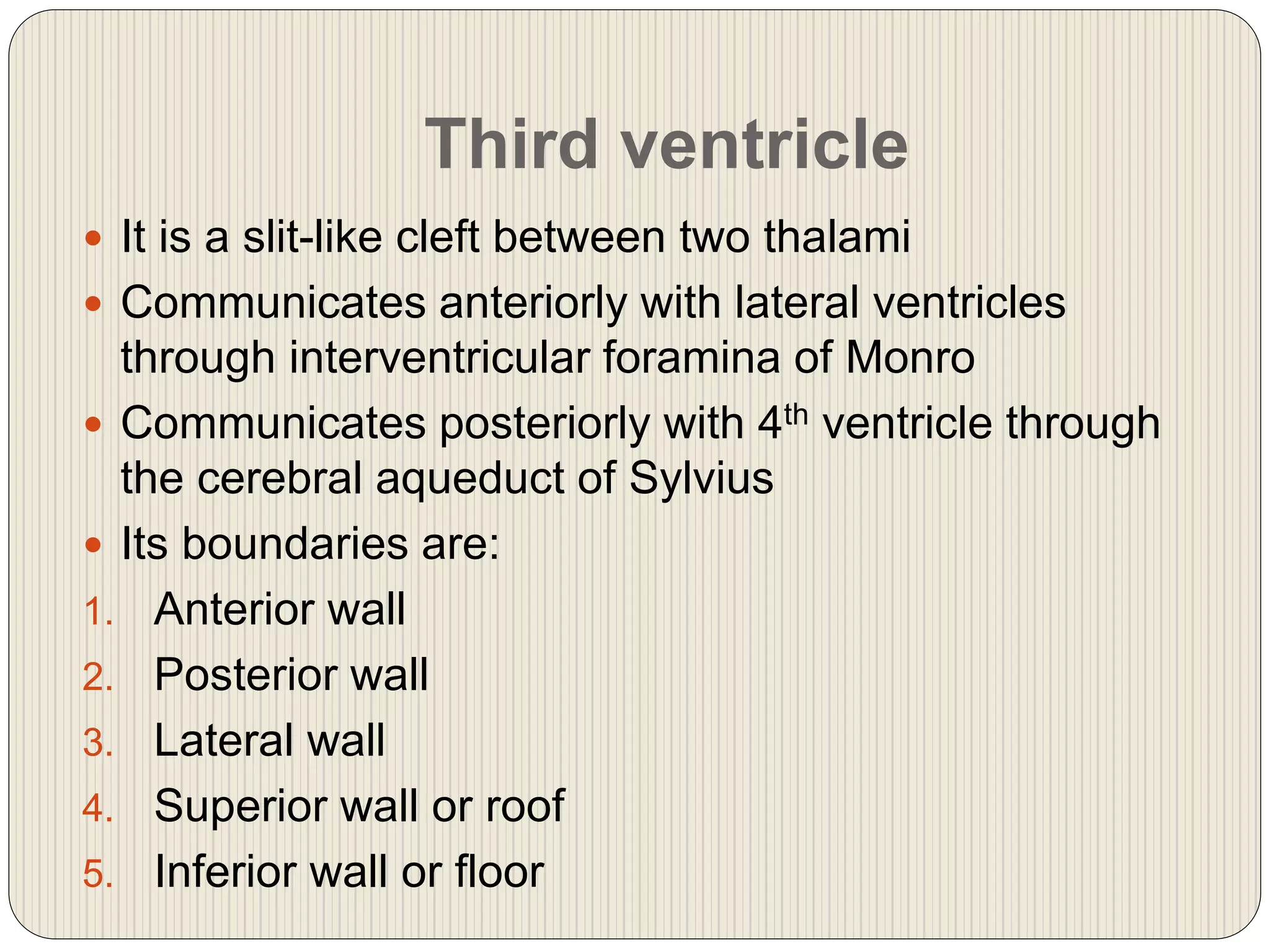
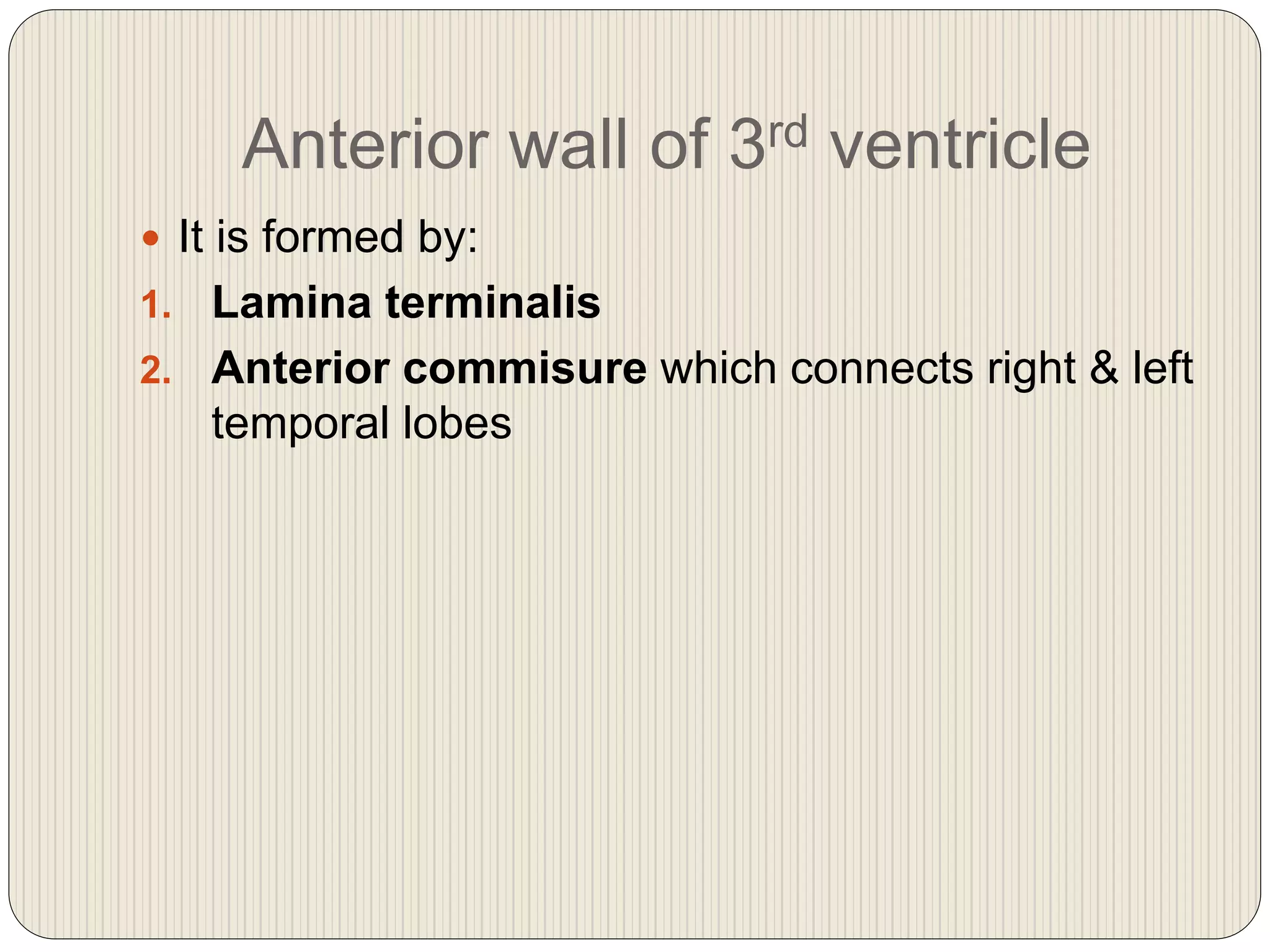
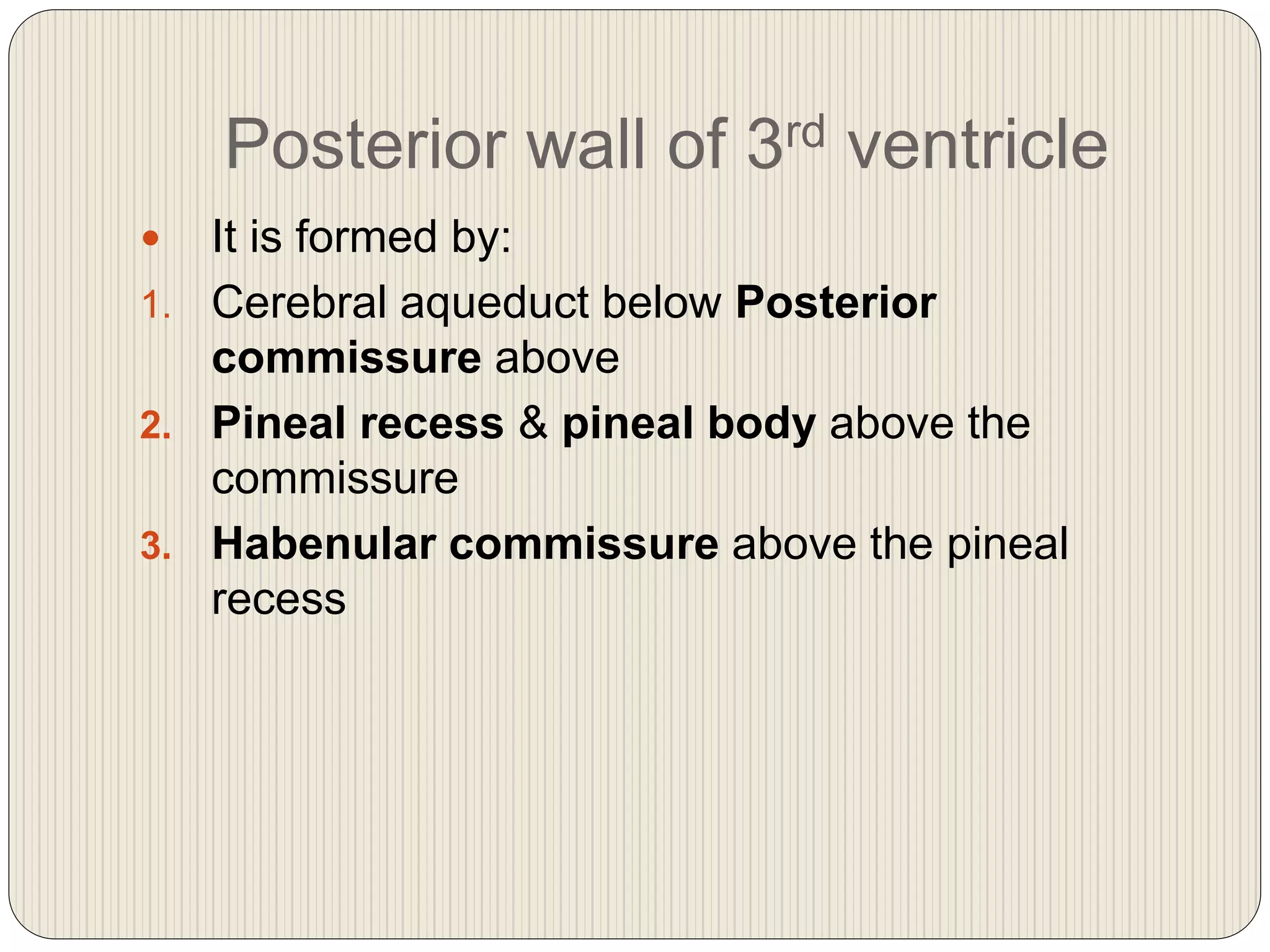
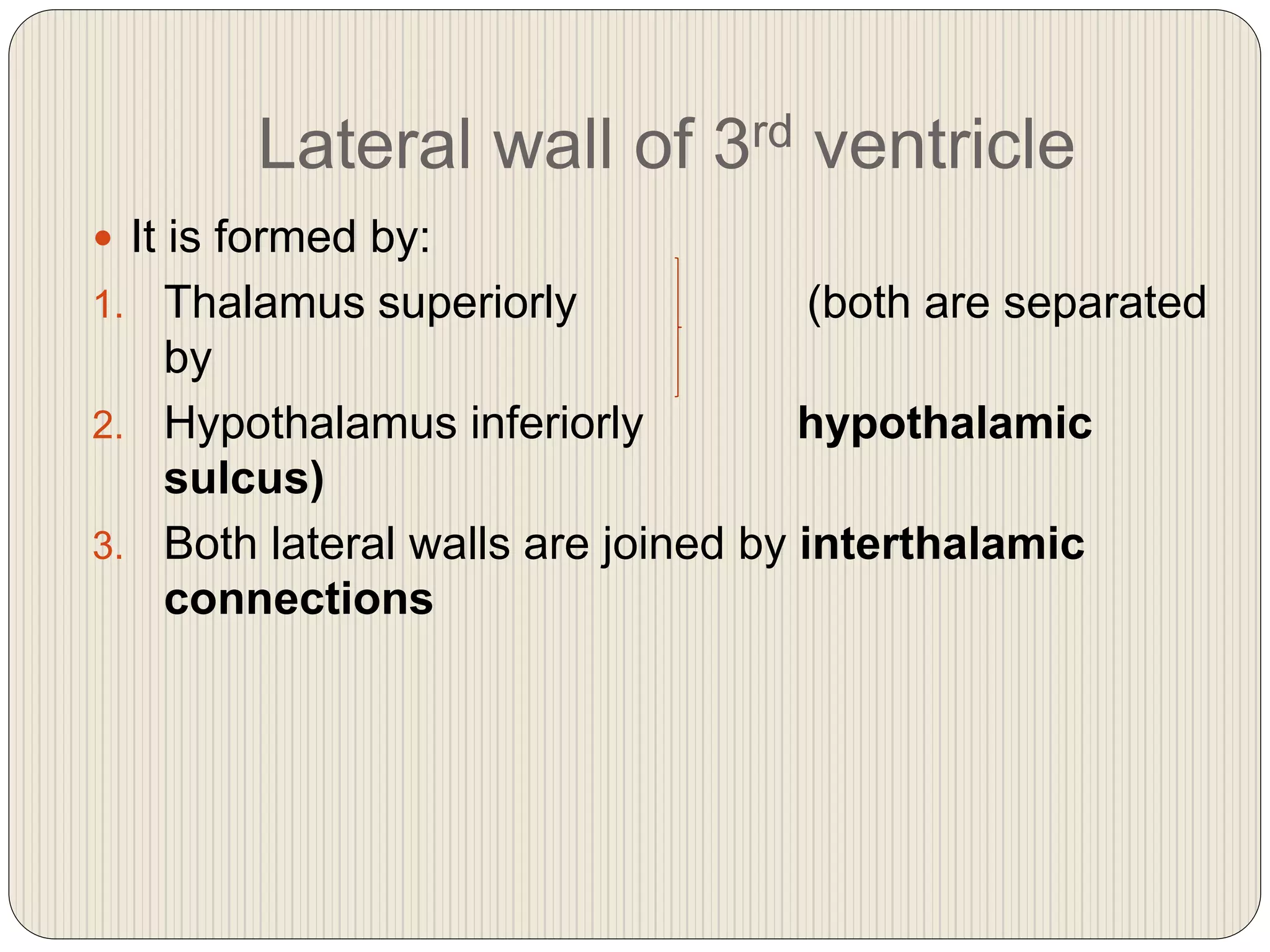
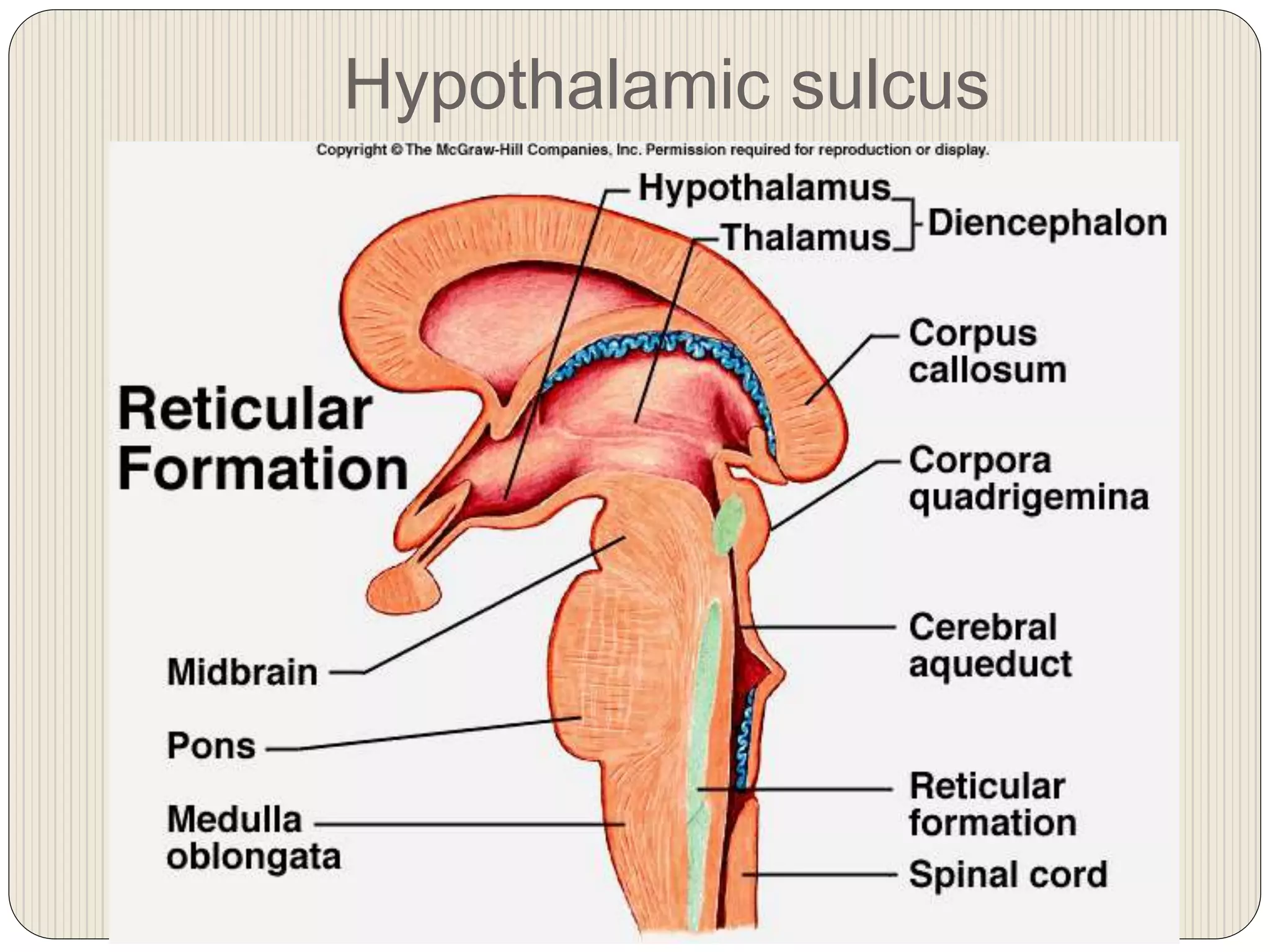
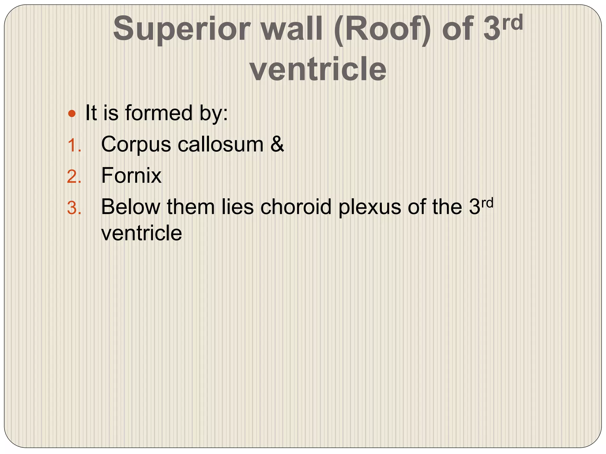
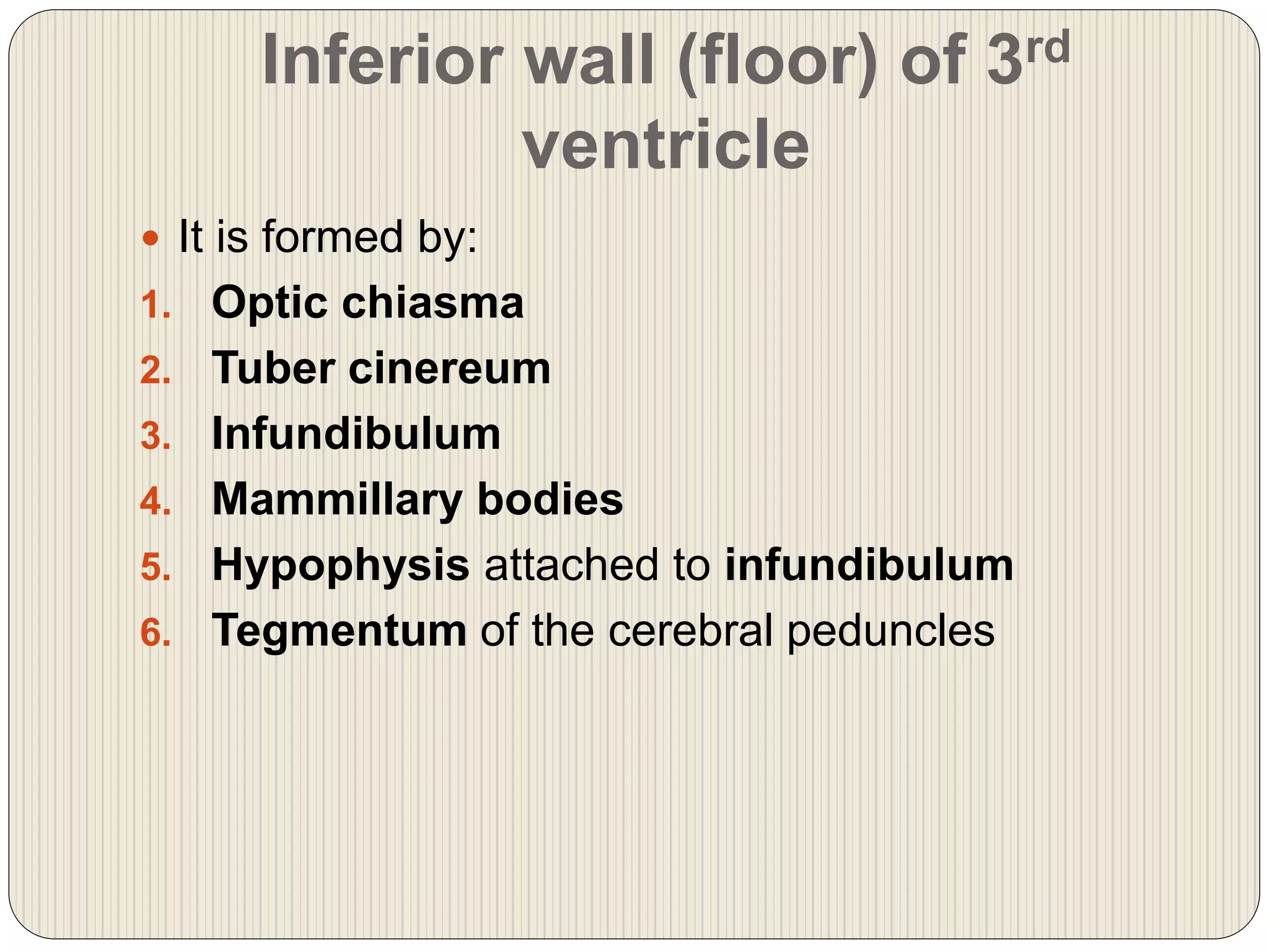
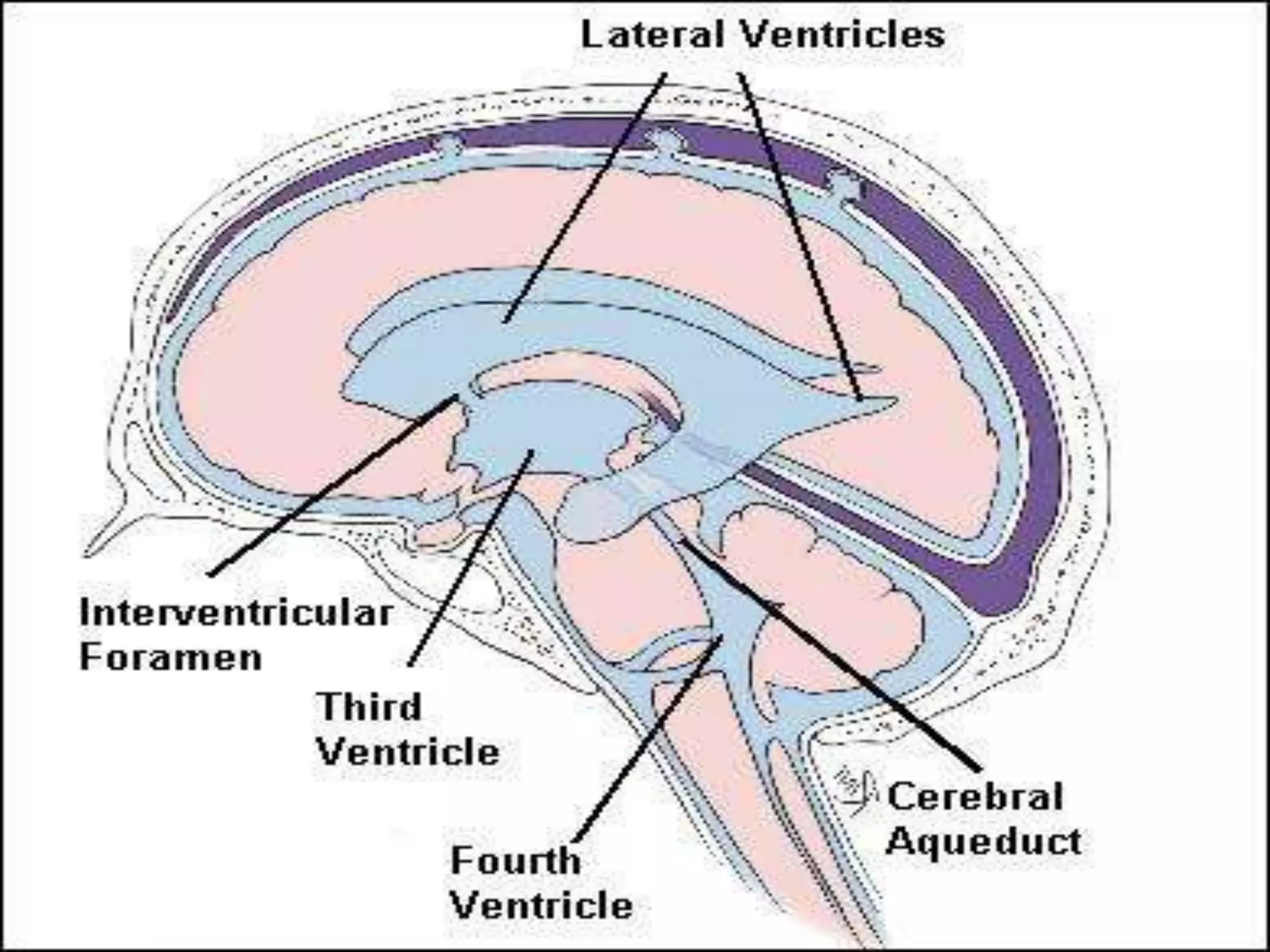
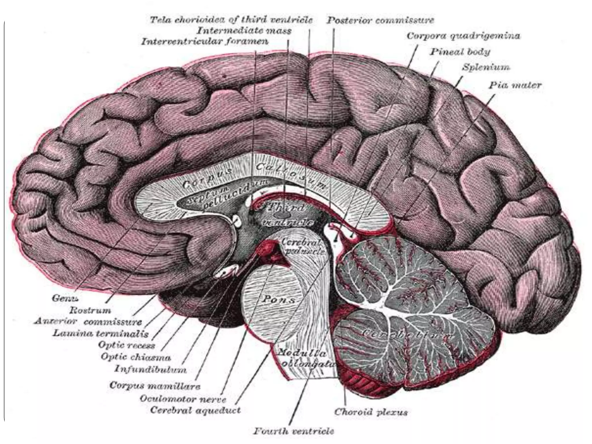
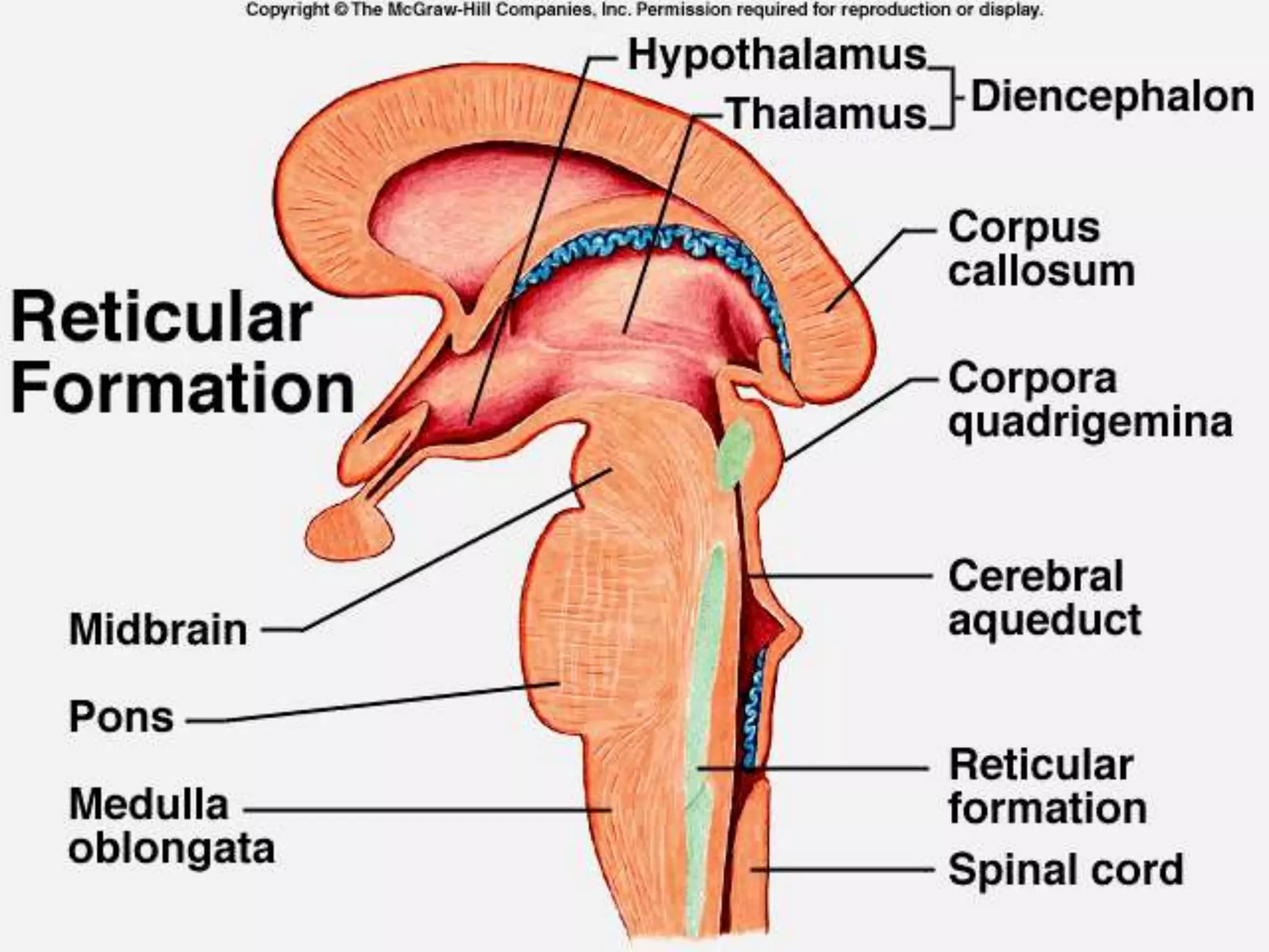
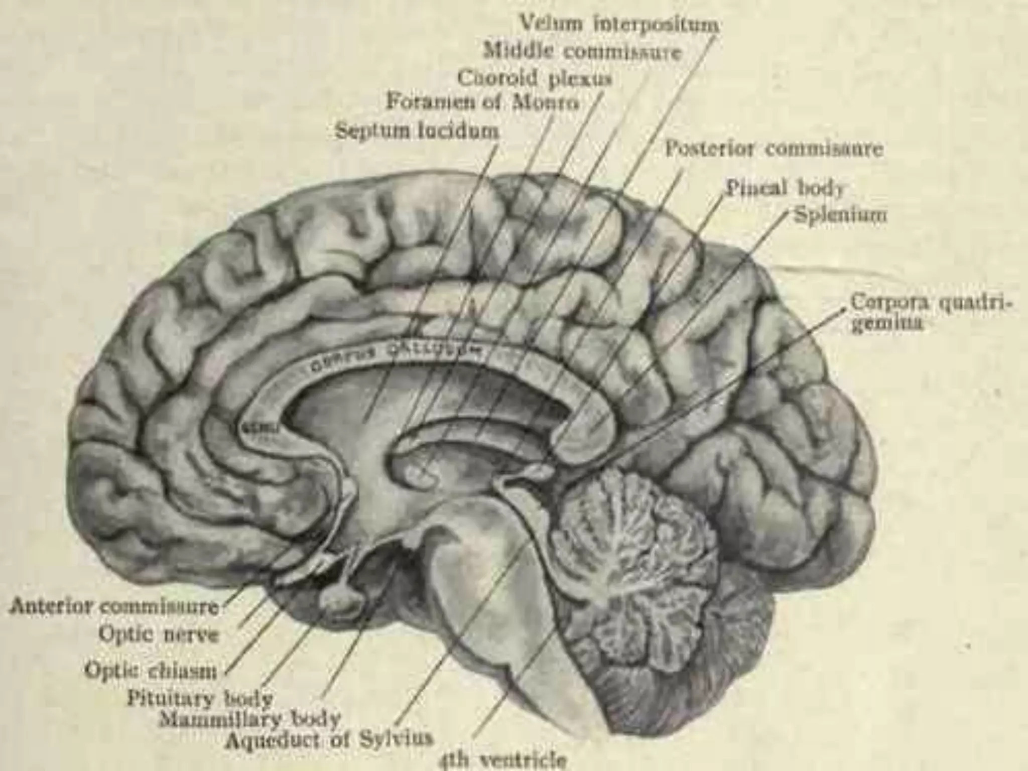
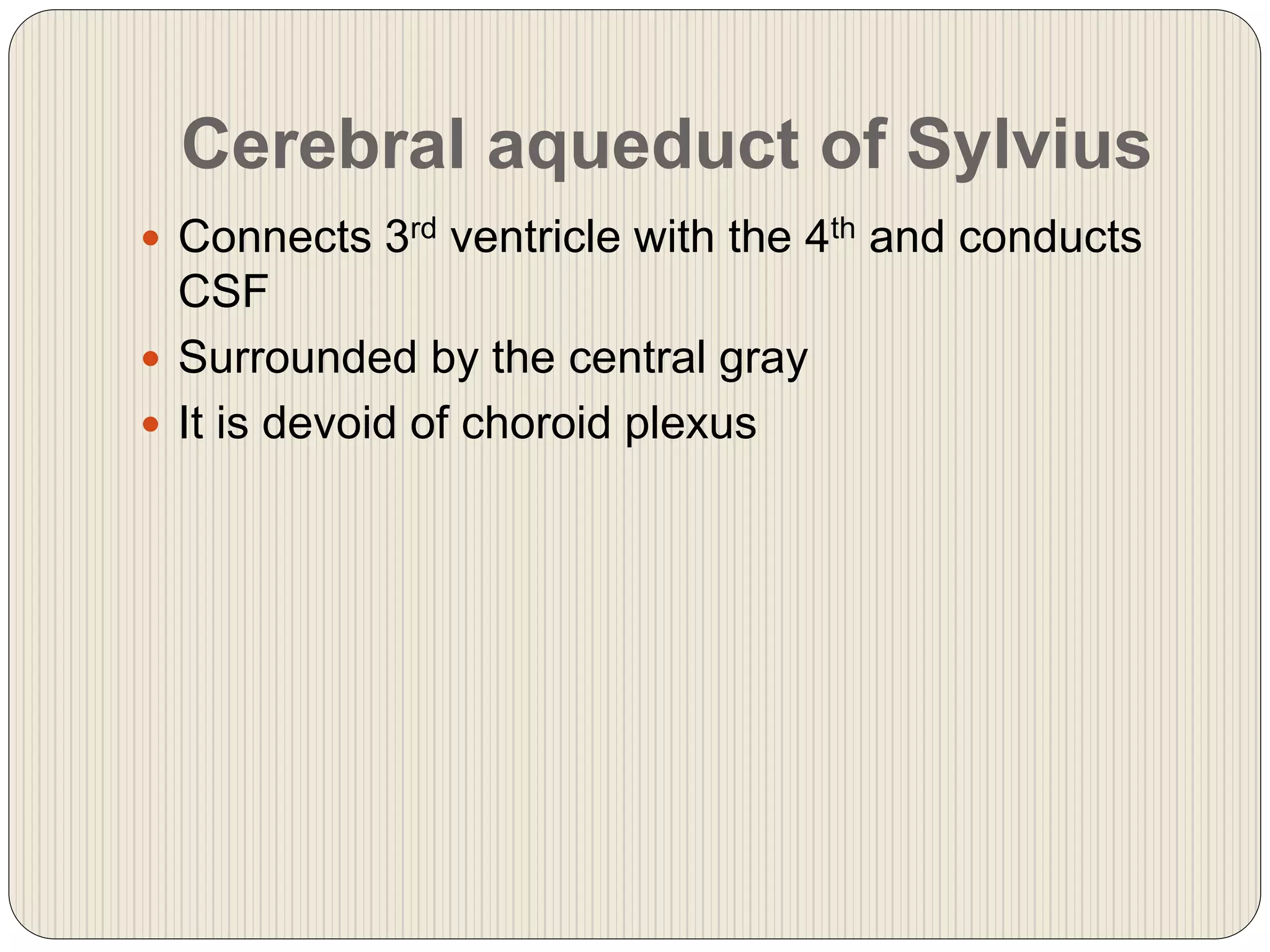
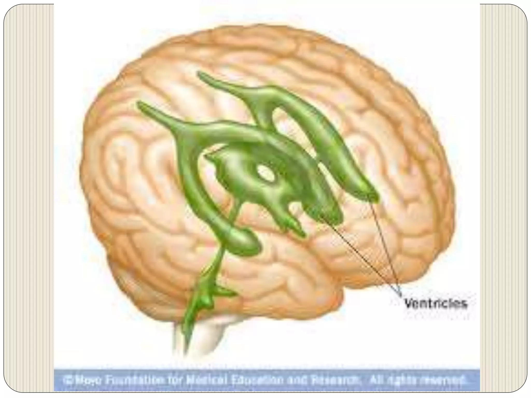
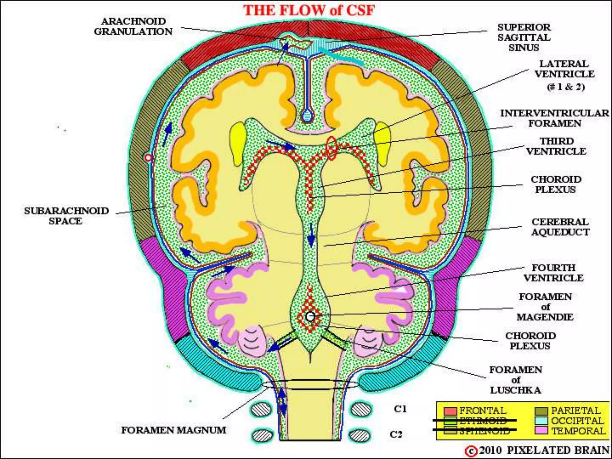
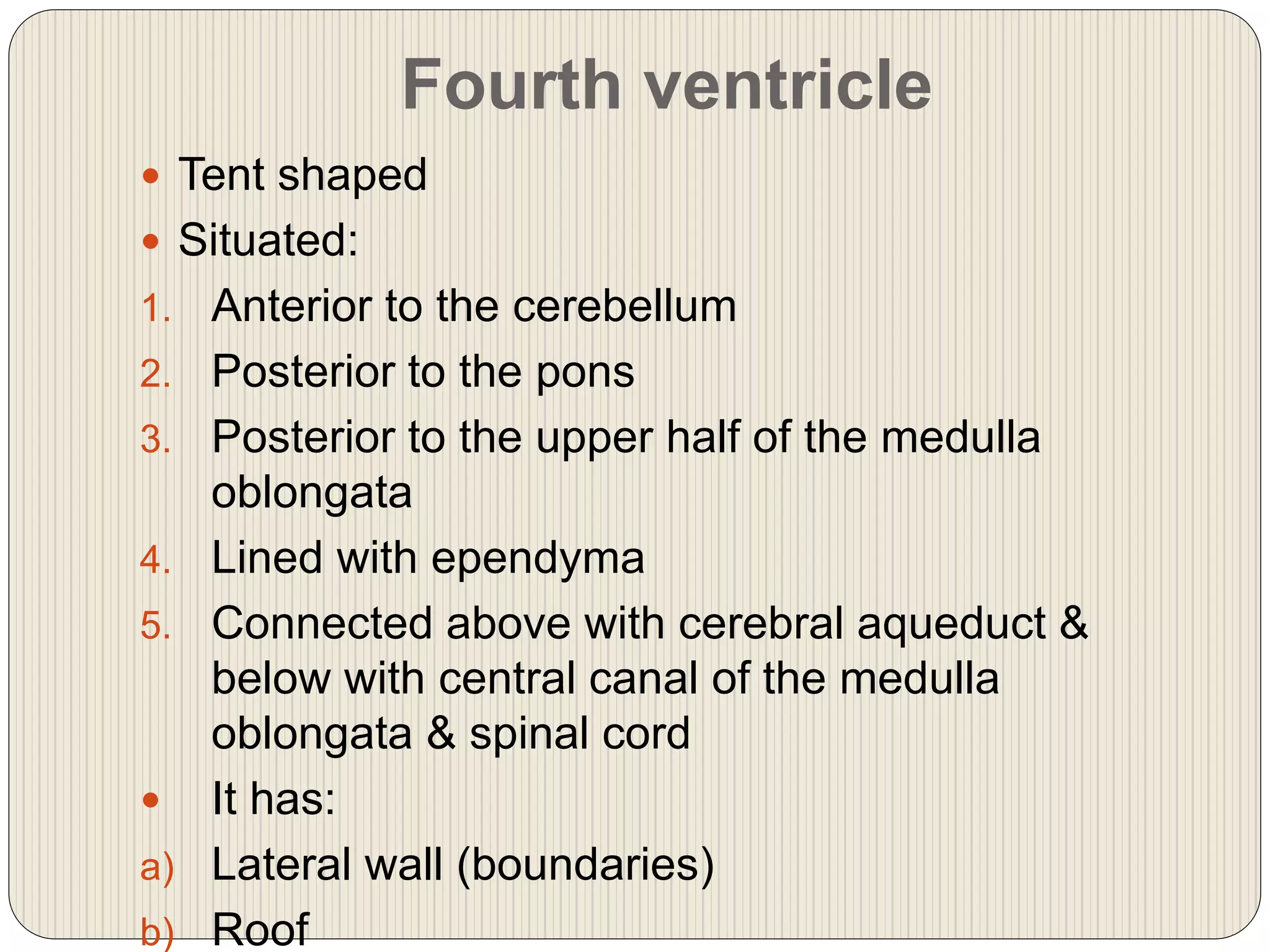
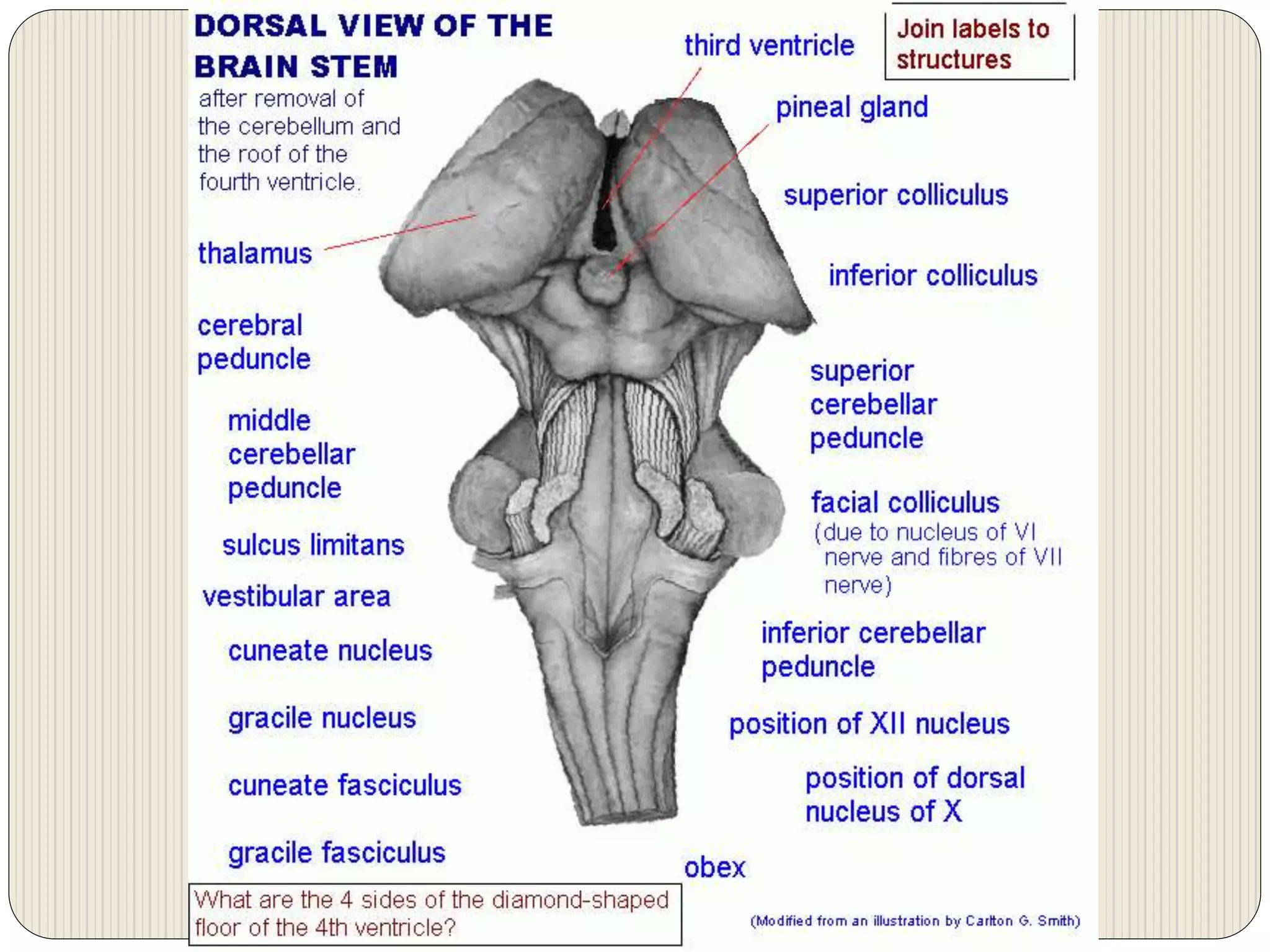
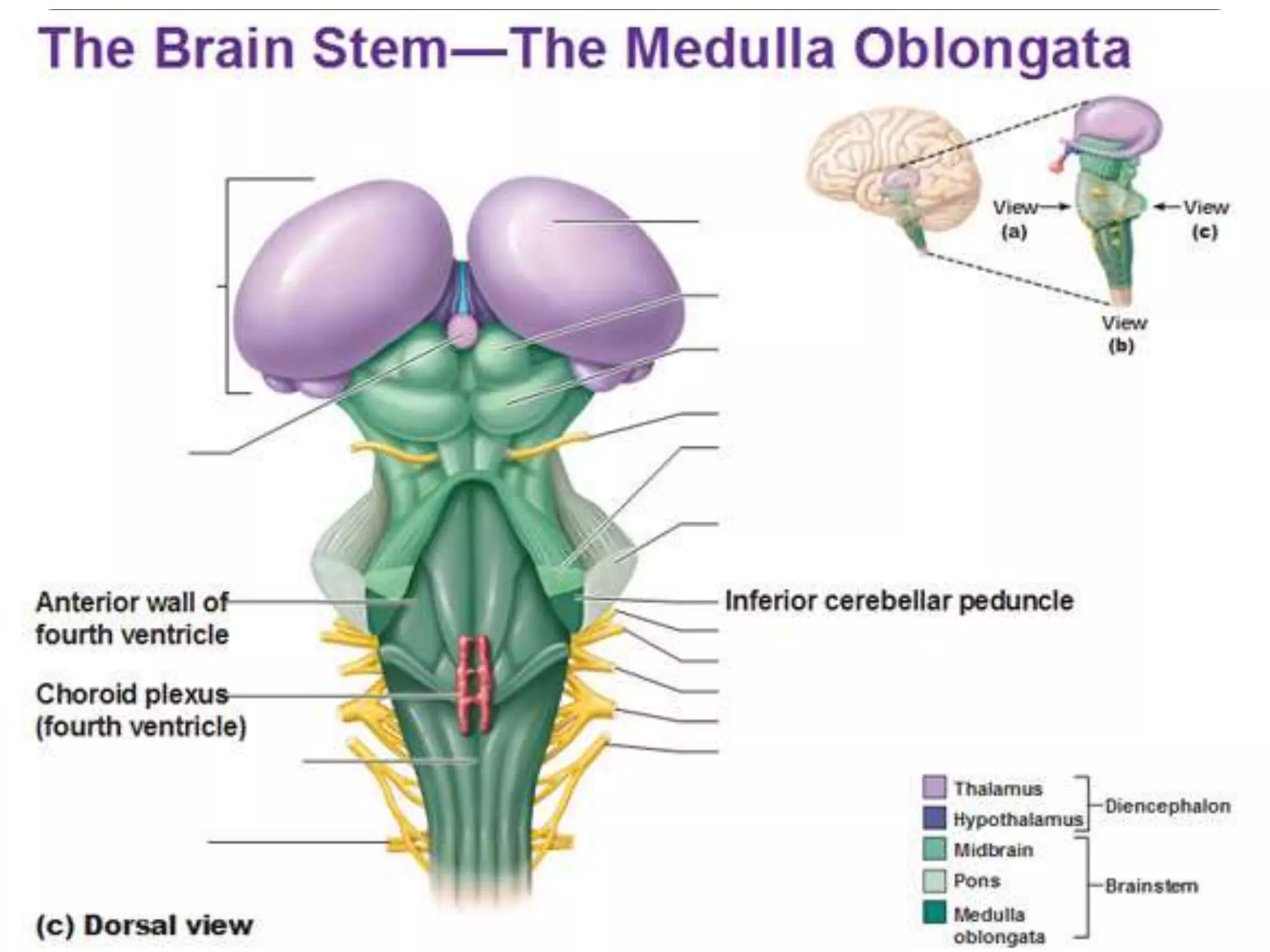
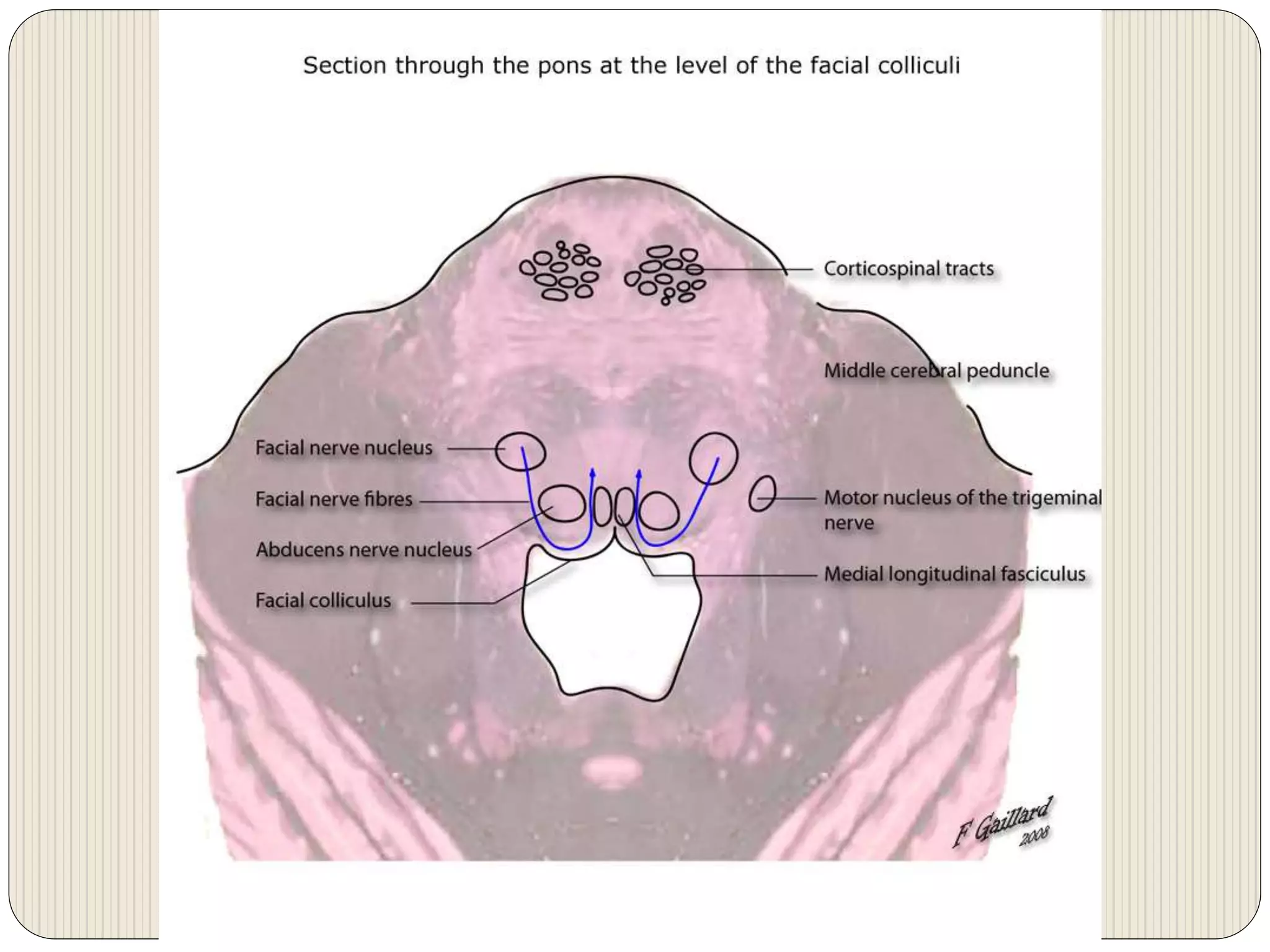
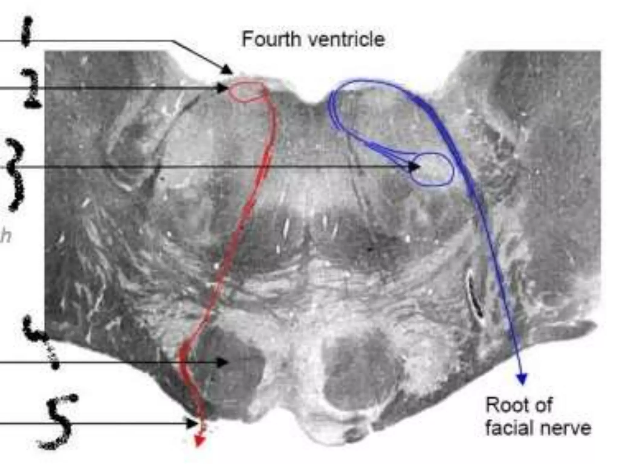
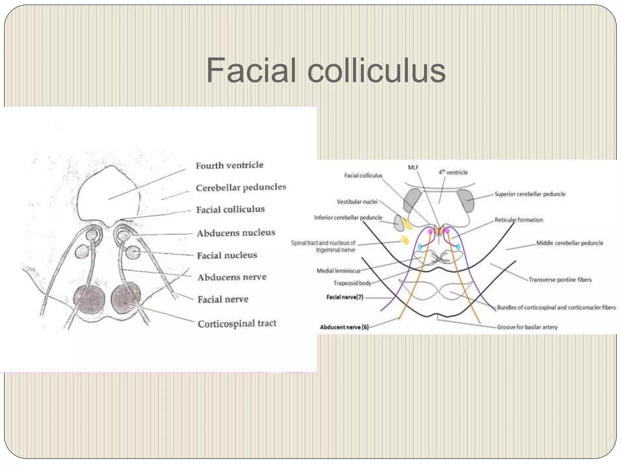
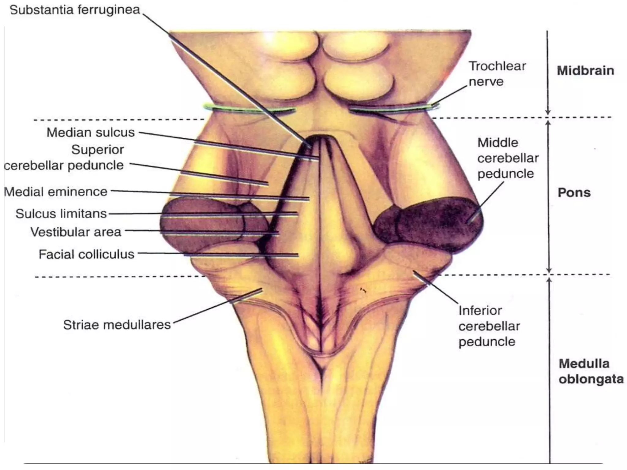
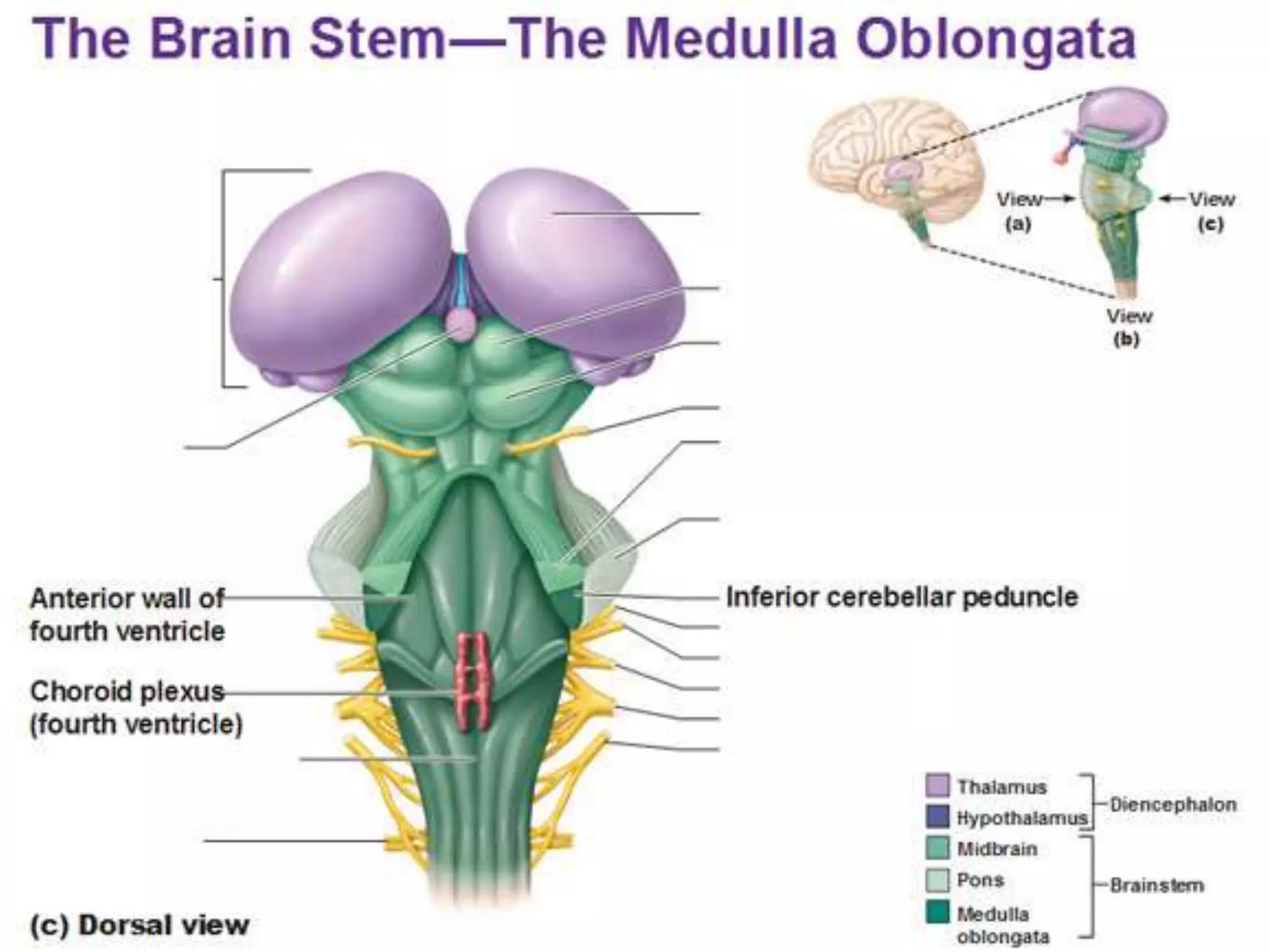
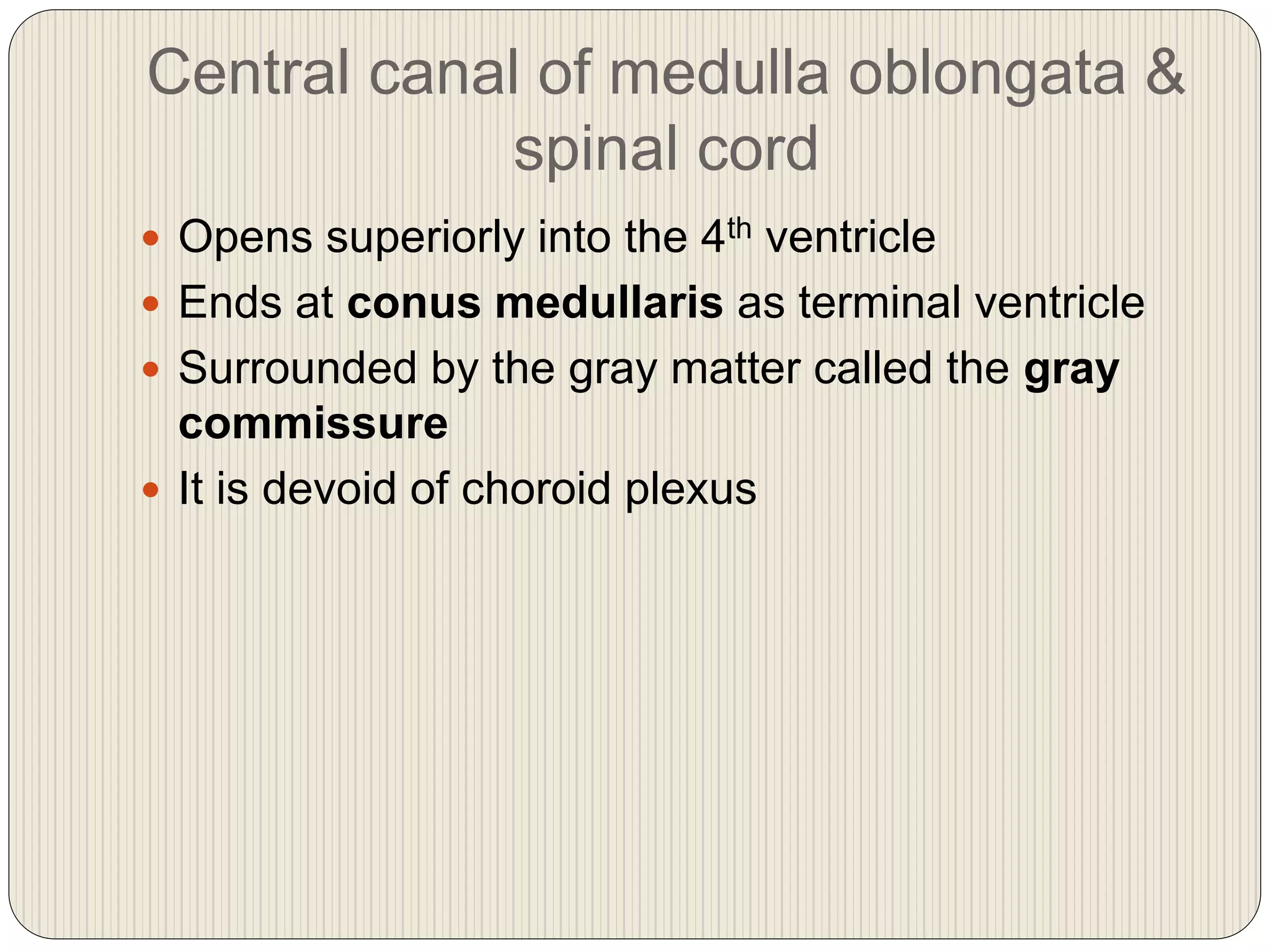
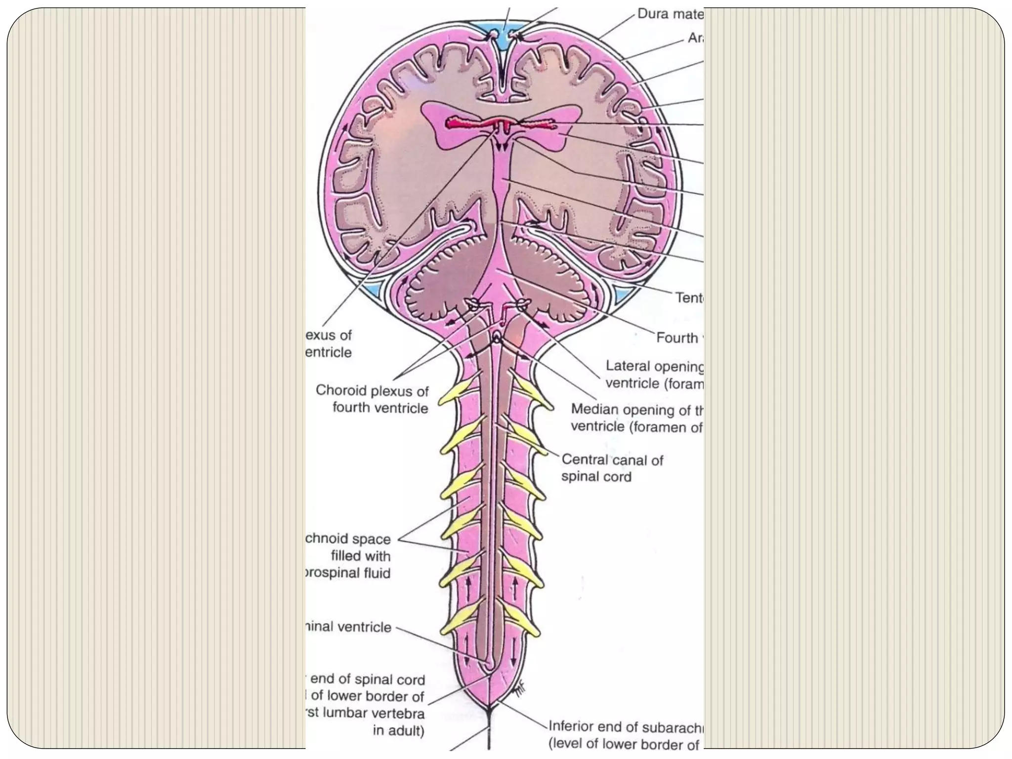
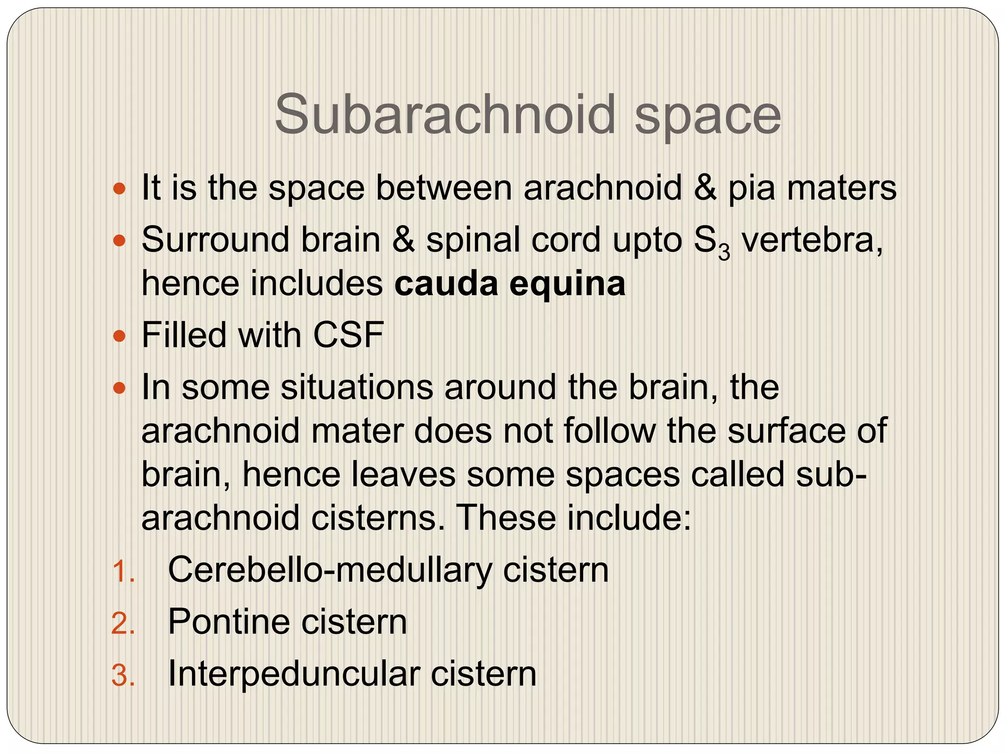
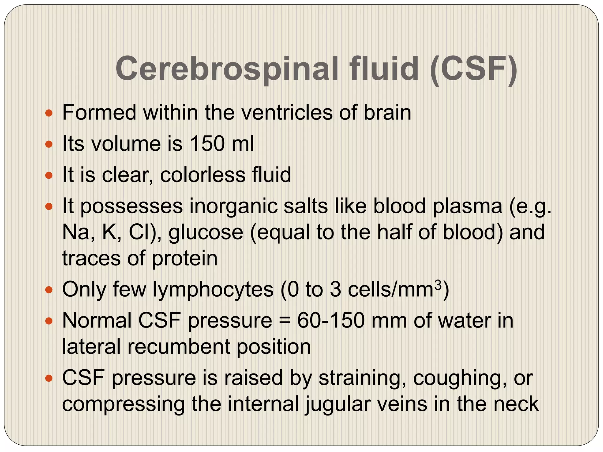
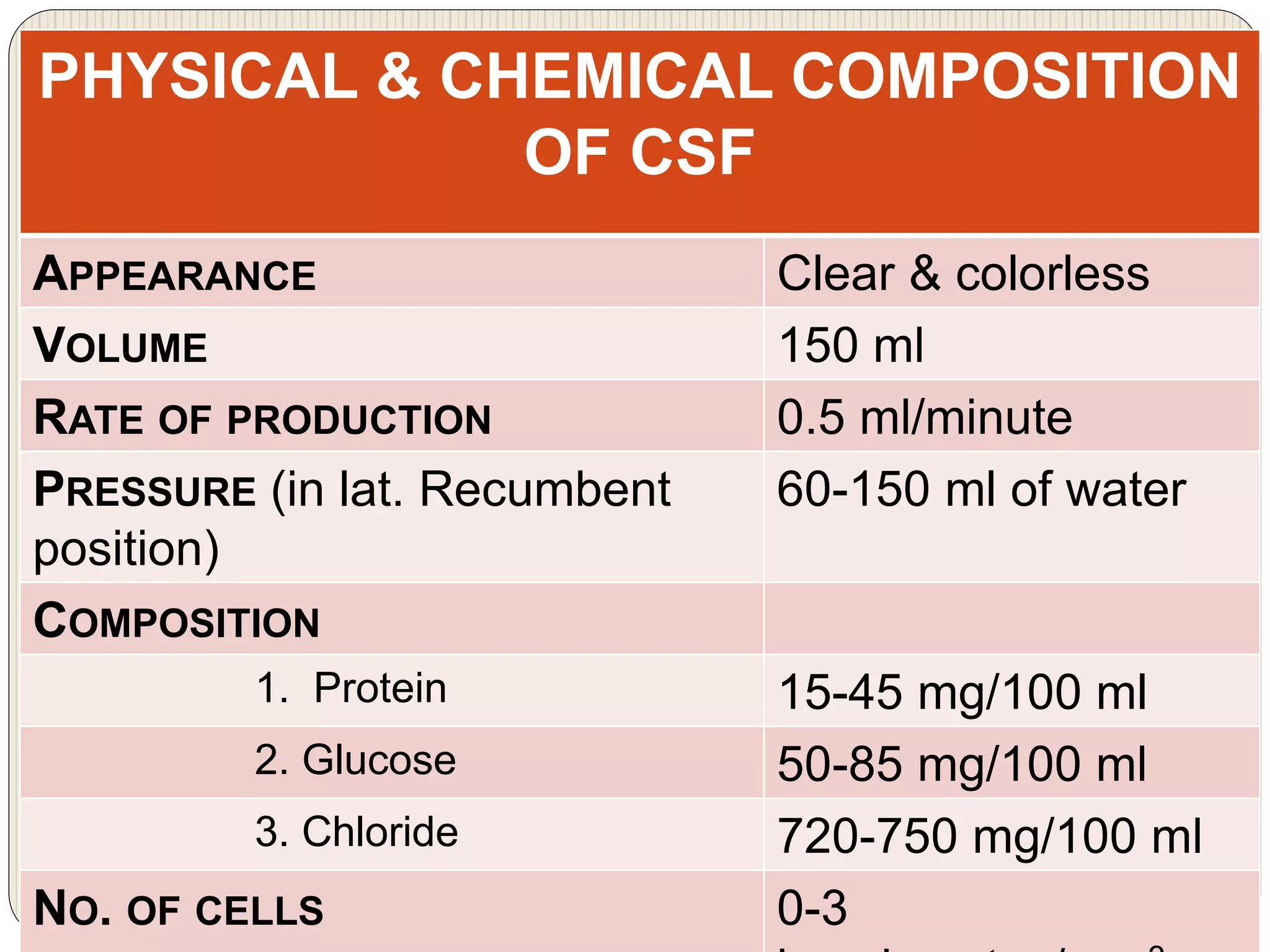
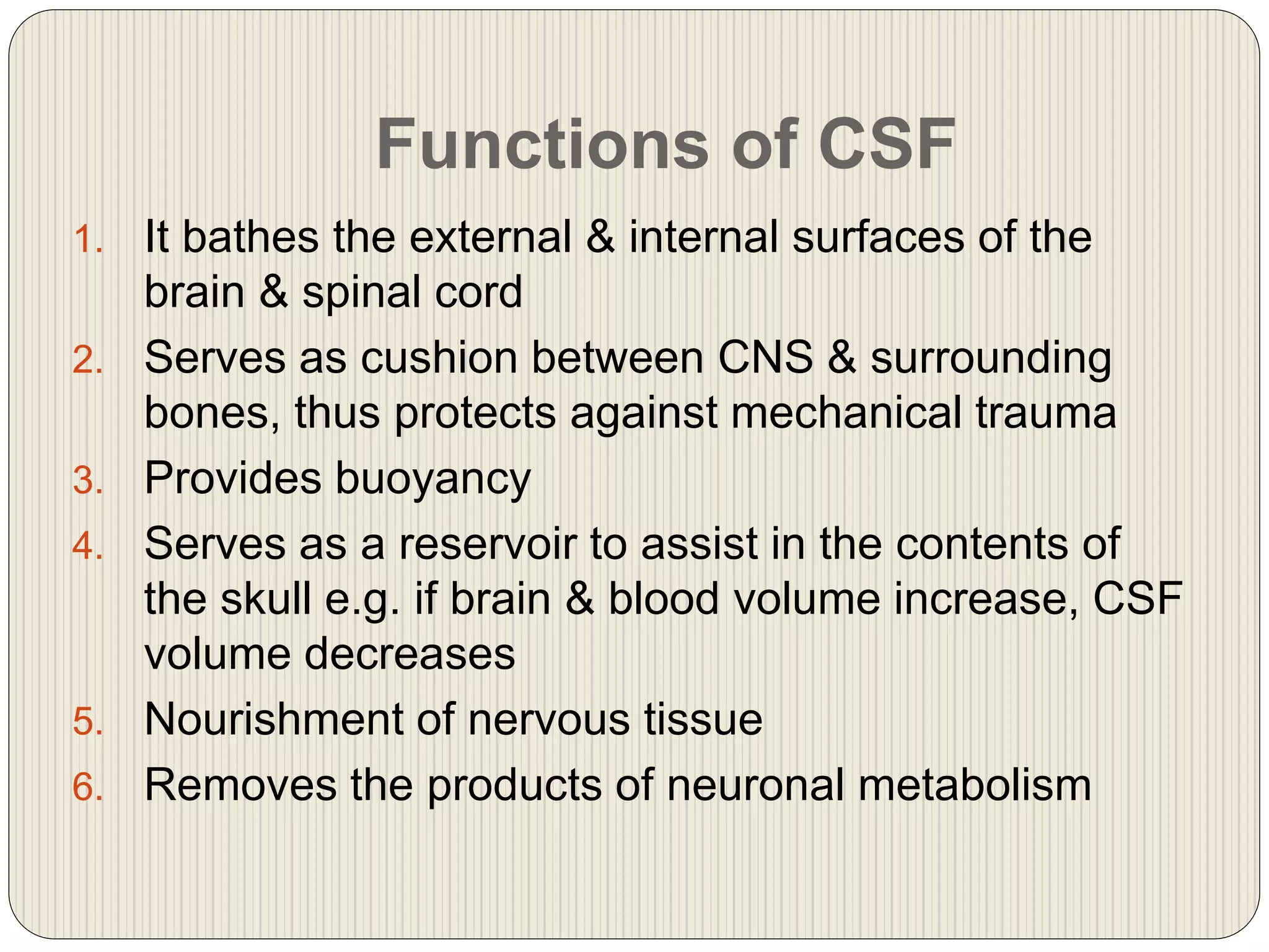
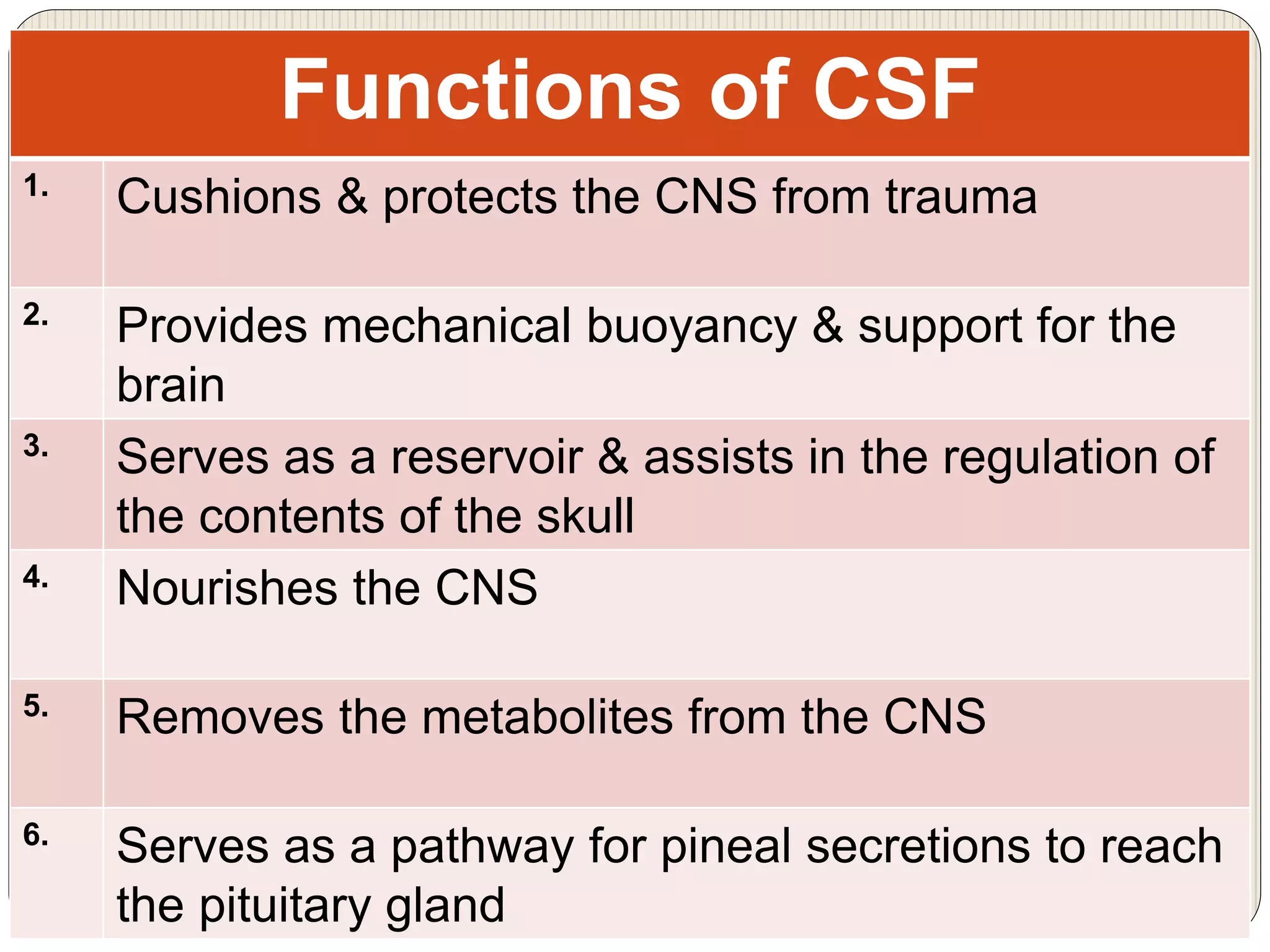
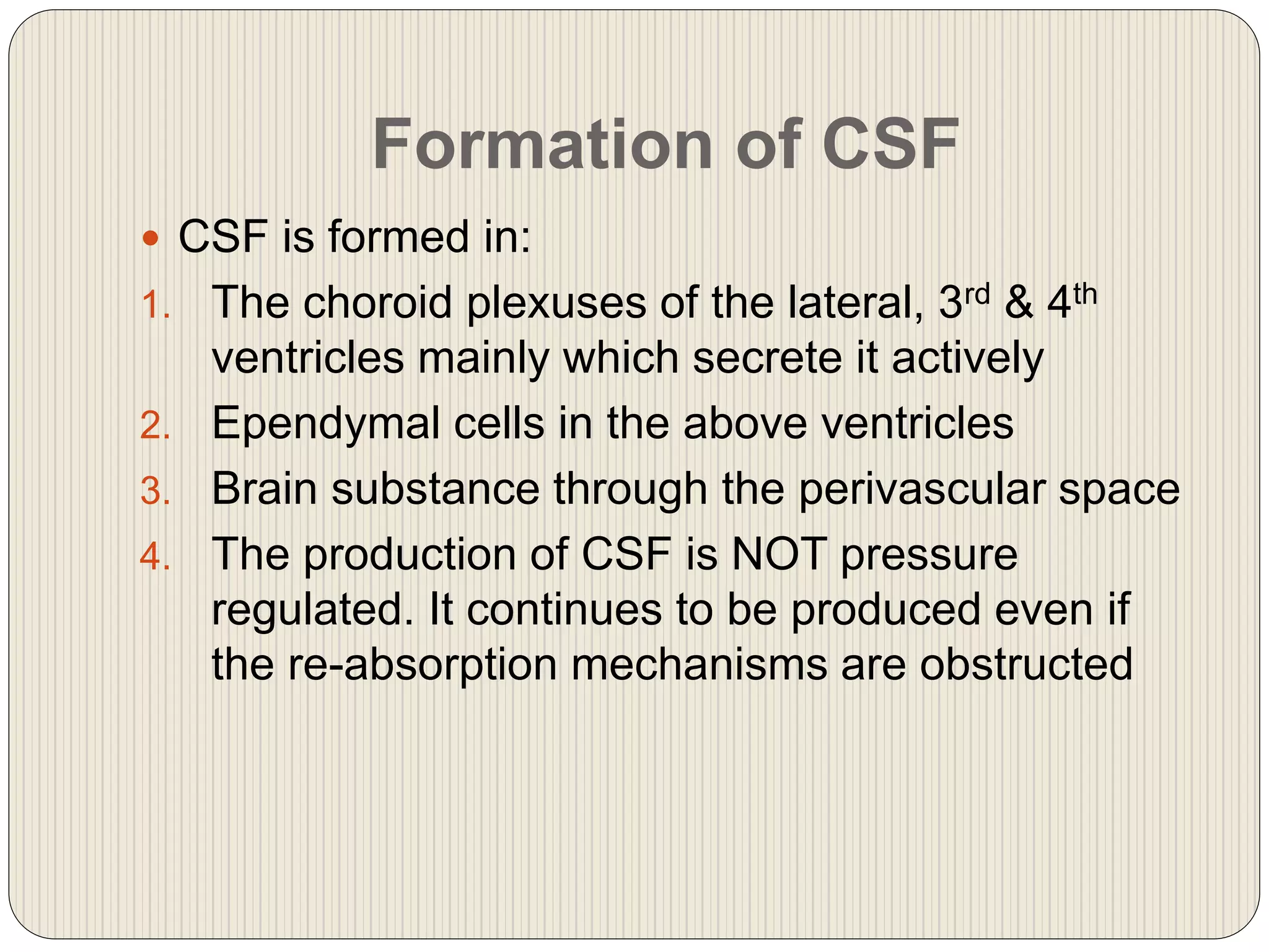
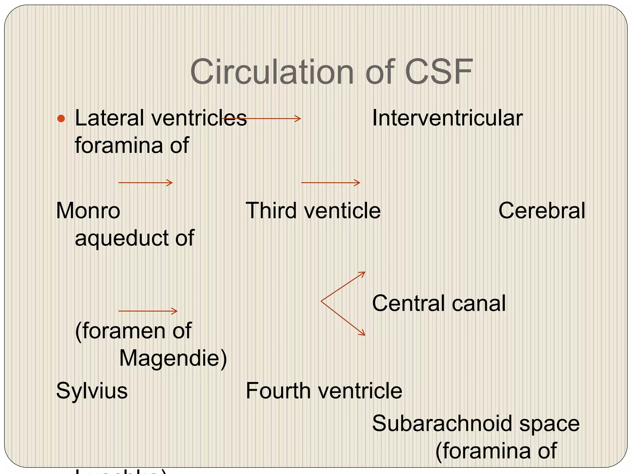
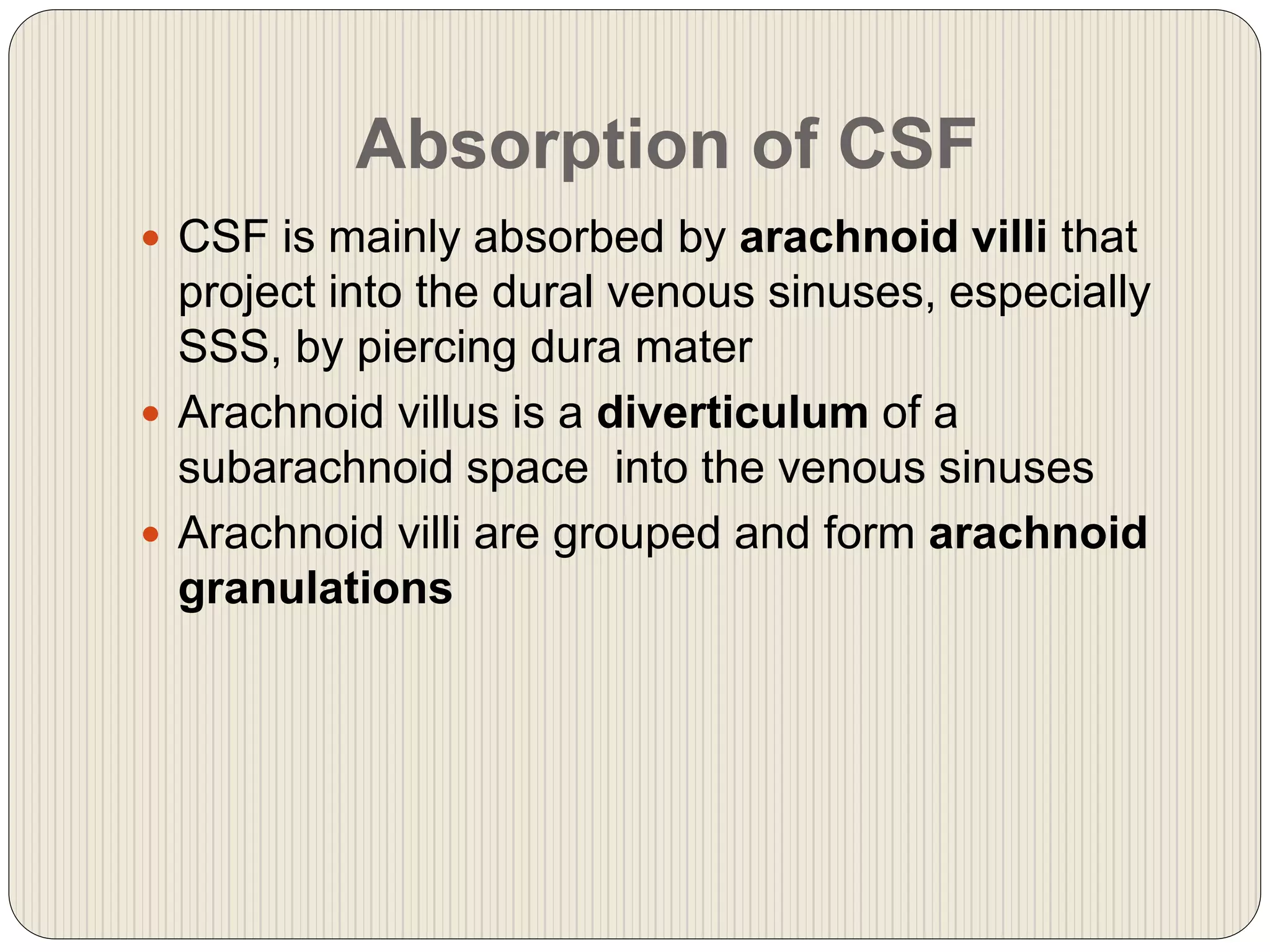
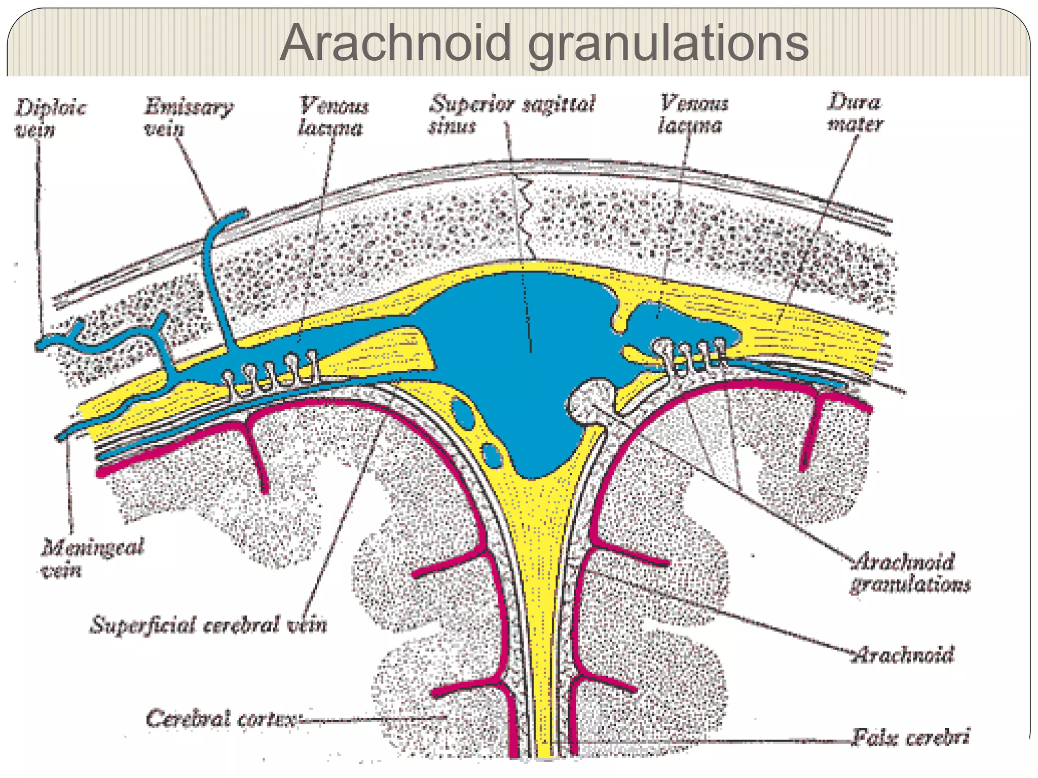
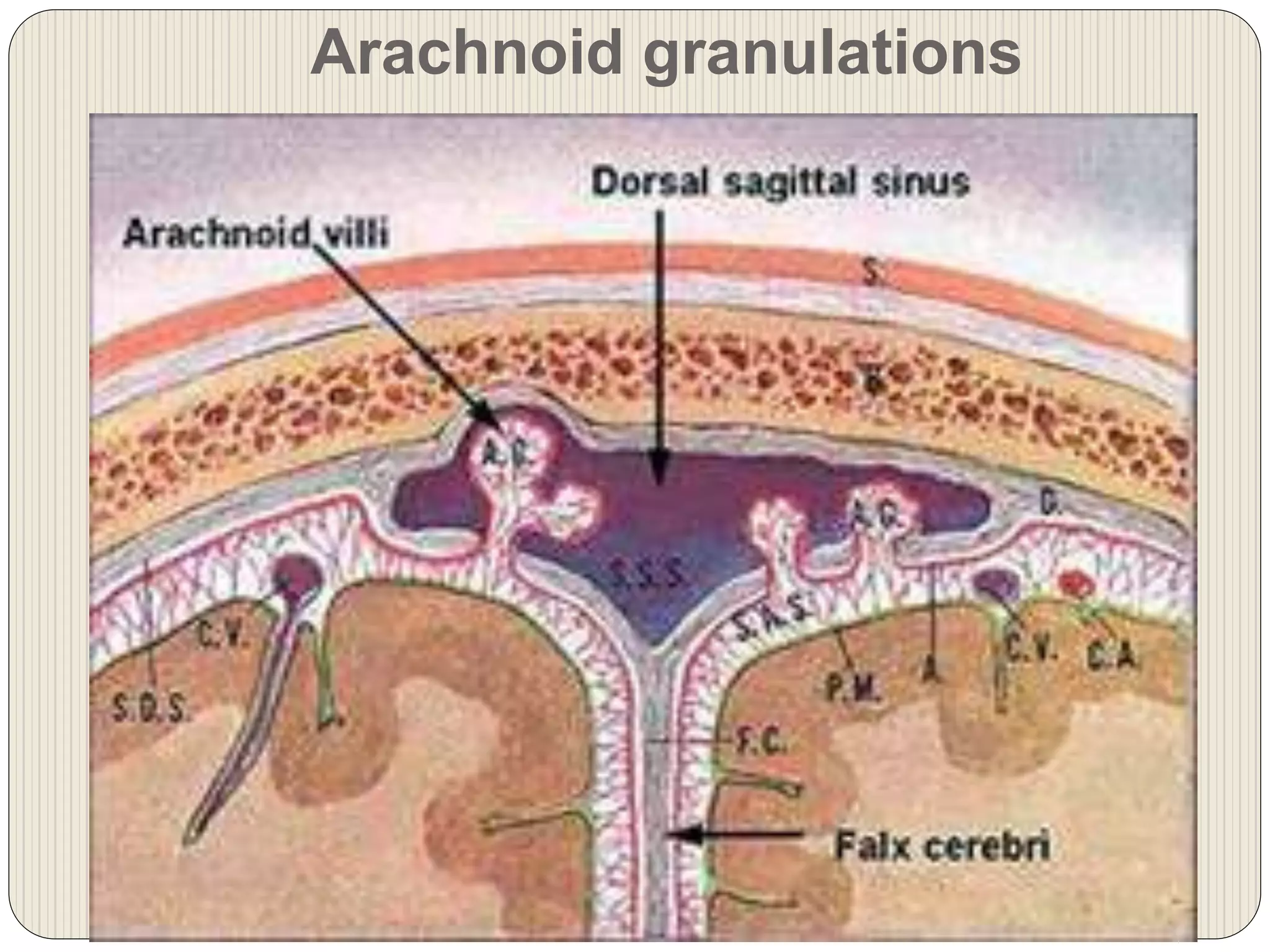
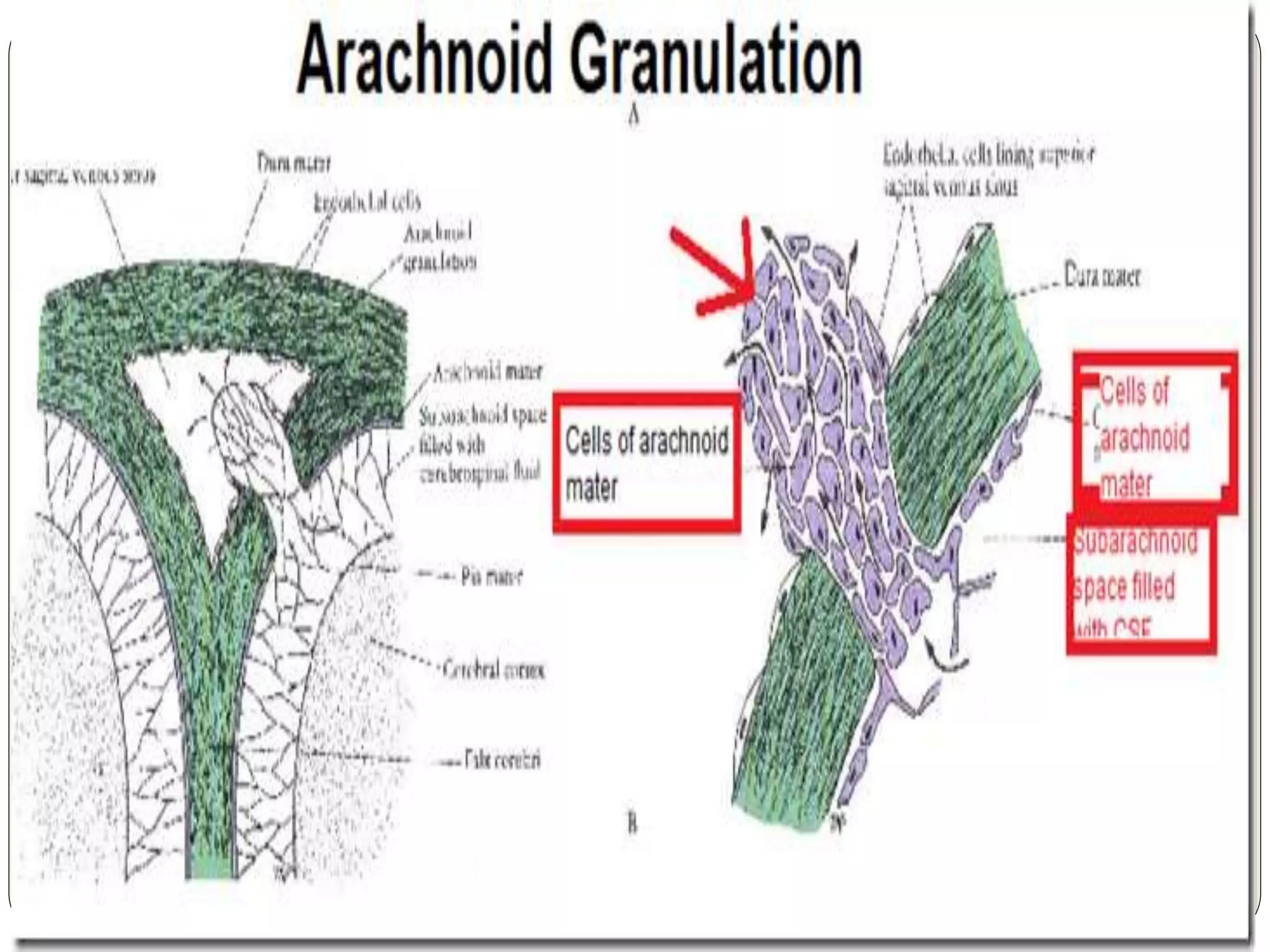
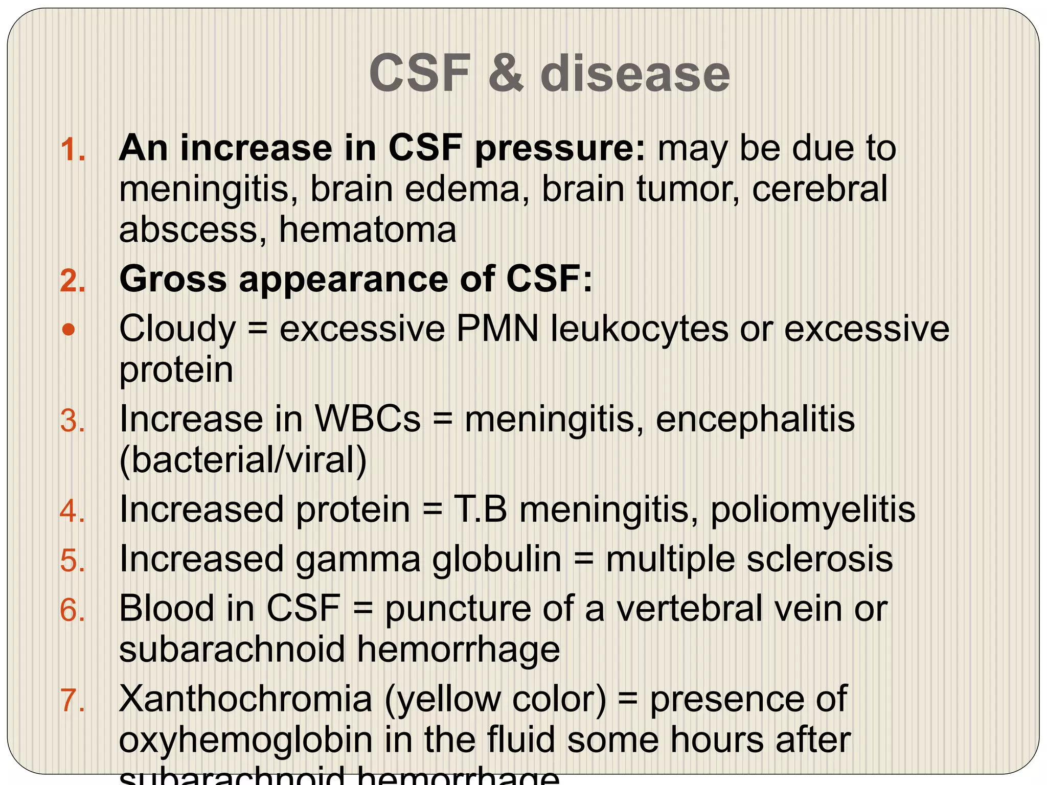
This document provides information about the ventricular system of the brain and cerebrospinal fluid (CSF). It details the four ventricles in the brain - two lateral ventricles, the third ventricle, and fourth ventricle. It describes the connections between the ventricles via foramina and discusses the choroid plexus which produces CSF. It then outlines the structures forming the walls and boundaries of each ventricle. Finally, it discusses the composition, circulation and functions of CSF, including cushioning and protecting the brain and spinal cord.
4 th ventricle- Anatomical and surgical perspectivesuresh Bishokarma
4th ventricle connects the entire ventricular system of brain. Its connection with cisterns magna and cerebella pontine cistern via foramen of magenta and Luschka. CSF absorbs into the arachnoid granulation.
4 th ventricle- Anatomical and surgical perspectivesuresh Bishokarma
4th ventricle connects the entire ventricular system of brain. Its connection with cisterns magna and cerebella pontine cistern via foramen of magenta and Luschka. CSF absorbs into the arachnoid granulation.
Lateral ventricle of Brain. By Dr.N.Mugunthan.M.Smgmcri1234
Lateral ventricle of brain. Lecture by Dr.N.Mugunthan.
Associate Professor,
Mahatma Gandhi Medical College & Research Institute,
Sri Balaji Vidyapeeth, Pondicherry.
You can watch the video on my you tube channel: https://youtu.be/I0FaX-iQfa0
Medulla oblongata or more simply medulla is part of brain stem which forms base of the brain stem. It contains pyramid, olive and above pyramidal structure, there is decussation of pyramids which explains why each part of brain controls opposite part of body. Adding to that medulla also has several nuclei which controls activity of cardiovascular system and respiratory system. Medulla also has nuclei for controlling reflexes of vomiting, swallowing, hiccuping, coughing and sneezing. It has also nuclei for test, hearing and balance. Medulla also contains nuclei of cranial nerve number VIII, IX, X, XI and XII.
Describe the location, function, and communications of ventricles of the brain
Name the parts and describe the boundaries of the lateral ventricle
Describe the third ventricle
Describe the fourth ventricle
Brain CT Anatomy and Basic Interpretation Part IISakher Alkhaderi
Detailed anatomy of the brain ventricles , CSF production and pathway and arterial supply and venous drainage of the brain and corresponding CT cross sectional anatomy and definition of sulcus and gyrus and fissure and the names of the important gyri .
Referred from different sources , here i present a very concise presentation on CRANIAL CAVITY . This presentation will give you complete knowledge of the topic cranial cavity with well elaborated and intellectual diagrams hand picked from F. Netter. ......... Do like and share , Leave your comments so as to get more stuff like this in future.
Neuroanatomy of ventricular system and CSF circulation also include
CSF composition
CSF production
CSF circulation
CSF absorption
Intracranial pressure
Raised ICP
Hydrocephalus
Similar to Ventricular system of brain & the CSF (20)
Hemostasis, Coagulation, Intrinsic, Extrinsic & common Pathways of Clotting, Common bleeding disorders & their investigations, BT, CT, PT, APTT, TT, Blood & its products, Blood transfusion & its complication.
Lung Cancer: Artificial Intelligence, Synergetics, Complex System Analysis, S...Oleg Kshivets
RESULTS: Overall life span (LS) was 2252.1±1742.5 days and cumulative 5-year survival (5YS) reached 73.2%, 10 years – 64.8%, 20 years – 42.5%. 513 LCP lived more than 5 years (LS=3124.6±1525.6 days), 148 LCP – more than 10 years (LS=5054.4±1504.1 days).199 LCP died because of LC (LS=562.7±374.5 days). 5YS of LCP after bi/lobectomies was significantly superior in comparison with LCP after pneumonectomies (78.1% vs.63.7%, P=0.00001 by log-rank test). AT significantly improved 5YS (66.3% vs. 34.8%) (P=0.00000 by log-rank test) only for LCP with N1-2. Cox modeling displayed that 5YS of LCP significantly depended on: phase transition (PT) early-invasive LC in terms of synergetics, PT N0—N12, cell ratio factors (ratio between cancer cells- CC and blood cells subpopulations), G1-3, histology, glucose, AT, blood cell circuit, prothrombin index, heparin tolerance, recalcification time (P=0.000-0.038). Neural networks, genetic algorithm selection and bootstrap simulation revealed relationships between 5YS and PT early-invasive LC (rank=1), PT N0—N12 (rank=2), thrombocytes/CC (3), erythrocytes/CC (4), eosinophils/CC (5), healthy cells/CC (6), lymphocytes/CC (7), segmented neutrophils/CC (8), stick neutrophils/CC (9), monocytes/CC (10); leucocytes/CC (11). Correct prediction of 5YS was 100% by neural networks computing (area under ROC curve=1.0; error=0.0).
CONCLUSIONS: 5YS of LCP after radical procedures significantly depended on: 1) PT early-invasive cancer; 2) PT N0--N12; 3) cell ratio factors; 4) blood cell circuit; 5) biochemical factors; 6) hemostasis system; 7) AT; 8) LC characteristics; 9) LC cell dynamics; 10) surgery type: lobectomy/pneumonectomy; 11) anthropometric data. Optimal diagnosis and treatment strategies for LC are: 1) screening and early detection of LC; 2) availability of experienced thoracic surgeons because of complexity of radical procedures; 3) aggressive en block surgery and adequate lymph node dissection for completeness; 4) precise prediction; 5) adjuvant chemoimmunoradiotherapy for LCP with unfavorable prognosis.
Anti ulcer drugs and their Advance pharmacology ||
Anti-ulcer drugs are medications used to prevent and treat ulcers in the stomach and upper part of the small intestine (duodenal ulcers). These ulcers are often caused by an imbalance between stomach acid and the mucosal lining, which protects the stomach lining.
||Scope: Overview of various classes of anti-ulcer drugs, their mechanisms of action, indications, side effects, and clinical considerations.
Factory Supply Best Quality Pmk Oil CAS 28578–16–7 PMK Powder in Stockrebeccabio
Factory Supply Best Quality Pmk Oil CAS 28578–16–7 PMK Powder in Stock
Telegram: bmksupplier
signal: +85264872720
threema: TUD4A6YC
You can contact me on Telegram or Threema
Communicate promptly and reply
Free of customs clearance, Double Clearance 100% pass delivery to USA, Canada, Spain, Germany, Netherland, Poland, Italy, Sweden, UK, Czech Republic, Australia, Mexico, Russia, Ukraine, Kazakhstan.Door to door service
Hot Selling Organic intermediates
The prostate is an exocrine gland of the male mammalian reproductive system
It is a walnut-sized gland that forms part of the male reproductive system and is located in front of the rectum and just below the urinary bladder
Function is to store and secrete a clear, slightly alkaline fluid that constitutes 10-30% of the volume of the seminal fluid that along with the spermatozoa, constitutes semen
A healthy human prostate measures (4cm-vertical, by 3cm-horizontal, 2cm ant-post ).
It surrounds the urethra just below the urinary bladder. It has anterior, median, posterior and two lateral lobes
It’s work is regulated by androgens which are responsible for male sex characteristics
Generalised disease of the prostate due to hormonal derangement which leads to non malignant enlargement of the gland (increase in the number of epithelial cells and stromal tissue)to cause compression of the urethra leading to symptoms (LUTS
Title: Sense of Taste
Presenter: Dr. Faiza, Assistant Professor of Physiology
Qualifications:
MBBS (Best Graduate, AIMC Lahore)
FCPS Physiology
ICMT, CHPE, DHPE (STMU)
MPH (GC University, Faisalabad)
MBA (Virtual University of Pakistan)
Learning Objectives:
Describe the structure and function of taste buds.
Describe the relationship between the taste threshold and taste index of common substances.
Explain the chemical basis and signal transduction of taste perception for each type of primary taste sensation.
Recognize different abnormalities of taste perception and their causes.
Key Topics:
Significance of Taste Sensation:
Differentiation between pleasant and harmful food
Influence on behavior
Selection of food based on metabolic needs
Receptors of Taste:
Taste buds on the tongue
Influence of sense of smell, texture of food, and pain stimulation (e.g., by pepper)
Primary and Secondary Taste Sensations:
Primary taste sensations: Sweet, Sour, Salty, Bitter, Umami
Chemical basis and signal transduction mechanisms for each taste
Taste Threshold and Index:
Taste threshold values for Sweet (sucrose), Salty (NaCl), Sour (HCl), and Bitter (Quinine)
Taste index relationship: Inversely proportional to taste threshold
Taste Blindness:
Inability to taste certain substances, particularly thiourea compounds
Example: Phenylthiocarbamide
Structure and Function of Taste Buds:
Composition: Epithelial cells, Sustentacular/Supporting cells, Taste cells, Basal cells
Features: Taste pores, Taste hairs/microvilli, and Taste nerve fibers
Location of Taste Buds:
Found in papillae of the tongue (Fungiform, Circumvallate, Foliate)
Also present on the palate, tonsillar pillars, epiglottis, and proximal esophagus
Mechanism of Taste Stimulation:
Interaction of taste substances with receptors on microvilli
Signal transduction pathways for Umami, Sweet, Bitter, Sour, and Salty tastes
Taste Sensitivity and Adaptation:
Decrease in sensitivity with age
Rapid adaptation of taste sensation
Role of Saliva in Taste:
Dissolution of tastants to reach receptors
Washing away the stimulus
Taste Preferences and Aversions:
Mechanisms behind taste preference and aversion
Influence of receptors and neural pathways
Impact of Sensory Nerve Damage:
Degeneration of taste buds if the sensory nerve fiber is cut
Abnormalities of Taste Detection:
Conditions: Ageusia, Hypogeusia, Dysgeusia (parageusia)
Causes: Nerve damage, neurological disorders, infections, poor oral hygiene, adverse drug effects, deficiencies, aging, tobacco use, altered neurotransmitter levels
Neurotransmitters and Taste Threshold:
Effects of serotonin (5-HT) and norepinephrine (NE) on taste sensitivity
Supertasters:
25% of the population with heightened sensitivity to taste, especially bitterness
Increased number of fungiform papillae
Flu Vaccine Alert in Bangalore Karnatakaaddon Scans
As flu season approaches, health officials in Bangalore, Karnataka, are urging residents to get their flu vaccinations. The seasonal flu, while common, can lead to severe health complications, particularly for vulnerable populations such as young children, the elderly, and those with underlying health conditions.
Dr. Vidisha Kumari, a leading epidemiologist in Bangalore, emphasizes the importance of getting vaccinated. "The flu vaccine is our best defense against the influenza virus. It not only protects individuals but also helps prevent the spread of the virus in our communities," he says.
This year, the flu season is expected to coincide with a potential increase in other respiratory illnesses. The Karnataka Health Department has launched an awareness campaign highlighting the significance of flu vaccinations. They have set up multiple vaccination centers across Bangalore, making it convenient for residents to receive their shots.
To encourage widespread vaccination, the government is also collaborating with local schools, workplaces, and community centers to facilitate vaccination drives. Special attention is being given to ensuring that the vaccine is accessible to all, including marginalized communities who may have limited access to healthcare.
Residents are reminded that the flu vaccine is safe and effective. Common side effects are mild and may include soreness at the injection site, mild fever, or muscle aches. These side effects are generally short-lived and far less severe than the flu itself.
Healthcare providers are also stressing the importance of continuing COVID-19 precautions. Wearing masks, practicing good hand hygiene, and maintaining social distancing are still crucial, especially in crowded places.
Protect yourself and your loved ones by getting vaccinated. Together, we can help keep Bangalore healthy and safe this flu season. For more information on vaccination centers and schedules, residents can visit the Karnataka Health Department’s official website or follow their social media pages.
Stay informed, stay safe, and get your flu shot today!
Ozempic: Preoperative Management of Patients on GLP-1 Receptor Agonists Saeid Safari
Preoperative Management of Patients on GLP-1 Receptor Agonists like Ozempic and Semiglutide
ASA GUIDELINE
NYSORA Guideline
2 Case Reports of Gastric Ultrasound
Ethanol (CH3CH2OH), or beverage alcohol, is a two-carbon alcohol
that is rapidly distributed in the body and brain. Ethanol alters many
neurochemical systems and has rewarding and addictive properties. It
is the oldest recreational drug and likely contributes to more morbidity,
mortality, and public health costs than all illicit drugs combined. The
5th edition of the Diagnostic and Statistical Manual of Mental Disorders
(DSM-5) integrates alcohol abuse and alcohol dependence into a single
disorder called alcohol use disorder (AUD), with mild, moderate,
and severe subclassifications (American Psychiatric Association, 2013).
In the DSM-5, all types of substance abuse and dependence have been
combined into a single substance use disorder (SUD) on a continuum
from mild to severe. A diagnosis of AUD requires that at least two of
the 11 DSM-5 behaviors be present within a 12-month period (mild
AUD: 2–3 criteria; moderate AUD: 4–5 criteria; severe AUD: 6–11 criteria).
The four main behavioral effects of AUD are impaired control over
drinking, negative social consequences, risky use, and altered physiological
effects (tolerance, withdrawal). This chapter presents an overview
of the prevalence and harmful consequences of AUD in the U.S.,
the systemic nature of the disease, neurocircuitry and stages of AUD,
comorbidities, fetal alcohol spectrum disorders, genetic risk factors, and
pharmacotherapies for AUD.
Pulmonary Thromboembolism - etilogy, types, medical- Surgical and nursing man...VarunMahajani
Disruption of blood supply to lung alveoli due to blockage of one or more pulmonary blood vessels is called as Pulmonary thromboembolism. In this presentation we will discuss its causes, types and its management in depth.
Explore natural remedies for syphilis treatment in Singapore. Discover alternative therapies, herbal remedies, and lifestyle changes that may complement conventional treatments. Learn about holistic approaches to managing syphilis symptoms and supporting overall health.
Prix Galien International 2024 Forum ProgramLevi Shapiro
June 20, 2024, Prix Galien International and Jerusalem Ethics Forum in ROME. Detailed agenda including panels:
- ADVANCES IN CARDIOLOGY: A NEW PARADIGM IS COMING
- WOMEN’S HEALTH: FERTILITY PRESERVATION
- WHAT’S NEW IN THE TREATMENT OF INFECTIOUS,
ONCOLOGICAL AND INFLAMMATORY SKIN DISEASES?
- ARTIFICIAL INTELLIGENCE AND ETHICS
- GENE THERAPY
- BEYOND BORDERS: GLOBAL INITIATIVES FOR DEMOCRATIZING LIFE SCIENCE TECHNOLOGIES AND PROMOTING ACCESS TO HEALTHCARE
- ETHICAL CHALLENGES IN LIFE SCIENCES
- Prix Galien International Awards Ceremony
2. Ventricular system
Four ventricles in the brain, among which:
1. Two lateral ventricles
2. A 3rd ventricle
3. A 4th ventricle
One terminal ventricle at the end of the central
canal within the spinal cord
3.
4. Foramina connecting the
ventricles
1. Inter-ventricular foramina (of Monro) in each
lateral ventricle connect them with the 3rd
ventricle
2. Cerebral aqueduct (of Sylvius) connects the
3rd ventricle with 4th ventricle
3. Foramen of Magendie connects the 4th
ventricle with central canal of the spinal cord
4. Foramina of Luschka on each side of the 4th
ventricle connects it to the subarachnoid space
around the brain & spinal cord
5.
6. Subarachnoid cisterns
At some areas around the base of brain, arachnoid
mater does not follow the surface of brain, hence forms
some spaces called sub-arachnoid cisterns, which also
contain CSF. These incluse:
1. Cerebello-medullary cistern (cisterna cerebello-
medullaris)
2. Pontine cistern (cisterna pontis)
3. Inter-peduncular cistern (cisterna inter-
peduncularis)
7. Lateral ventricles
These are two irregular cavities lying within each
cerebral hemisphere
It is roughly C-shaped & consist of:
1. A central part or body
2. Three horns: anterior, posterior and inferior, each
extending to frontal, occipital & frontal lobes
respectively
3. Each lateral ventricle communicates with 3rd
ventricle through inter-ventricular foramen of
Monro
9. Body (central part) of lateral
ventricle
Extends from inter-ventricular foramen posteriorly
as far as the posterior end of thalamus
Here it is continuous with the posterior & inferior
horns
The body of lateral ventricle has:
1. Roof
2. Floor
3. Medial wall
10. Roof of body of lateral
ventricle
It is formed by
the under
surface of
corpus
callosum
11.
12. Floor of body of lateral
ventricle
It is formed by the:
1. Caudate nucleus
2. Lateral margin of the thalamus
3. Choroidal fissure is a slit like gap between the
body of fornix and superior surface of thalamus
through which choroid plexus of the ventricle
projects into the body of the ventricle
13.
14. Medial wall of body of lateral
ventricle
It is formed by the septum pellucidum anteriorly
The roof & the floor come together posteriorly to
meet the medial wall
15. Anterior horn of lateral
ventricle
Lies within frontal lobe of brain
Continues with body at interventricular foramen
Anterior horn has:
1. Roof
2. Floor
3. Medial wall
16. Roof of anterior horn of lateral
ventricle
It is formed by the undersurface of the corpus
callosum
Anterior horn is limited anteriorly by the genu of
corpus callosum
17. Floor of anterior horn of lateral
ventricle
Its floor is formed by:
1. Head of caudate nucleus
2. Rostrum of corpus callosum medially having a
small portion
18. Medial wall of anterior horn of
lateral ventricle
It formed by the:
1. Septum pellucidum
2. Anterior column of the fornix
20. Posterior horn of lateral
ventricle
Lies into the occipital lobe of brain
It has:
1. Roof
2. Lateral wall
3. Medial wall
21. Roof & lateral wall of posterior horn
of lateral ventricle
Both are formed by the tapetum of the corpus
callosum
Lateral to the tapetum are the optic radiation
22. Medial wall of posterior horn of
lateral ventricle
It has two elevations (swellings)
Superior swelling (also called bulb of the posterior
horn) is formed by the splenial fibers of corpus
callosum which is called forceps major
Inferior swelling is formed by the calcarine sulcus
and is called calcar avis
23. Inferior horn of lateral ventricle
It lies in temporal lobe of brain
It has
1. roof
2. floor
24. Roof of Inferior horn of lateral
ventricle
It is formed by:
1. Inferior surface of the tapetum of the corpus
callosum and
2. Tail of caudate nucleus
25. Floor of Inferior horn of lateral
ventricle
It is formed by:
1. Collateral eminence laterally
2. Hippocampus medially
26. Choroid plexus of lateral
ventricle
It is composed of highly vascular pia mater in two
layers, lined with ependymal cells
It is situated between fornix superiorly and
thanlamus inferiorly
It produces CSF
30. Third ventricle
It is a slit-like cleft between two thalami
Communicates anteriorly with lateral ventricles
through interventricular foramina of Monro
Communicates posteriorly with 4th ventricle through
the cerebral aqueduct of Sylvius
Its boundaries are:
1. Anterior wall
2. Posterior wall
3. Lateral wall
4. Superior wall or roof
5. Inferior wall or floor
31. Anterior wall of 3rd ventricle
It is formed by:
1. Lamina terminalis
2. Anterior commisure which connects right & left
temporal lobes
32. Posterior wall of 3rd ventricle
It is formed by:
1. Cerebral aqueduct below Posterior
commissure above
2. Pineal recess & pineal body above the
commissure
3. Habenular commissure above the pineal
recess
33. Lateral wall of 3rd ventricle
It is formed by:
1. Thalamus superiorly (both are separated
by
2. Hypothalamus inferiorly hypothalamic
sulcus)
3. Both lateral walls are joined by interthalamic
connections
35. Superior wall (Roof) of 3rd
ventricle
It is formed by:
1. Corpus callosum &
2. Fornix
3. Below them lies choroid plexus of the 3rd
ventricle
36. Inferior wall (floor) of 3rd
ventricle
It is formed by:
1. Optic chiasma
2. Tuber cinereum
3. Infundibulum
4. Mammillary bodies
5. Hypophysis attached to infundibulum
6. Tegmentum of the cerebral peduncles
37.
38.
39.
40.
41. Cerebral aqueduct of Sylvius
Connects 3rd ventricle with the 4th and conducts
CSF
Surrounded by the central gray
It is devoid of choroid plexus
42.
43.
44. Fourth ventricle
Tent shaped
Situated:
1. Anterior to the cerebellum
2. Posterior to the pons
3. Posterior to the upper half of the medulla
oblongata
4. Lined with ependyma
5. Connected above with cerebral aqueduct &
below with central canal of the medulla
oblongata & spinal cord
It has:
a) Lateral wall (boundaries)
b) Roof
45.
46. Lateral wall (boundaries) of 4th ventricle
It is formed by:
1. Superior cerebellar peduncle above
2. Inferior cerebellar peduncle below
47. Roof (posterior wall) of 4th
ventricle
It is tent-shaped & projects into the cerebellum behind
It communicates with subarachnoid space through 3
foramina
It is formed by:
1. Two superior cerebellar peduncles above which are
connected by superior medullary vellum
2. Two inferior cerebellar peduncles below which are
connected by inferior medullary vellum
Inferiorly, the roof is pierced by foramen of
Magendie
Laterally on each side, it has an opening, the
foramina of Luschka
48.
49. Floor (Rhomboid fossa) of 4th
ventricle
It is diamond-shaped & formed by:
1. Posterior surface of pons &
2. Cranial (upper) half of medulla oblongata
The floor is divided into symmetrical halves by median
sulcus
On each side of this sulcus, there is an elevation, the
medial eminence
Lateral to the eminence, there is another sulcus, the
sulcus limitans
Lateral to the sulcus limitans lies vestibular area
Facial colliculus is a slight swelling at the inferior end
of the medial eminence that is produced by the fibers
from the motor nucleus of the facial nerve looping over
the abducens nucleus
55. Floor (Rhomboid fossa) of 4th ventricle
Stria medullaris emerge from the median sulcus and
pass laterally on each side over the medial eminence &
the vestibular area and enter the inferior cerebellar
peduncle to reach the cerebellum
Below the stria medullaris following structures lie in the
floor of the ventricle
1. Hypoglossal triangle: which indicates the position of
underlying hypoglossal nucleus
2. Vagal triangle: beneath which lies the dorsal motor
nucleus of the vagus
3. Area postrema: is a narrow area between the vagal
triangle & the lateral margin of the ventricle
56.
57. Choroid plexus of 4th ventricle
It is T-shaped
Lies in the lower half of the roof of 4th ventricle
Produces CSF
58.
59. Central canal of medulla oblongata &
spinal cord
Opens superiorly into the 4th ventricle
Ends at conus medullaris as terminal ventricle
Surrounded by the gray matter called the gray
commissure
It is devoid of choroid plexus
60.
61. Subarachnoid space
It is the space between arachnoid & pia maters
Surround brain & spinal cord upto S3 vertebra,
hence includes cauda equina
Filled with CSF
In some situations around the brain, the
arachnoid mater does not follow the surface of
brain, hence leaves some spaces called sub-
arachnoid cisterns. These include:
1. Cerebello-medullary cistern
2. Pontine cistern
3. Interpeduncular cistern
63. Cerebrospinal fluid (CSF)
Formed within the ventricles of brain
Its volume is 150 ml
It is clear, colorless fluid
It possesses inorganic salts like blood plasma (e.g.
Na, K, Cl), glucose (equal to the half of blood) and
traces of protein
Only few lymphocytes (0 to 3 cells/mm3)
Normal CSF pressure = 60-150 mm of water in
lateral recumbent position
CSF pressure is raised by straining, coughing, or
compressing the internal jugular veins in the neck
64. PHYSICAL & CHEMICAL COMPOSITION
OF CSF
APPEARANCE Clear & colorless
VOLUME 150 ml
RATE OF PRODUCTION 0.5 ml/minute
PRESSURE (in lat. Recumbent
position)
60-150 ml of water
COMPOSITION
1. Protein 15-45 mg/100 ml
2. Glucose 50-85 mg/100 ml
3. Chloride 720-750 mg/100 ml
NO. OF CELLS 0-3
65. Functions of CSF
1. It bathes the external & internal surfaces of the
brain & spinal cord
2. Serves as cushion between CNS & surrounding
bones, thus protects against mechanical trauma
3. Provides buoyancy
4. Serves as a reservoir to assist in the contents of
the skull e.g. if brain & blood volume increase, CSF
volume decreases
5. Nourishment of nervous tissue
6. Removes the products of neuronal metabolism
66. Functions of CSF
1.
Cushions & protects the CNS from trauma
2.
Provides mechanical buoyancy & support for the
brain
3.
Serves as a reservoir & assists in the regulation of
the contents of the skull
4.
Nourishes the CNS
5.
Removes the metabolites from the CNS
6.
Serves as a pathway for pineal secretions to reach
the pituitary gland
67. Formation of CSF
CSF is formed in:
1. The choroid plexuses of the lateral, 3rd & 4th
ventricles mainly which secrete it actively
2. Ependymal cells in the above ventricles
3. Brain substance through the perivascular space
4. The production of CSF is NOT pressure
regulated. It continues to be produced even if
the re-absorption mechanisms are obstructed
68. Circulation of CSF
Lateral ventricles Interventricular
foramina of
Monro Third venticle Cerebral
aqueduct of
Central canal
(foramen of
Magendie)
Sylvius Fourth ventricle
Subarachnoid space
(foramina of
69. Absorption of CSF
CSF is mainly absorbed by arachnoid villi that
project into the dural venous sinuses, especially
SSS, by piercing dura mater
Arachnoid villus is a diverticulum of a
subarachnoid space into the venous sinuses
Arachnoid villi are grouped and form arachnoid
granulations
73. Absorption of CSF
If venous pressure rises & exceeds the CSF
pressure, compression of the tips of villi closes
the tubules & prevents the reflux of blood into the
subarachnoid space. Thus aracnoid villi serve as
valves
As the production of CSF is constant, hence the
rate of absorption of it controls the CSF pressure
75. Papilloedema
Optic disc will show bulging in raised intracranial
pressure e.g. by tumor, because optic nerve
carries the meninges around it into the eye
76. Hydrocephalus
An abnormal increase in the volume of CSF
within the skull
Intracranial pressure will be raised
Causes may be:
1. Increased formation of CSF
2. Decreased absorption of CSF
3. Blockage of the circulation of CSF
Two varieties:
1. Non-communication hydrocephalus
2. Communicating hydrocephalus
77. Non-communicating
hydrocephalus
The raised CSF pressure will be due to blockage
at some point between its formation at the
choroid plexuses and its exit through the foramina
in the roof of the 4th ventricle
80. Intracranial pneumography
(Ventriculogram)
Air or oxygen is introduced into the lateral
ventricle through a needle inserted through a hole
in the skull (or through a suture in a young child)
Then radiograph of the skull is made
Only ventricles are visualized
82. CSF & disease
1. An increase in CSF pressure: may be due to
meningitis, brain edema, brain tumor, cerebral
abscess, hematoma
2. Gross appearance of CSF:
Cloudy = excessive PMN leukocytes or excessive
protein
3. Increase in WBCs = meningitis, encephalitis
(bacterial/viral)
4. Increased protein = T.B meningitis, poliomyelitis
5. Increased gamma globulin = multiple sclerosis
6. Blood in CSF = puncture of a vertebral vein or
subarachnoid hemorrhage
7. Xanthochromia (yellow color) = presence of
oxyhemoglobin in the fluid some hours after
83. Tumors of the 4th ventricle
(Ependymomas)
May invade the cerebellum, hence s/s of
cerebellar deficiency
May compress the nuclear centers situated
beneath the floor of 4th ventricle i.e. hypoglossal,
vagus. Therefore, s/s of movements of tongue,
swallowing, respiration, heart rate, blood pressure
etc.