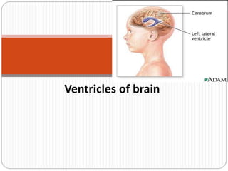
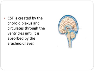
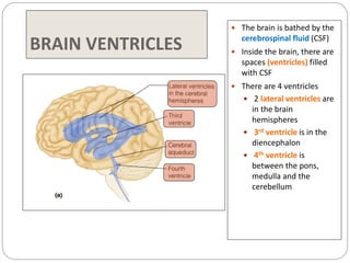
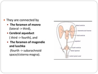
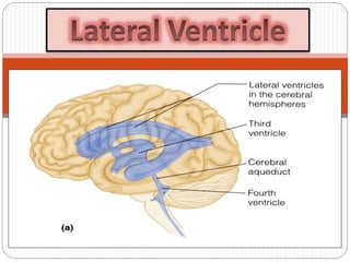
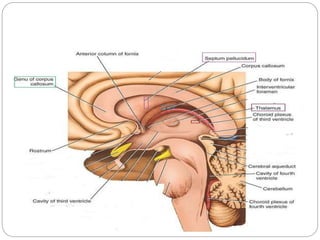
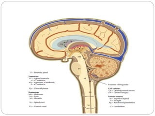
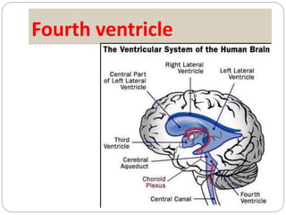
Ventricles of brain
The brain has a
series of ventricles
that hold CSF
(Cerebrospinal
Fluid).
• CSF is created by the
choroid plexus and
circulates through the
ventricles until it is
absorbed by the
arachnoid layer.
• CSF seems to
1. act as a fluid cushion for the brain
2. transports some substances into/out of the brain
3. maintains pressure around the brain.
BRAIN VENTRICLES
 The brain is bathed by the
cerebrospinal fluid (CSF)
 Inside the brain, there are
spaces (ventricles) filled
with CSF
 There are 4 ventricles
 2 lateral ventricles are
in the brain
hemispheres
 3rd ventricle is in the
diencephalon
 4th ventricle is
between the pons,
medulla and the
cerebellum
 They are connected by
 The foramen of monro
(lateral -> third),
 Cerebral aqueduct
( third -> fourth), and
 The foramen of magendie
and luschka
(fourth -> subarachnoid
space/cisterna magna).
Lateral Ventricle
 Definition :
 It is the cavity of
the cerebral
hemisphere.
 It is C-shaped.
 It has 3 horns &
central part.
 Anterior Horn: in
the frontal lobe.
 Posterior horn:
in the occipital
lobe
 Inferior horn: in
temporal lobe.
 Central part or
body: in the
parietal lobe.
Lateral Ventricle
 Superior view of the
ventricular system.
 Lateral ventricle
 Anterior horn in the frontal
lobe.
 Posterior horn in the
occipital lobe.
 Inferior horn in the temporal
lobe.
 Body: In the parietal lobe.
 The inferior and posterior
horns are connected in the
trigon.
Relationship of corpus callosum and
third ventricle
Suprolateral surface of brain
Relationship of caudate nucleusand
third ventricle
Anterior Horn
 In the frontal lobe.
 Roof:
 Corpus callosum
(trunk)
 Floor:
 Corpus callosum
(Rostrum)
 Caudate nucleus
head
 Anterior:
 Corpus callosum
(Genu)
 Medially:
 Septum pellucidum.
Body or Central part
 Lies in the parietal lobe.
 Roof:
 Corpus callosum (Trunk).
 Floor:
 Sloping, From lateral to
medial it is formed by:
 Body of caudate nucleus,
 Upper surface of
thalamus
 Choroid plexus,
 Body of fornix.
 Medial wall:
 Septum pellucidum.
 Lateral wall:
 narrow area at the
meeting of roof & floor.
Posterior Horn
 In the occipital lobe.
 Roof, lateral wall:
 Are formed by the
Tapetum of the corpus
callosum.
 Medially:
 There are 2 elevations:
Bulb of posterior horn
(formed by forceps
major-2-).
Calcar avis: produced by
calcarine sulcus-3-.
Inferior Horn
 It lies in the temporal lobe.
 Roof:
 Tapetum,
 Tail of caudate nucleus,
 Amygdaloid nucleus
 Stria terminalis.
 Floor:
 Hippocampus,
 Fimbria of hippocampus &
Collateral eminence.
Choroid Plexus of the Lateral Ventricle
 is a vascular fringe of pia mater
covered with the ependymal lining
of the ventricular cavity
 The choroid plexus projects into
the ventricle on its medial aspect

 At the junction of the body of the
lateral ventricle and the inferior
horn, the choroid plexus is
continued into the inferior horn.
Fourth ventricle
Fourth Ventricle
 A tent-shaped cavity filled
with cerebrospinal fluid.
 lined with ependyma
 continuous above with the
cerebral aqueduct of the
midbrain and below with the
central canal of the medulla
oblongata and the spinal cord

Situated
 anterior to the cerebellum
and
 posterior to the pons and
the superior half of the
medulla oblongata
 The fourth ventricle possesses
1. Lateral boundaries
2. Roof, and
3. Rhomboid-shaped floor.
Lateral Boundaries
 The caudal part .... the inferior cerebellar peduncle
 The cranial part ..... the superior cerebellar peduncle.
Roof or Posterior Wall
 The tent-shaped roof projects into
the cerebellum
 The superior part....medial
borders of the two superior
cerebellar peduncles and a
connecting sheet of white matter
called the superior medullary
velum
 The inferior part ..... the inferior
medullary velum, which consists of
a thin sheet devoid of nervous
tissue and formed by the
ventricular ependyma and its
posterior covering of pia mater
Fourth ventricle communicates with the subarachnoid
space through a single median and two lateral
apertures.
 In the midline ,the roof is
pierced by a large aperture, the
median aperture or foramen of
Magendie .
 Lateral recesses extend laterally
around the sides of the medulla
and open anteriorly as the
lateral openings of the fourth
ventricle, or the foramina of
Luschka .
Floor or Rhomboid Fossa
 The diamond-shaped floor
 formed by the posterior surface of the pons and the
cranial half of the medulla oblongata
 is divided into symmetrical halves by the median
sulcus.
 the medial eminence, ...
 the sulcus limitans.
 vestibular area ..vestibular nuclei
Nerve nuclei in floor of fourth ventricle
The facial colliculus
 the inferior end of the medial eminence
 produced by the fibers from the motor nucleus of the
facial nerve looping over the abducens nucleus
Substantia ferruginea
 Lies at the superior end of the sulcus limitans, there is a
bluish-gray area, nerve cells contain melanin pigment.
Stria medullaris
 Strands of nerve fibers derived from the arcuate nuclei,
emerge from the median sulcus and pass laterally over the
medial eminence and the vestibular area and enter the
inferior cerebellar peduncle to reach the cerebellum
Facial colliculus
Choroid Plexus of the Fourth Ventricle is formed
the posterior inferior cerebellar arteries.
 The choroid plexus has a T
shape
 The vertical part of the T
is double .
 Is formed from the highly
vascular tela choroidea.
 The tela choroidea is a
two-layered fold of pia
mater that projects
through the roof of the
ventricle and is covered by
ependyma.
ventricle

ventricle

  • 1.
  • 2.
    The brain hasa series of ventricles that hold CSF (Cerebrospinal Fluid).
  • 3.
    • CSF iscreated by the choroid plexus and circulates through the ventricles until it is absorbed by the arachnoid layer.
  • 4.
    • CSF seemsto 1. act as a fluid cushion for the brain 2. transports some substances into/out of the brain 3. maintains pressure around the brain.
  • 5.
    BRAIN VENTRICLES  Thebrain is bathed by the cerebrospinal fluid (CSF)  Inside the brain, there are spaces (ventricles) filled with CSF  There are 4 ventricles  2 lateral ventricles are in the brain hemispheres  3rd ventricle is in the diencephalon  4th ventricle is between the pons, medulla and the cerebellum
  • 6.
     They areconnected by  The foramen of monro (lateral -> third),  Cerebral aqueduct ( third -> fourth), and  The foramen of magendie and luschka (fourth -> subarachnoid space/cisterna magna).
  • 8.
    Lateral Ventricle  Definition:  It is the cavity of the cerebral hemisphere.  It is C-shaped.  It has 3 horns & central part.  Anterior Horn: in the frontal lobe.  Posterior horn: in the occipital lobe  Inferior horn: in temporal lobe.  Central part or body: in the parietal lobe.
  • 9.
    Lateral Ventricle  Superiorview of the ventricular system.  Lateral ventricle  Anterior horn in the frontal lobe.  Posterior horn in the occipital lobe.  Inferior horn in the temporal lobe.  Body: In the parietal lobe.  The inferior and posterior horns are connected in the trigon.
  • 11.
    Relationship of corpuscallosum and third ventricle
  • 12.
  • 13.
    Relationship of caudatenucleusand third ventricle
  • 14.
    Anterior Horn  Inthe frontal lobe.  Roof:  Corpus callosum (trunk)  Floor:  Corpus callosum (Rostrum)  Caudate nucleus head  Anterior:  Corpus callosum (Genu)  Medially:  Septum pellucidum.
  • 16.
    Body or Centralpart  Lies in the parietal lobe.  Roof:  Corpus callosum (Trunk).  Floor:  Sloping, From lateral to medial it is formed by:  Body of caudate nucleus,  Upper surface of thalamus  Choroid plexus,  Body of fornix.  Medial wall:  Septum pellucidum.  Lateral wall:  narrow area at the meeting of roof & floor.
  • 17.
    Posterior Horn  Inthe occipital lobe.  Roof, lateral wall:  Are formed by the Tapetum of the corpus callosum.  Medially:  There are 2 elevations: Bulb of posterior horn (formed by forceps major-2-). Calcar avis: produced by calcarine sulcus-3-.
  • 19.
    Inferior Horn  Itlies in the temporal lobe.  Roof:  Tapetum,  Tail of caudate nucleus,  Amygdaloid nucleus  Stria terminalis.  Floor:  Hippocampus,  Fimbria of hippocampus & Collateral eminence.
  • 20.
    Choroid Plexus ofthe Lateral Ventricle  is a vascular fringe of pia mater covered with the ependymal lining of the ventricular cavity  The choroid plexus projects into the ventricle on its medial aspect   At the junction of the body of the lateral ventricle and the inferior horn, the choroid plexus is continued into the inferior horn.
  • 22.
  • 24.
    Fourth Ventricle  Atent-shaped cavity filled with cerebrospinal fluid.  lined with ependyma  continuous above with the cerebral aqueduct of the midbrain and below with the central canal of the medulla oblongata and the spinal cord 
  • 25.
    Situated  anterior tothe cerebellum and  posterior to the pons and the superior half of the medulla oblongata
  • 26.
     The fourthventricle possesses 1. Lateral boundaries 2. Roof, and 3. Rhomboid-shaped floor.
  • 27.
    Lateral Boundaries  Thecaudal part .... the inferior cerebellar peduncle  The cranial part ..... the superior cerebellar peduncle.
  • 28.
    Roof or PosteriorWall  The tent-shaped roof projects into the cerebellum  The superior part....medial borders of the two superior cerebellar peduncles and a connecting sheet of white matter called the superior medullary velum  The inferior part ..... the inferior medullary velum, which consists of a thin sheet devoid of nervous tissue and formed by the ventricular ependyma and its posterior covering of pia mater
  • 29.
    Fourth ventricle communicateswith the subarachnoid space through a single median and two lateral apertures.  In the midline ,the roof is pierced by a large aperture, the median aperture or foramen of Magendie .  Lateral recesses extend laterally around the sides of the medulla and open anteriorly as the lateral openings of the fourth ventricle, or the foramina of Luschka .
  • 30.
    Floor or RhomboidFossa  The diamond-shaped floor  formed by the posterior surface of the pons and the cranial half of the medulla oblongata  is divided into symmetrical halves by the median sulcus.  the medial eminence, ...  the sulcus limitans.  vestibular area ..vestibular nuclei
  • 33.
    Nerve nuclei infloor of fourth ventricle
  • 34.
    The facial colliculus the inferior end of the medial eminence  produced by the fibers from the motor nucleus of the facial nerve looping over the abducens nucleus Substantia ferruginea  Lies at the superior end of the sulcus limitans, there is a bluish-gray area, nerve cells contain melanin pigment. Stria medullaris  Strands of nerve fibers derived from the arcuate nuclei, emerge from the median sulcus and pass laterally over the medial eminence and the vestibular area and enter the inferior cerebellar peduncle to reach the cerebellum
  • 35.
  • 36.
    Choroid Plexus ofthe Fourth Ventricle is formed the posterior inferior cerebellar arteries.  The choroid plexus has a T shape  The vertical part of the T is double .  Is formed from the highly vascular tela choroidea.  The tela choroidea is a two-layered fold of pia mater that projects through the roof of the ventricle and is covered by ependyma.