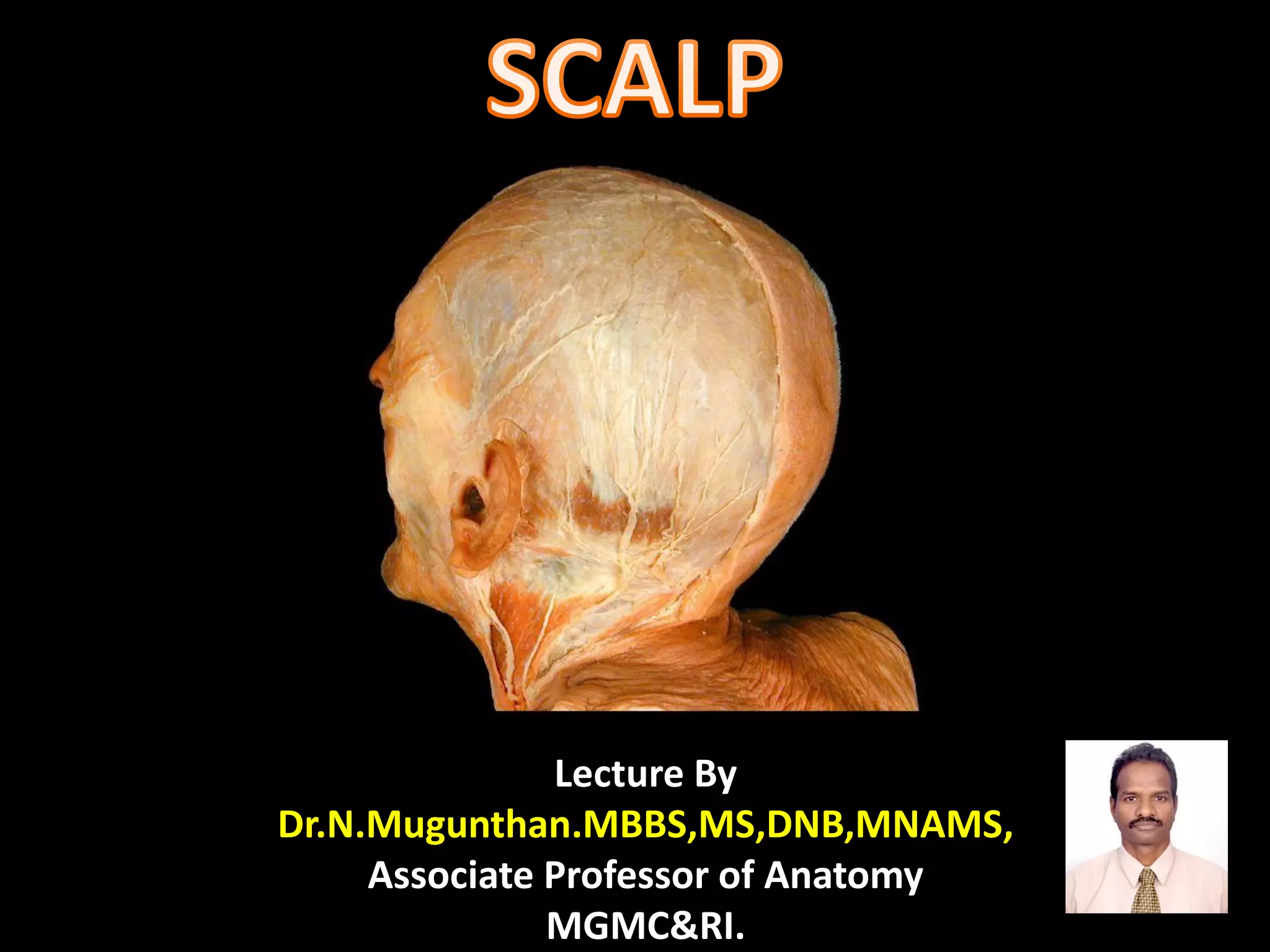
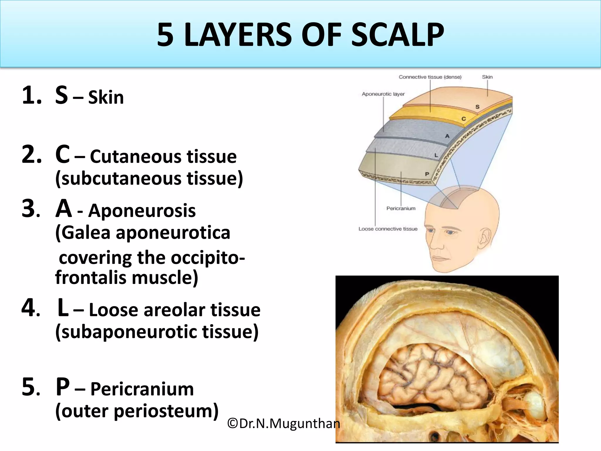
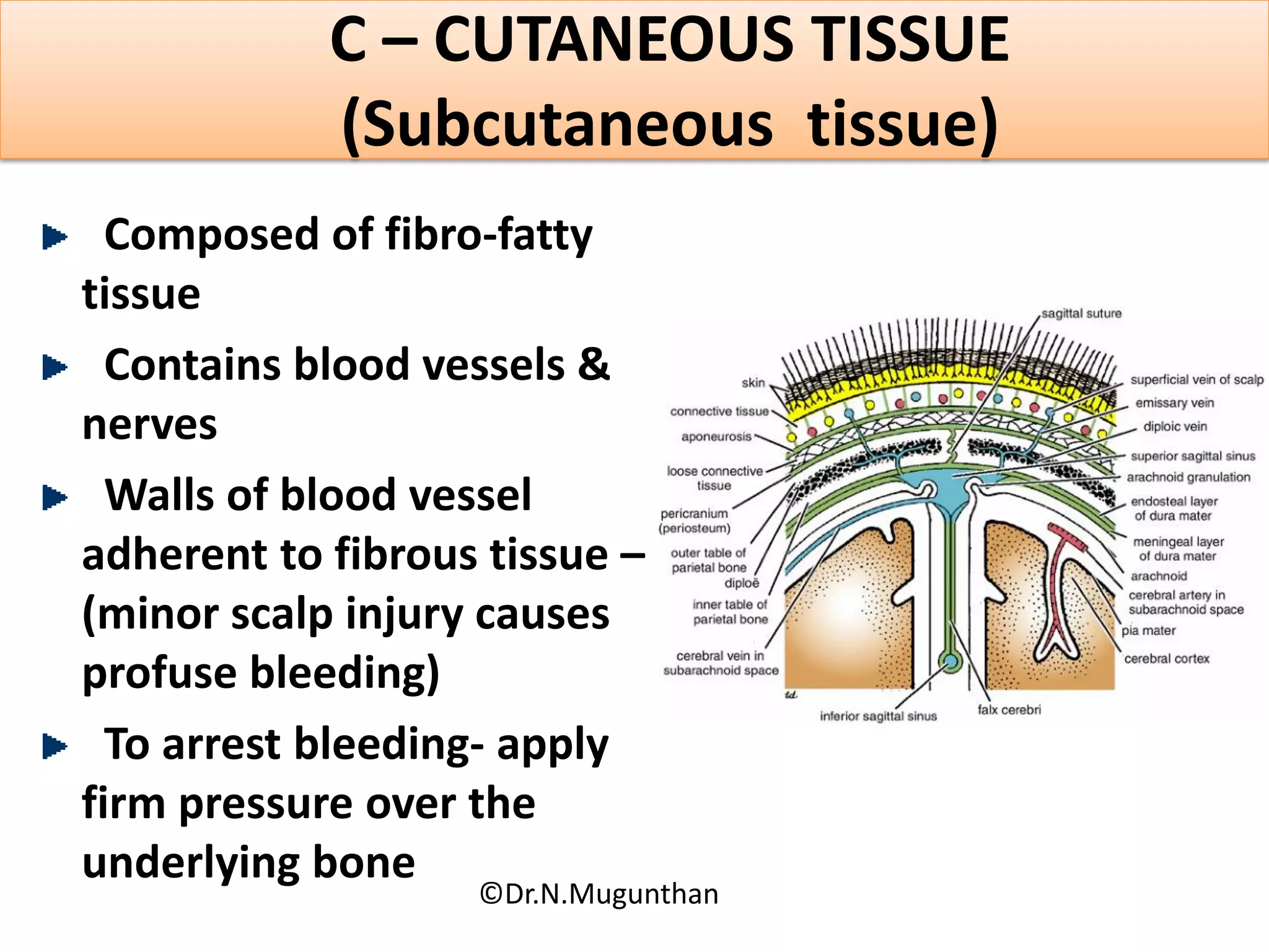
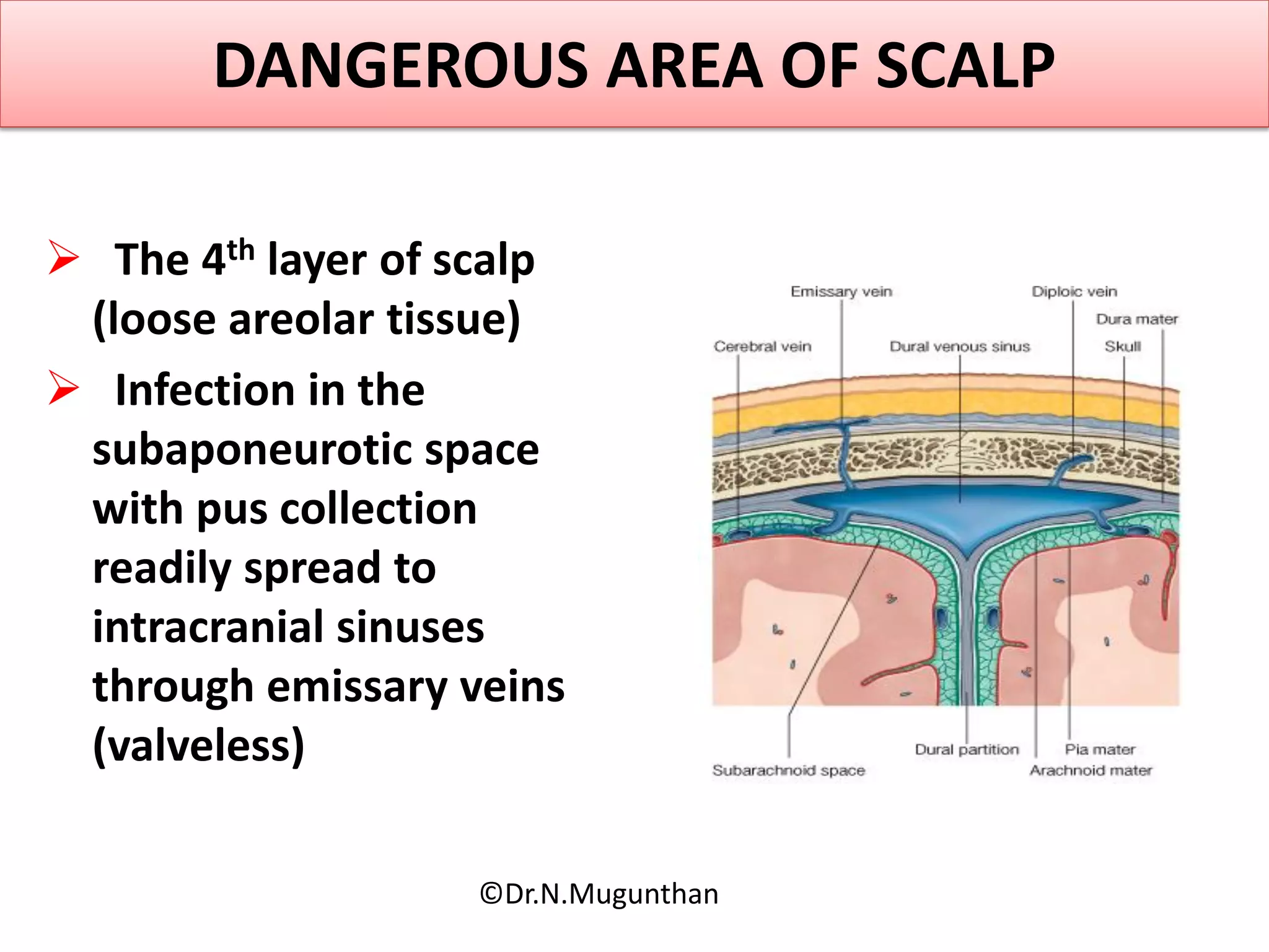
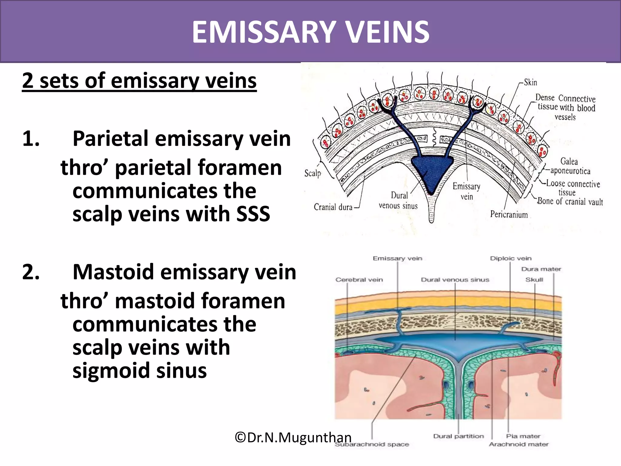
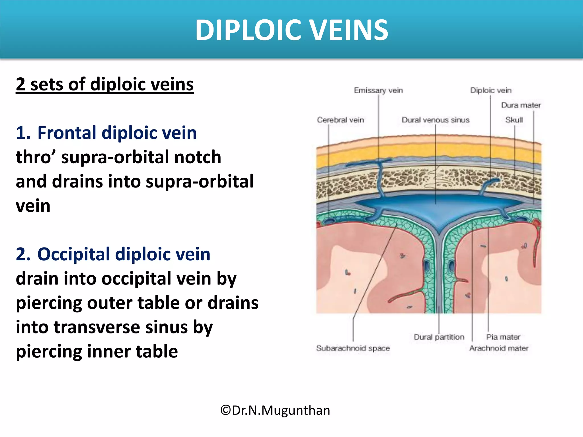
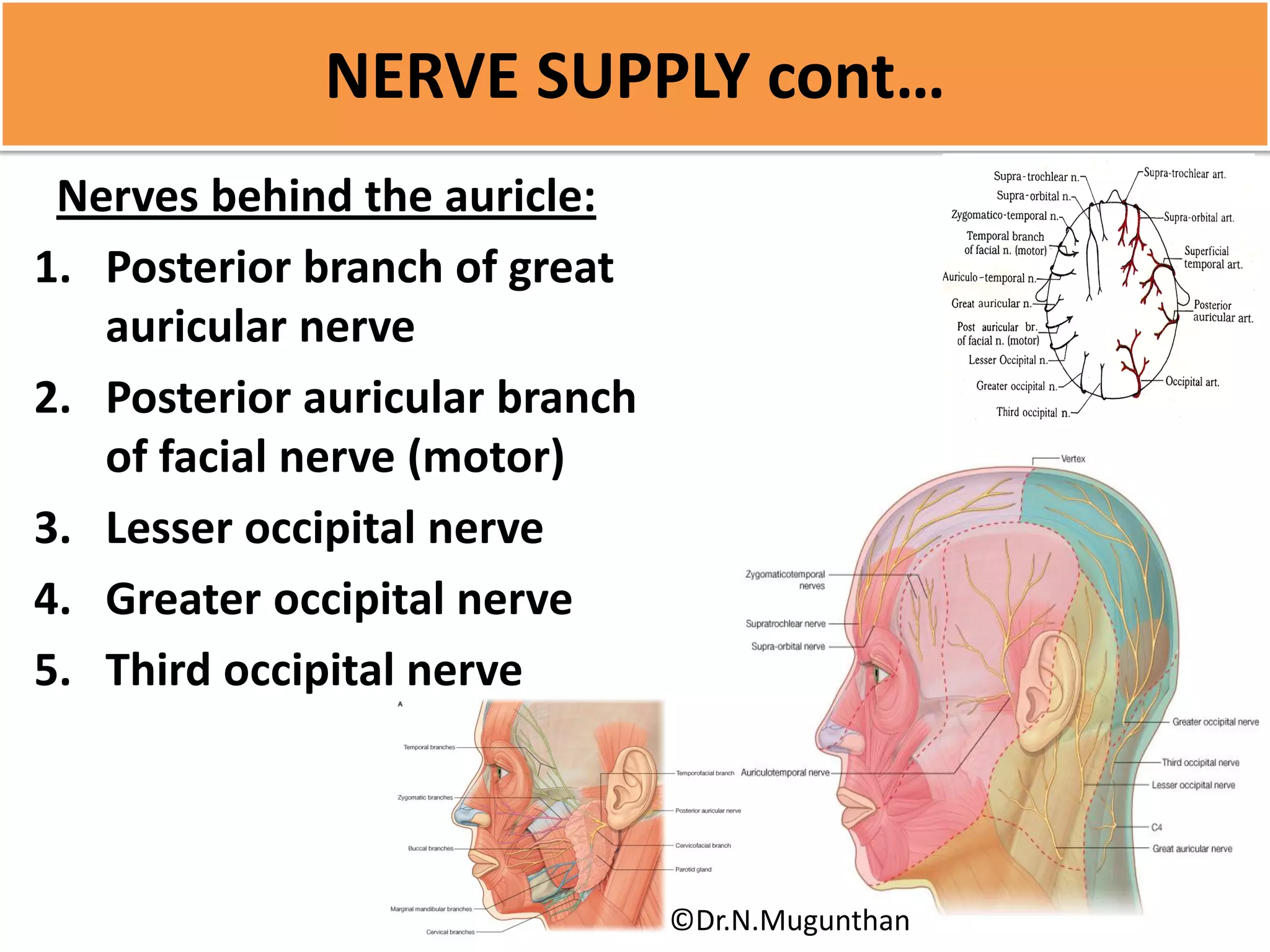
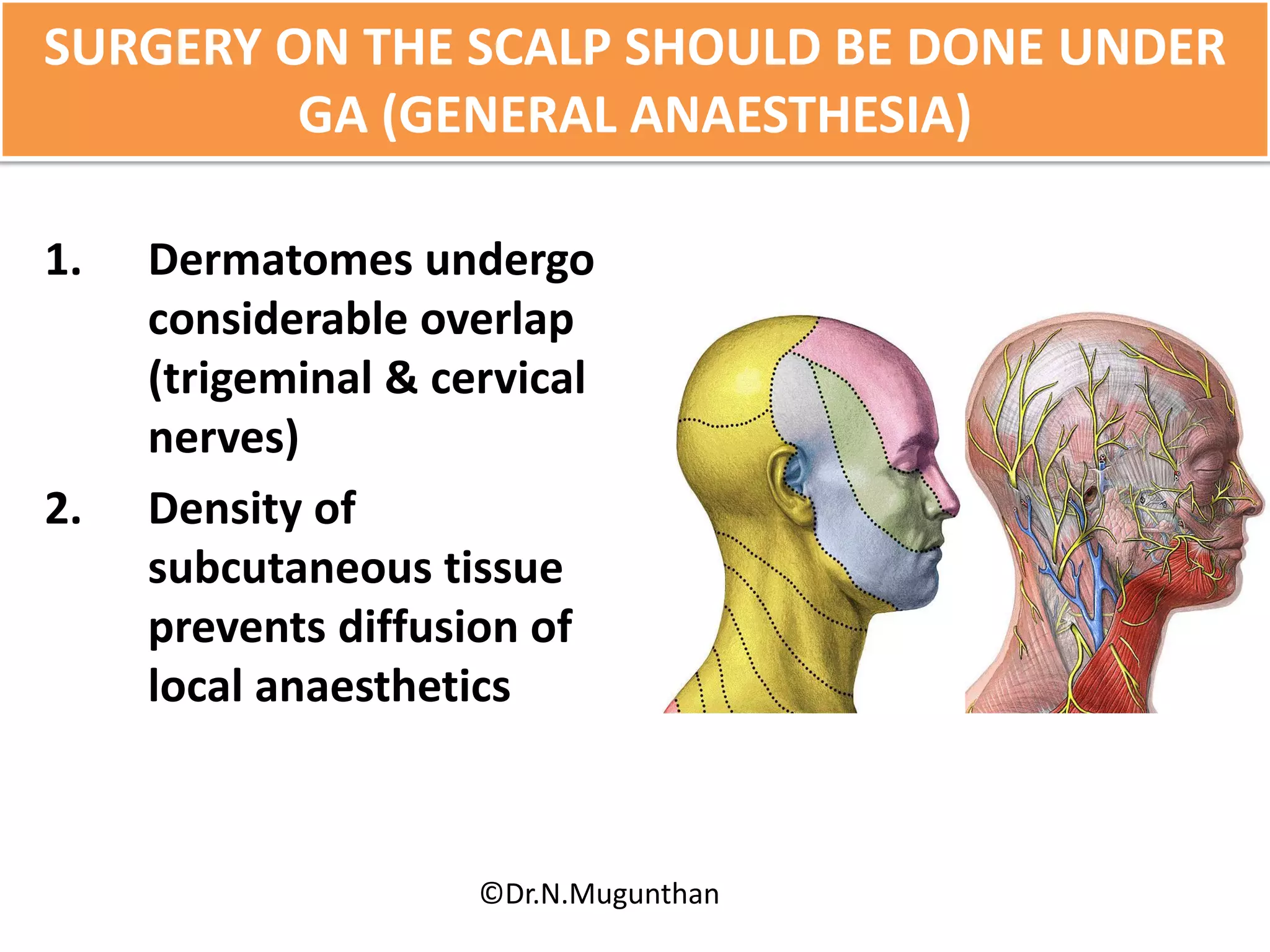
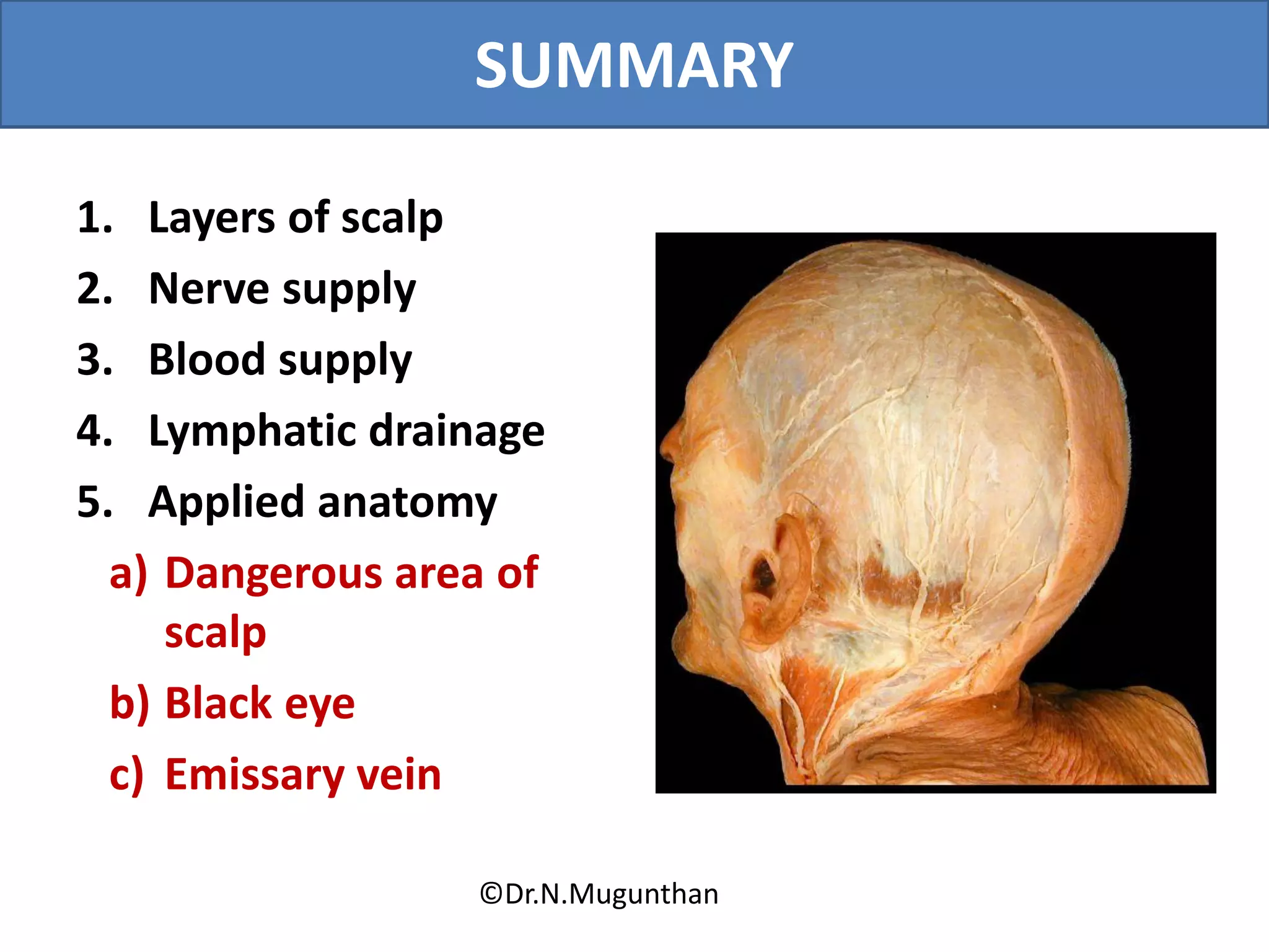
Lecture By
Dr.N.Mugunthan.MBBS,MS,DNB,MNAMS,
Associate Professor of Anatomy
MGMC&RI.
OBJECTIVES
1. Layers of scalp
2. Nerve supply
3. Blood supply
4. Lymphatic drainage
5. Applied anatomy
a) Dangerous area of scalp
b) Black eye
c) Emissary vein
©Dr.N.Mugunthan
SCALP
Soft tissue which
covers the calvaria of
skull
©Dr.N.Mugunthan
EXTENT
Front :
supraorbital margin
Behind :
external occipital
protuberance & superior
nuchal line
On each side :
zygomatic arch
©Dr.N.Mugunthan
5 LAYERS OF SCALP
1. S – Skin
2. C – Cutaneous tissue
(subcutaneous tissue)
3. A - Aponeurosis
(Galea aponeurotica
covering the occipito-
frontalis muscle)
4. L – Loose areolar tissue
(subaponeurotic tissue)
5. P – Pericranium
(outer periosteum)
©Dr.N.Mugunthan
S - SKIN
Hairs
Sebaceous gland
Sweat glands
©Dr.N.Mugunthan
C – CUTANEOUS TISSUE
(Subcutaneous tissue)
Composed of fibro-fatty
tissue
Contains blood vessels &
nerves
Walls of blood vessel
adherent to fibrous tissue –
(minor scalp injury causes
profuse bleeding)
To arrest bleeding- apply
firm pressure over the
underlying bone
©Dr.N.Mugunthan
Subcutaneous
haemorrhage – localised
in extent
Inflammation painful
Good healing
C – CUTANEOUS TISSUE cont…
©Dr.N.Mugunthan
A - APONEUROSIS
(Galea aponeurotica covering
the occipito-frontalis
muscle)
Epicranius muscle & it’s
aponeurosis
 Occipito – frontalis
 Temporo- parietalis
©Dr.N.Mugunthan
OCCIPITO - FRONTALIS
©Dr.N.Mugunthan
TEMPORO- PARIETALIS
©Dr.N.Mugunthan
Galea aponeurotica
(epicranial aponeurosis)
 Sheet of fibrous tissue
 Connects occipitalis &
frontalis muscles
Extent:
Behind : – ext.occi.protub
&highest nuchal line
Front :– blends with
subcutaneous tissue of root
of nose
Sides :- zygomatic arch
A – APONEUROSIS cont…
©Dr.N.Mugunthan
Galea aponeurotica
Applied:
If cut trasversely produce
wounds of the scalp gape
A – APONEUROSIS cont…
©Dr.N.Mugunthan
L – LOOSE AREOLAR TISSUE
 Loose subaponeurotic
tissue
 Potential space contains
emissary vein
 Emissary vein – are
devoid of valves and
communicates the veins of
scalp with intracranial
venous sinuses
©Dr.N.Mugunthan
DANGEROUS AREA OF SCALP
 The 4th layer of scalp
(loose areolar tissue)
 Infection in the
subaponeurotic space
with pus collection
readily spread to
intracranial sinuses
through emissary veins
(valveless)
©Dr.N.Mugunthan
BLACK EYE
 Blow on the skull
 Collection of blood in the
subaponeurotic space
produce generalised swelling
affecting the whole dome of
skull
 Blood slowly gravitates into
the eye lids because the
frontalis has no bony
attachments
©Dr.N.Mugunthan
SAFETY - VALVE HAEMATOMA
# of cranial vault in children
with tearing of duramatter &
pericranium
Blood from Intra cranial
haemorrhage communicate
with subaponeurotic space (4th
layer) through the line of #
Cerebral compression do not
develop
Traumatic cephalo-hydrocele
(CSF)
©Dr.N.Mugunthan
CAPUT SUCCEDANEUM
Temporary swelling in
newborn
Oedematous condtion
of a portion of scalp
Due to interference of
venous return while
passage through birth
canal
©Dr.N.Mugunthan
P-PERICRANIUM
Outer peroisteum of
skull
Loosely covers the
bone
At the sutures it is
continuous with
endocranium through
sutural membrane
©Dr.N.Mugunthan
CEPHALHAEMATOMA
Collection of blood
beneath the
pericranium
Localised swelling
which assumes the
shape of related bones
Traumatic
cephalohydrocele
©Dr.N.Mugunthan
BLOOD SUPPLY
©Dr.N.Mugunthan
ARTERIAL SUPPLY
5 sets of arteries on each
side of scalp
3 in front of auricle
2 behind the auricle
Out of 5, 2 arteries
(indirectly) from ICA
3 arteries (directly) from
ECA
©Dr.N.Mugunthan
ARTERIES IN FRONT OF AURICLE
1. Supra-trochlear
artery(Ophthalmic-
ICA)
2. Supra-orbital artery
(Ophthalmic-ICA)
3. Superficial temporal
artery (ECA)
©Dr.N.Mugunthan
ARTERIES BEHIND THE AURICLE
1. Posterior auricular
artery(ECA)
2. Occipital artery(ECA)
 Scalp is the site of free
anastomosis between
the branches of internal
& external carotid
arteries
©Dr.N.Mugunthan
VENOUS DRAINAGE
1. Supra-trochlear vein
2. Supra-orbital vein
3. Superficial temporal vein
4. Posterior auricular vein
5. Occipital vein
©Dr.N.Mugunthan
EMISSARY VEINS
2 sets of emissary veins
1. Parietal emissary vein
thro’ parietal foramen
communicates the
scalp veins with SSS
2. Mastoid emissary vein
thro’ mastoid foramen
communicates the
scalp veins with
sigmoid sinus
©Dr.N.Mugunthan
DIPLOIC VEINS
2 sets of diploic veins
1. Frontal diploic vein
thro’ supra-orbital notch
and drains into supra-orbital
vein
2. Occipital diploic vein
drain into occipital vein by
piercing outer table or drains
into transverse sinus by
piercing inner table
©Dr.N.Mugunthan
NERVE SUPPLY
10 nerves on each side of
scalp
5 infront of auricle
5 behind the auricle
4 is sensory
1 is motor
©Dr.N.Mugunthan
Nerves infront of auricle:
1. Supra-trochlear nerve
2. Supra-orbital nerve
3. Zygomatico-temporal
nerve
4. Temporal branch of
facial nerve (motor)
5. Auriculo-temporal
nerve
NERVE SUPPLY cont…
©Dr.N.Mugunthan
Nerves behind the auricle:
1. Posterior branch of great
auricular nerve
2. Posterior auricular branch
of facial nerve (motor)
3. Lesser occipital nerve
4. Greater occipital nerve
5. Third occipital nerve
NERVE SUPPLY cont…
©Dr.N.Mugunthan
SURGERY ON THE SCALP SHOULD BE DONE UNDER
GA (GENERAL ANAESTHESIA)
1. Dermatomes undergo
considerable overlap
(trigeminal & cervical
nerves)
2. Density of
subcutaneous tissue
prevents diffusion of
local anaesthetics
©Dr.N.Mugunthan
LYMPHATIC DRAINAGE OF SCALP
Anterior part of scalp -
preauricular lymph nodes
(superficial parotid
nodes)
Posterior part of scalp
– post auricular lymph
nodes or mastoid group
of nodes and occipital
nodes
©Dr.N.Mugunthan
SUMMARY
1. Layers of scalp
2. Nerve supply
3. Blood supply
4. Lymphatic drainage
5. Applied anatomy
a) Dangerous area of
scalp
b) Black eye
c) Emissary vein
©Dr.N.Mugunthan
THANK YOU

Scalp

  • 1.
  • 2.
    OBJECTIVES 1. Layers ofscalp 2. Nerve supply 3. Blood supply 4. Lymphatic drainage 5. Applied anatomy a) Dangerous area of scalp b) Black eye c) Emissary vein ©Dr.N.Mugunthan
  • 3.
    SCALP Soft tissue which coversthe calvaria of skull ©Dr.N.Mugunthan
  • 4.
    EXTENT Front : supraorbital margin Behind: external occipital protuberance & superior nuchal line On each side : zygomatic arch ©Dr.N.Mugunthan
  • 5.
    5 LAYERS OFSCALP 1. S – Skin 2. C – Cutaneous tissue (subcutaneous tissue) 3. A - Aponeurosis (Galea aponeurotica covering the occipito- frontalis muscle) 4. L – Loose areolar tissue (subaponeurotic tissue) 5. P – Pericranium (outer periosteum) ©Dr.N.Mugunthan
  • 6.
    S - SKIN Hairs Sebaceousgland Sweat glands ©Dr.N.Mugunthan
  • 7.
    C – CUTANEOUSTISSUE (Subcutaneous tissue) Composed of fibro-fatty tissue Contains blood vessels & nerves Walls of blood vessel adherent to fibrous tissue – (minor scalp injury causes profuse bleeding) To arrest bleeding- apply firm pressure over the underlying bone ©Dr.N.Mugunthan
  • 8.
    Subcutaneous haemorrhage – localised inextent Inflammation painful Good healing C – CUTANEOUS TISSUE cont… ©Dr.N.Mugunthan
  • 9.
    A - APONEUROSIS (Galeaaponeurotica covering the occipito-frontalis muscle) Epicranius muscle & it’s aponeurosis  Occipito – frontalis  Temporo- parietalis ©Dr.N.Mugunthan
  • 10.
  • 11.
  • 12.
    Galea aponeurotica (epicranial aponeurosis) Sheet of fibrous tissue  Connects occipitalis & frontalis muscles Extent: Behind : – ext.occi.protub &highest nuchal line Front :– blends with subcutaneous tissue of root of nose Sides :- zygomatic arch A – APONEUROSIS cont… ©Dr.N.Mugunthan
  • 13.
    Galea aponeurotica Applied: If cuttrasversely produce wounds of the scalp gape A – APONEUROSIS cont… ©Dr.N.Mugunthan
  • 14.
    L – LOOSEAREOLAR TISSUE  Loose subaponeurotic tissue  Potential space contains emissary vein  Emissary vein – are devoid of valves and communicates the veins of scalp with intracranial venous sinuses ©Dr.N.Mugunthan
  • 15.
    DANGEROUS AREA OFSCALP  The 4th layer of scalp (loose areolar tissue)  Infection in the subaponeurotic space with pus collection readily spread to intracranial sinuses through emissary veins (valveless) ©Dr.N.Mugunthan
  • 16.
    BLACK EYE  Blowon the skull  Collection of blood in the subaponeurotic space produce generalised swelling affecting the whole dome of skull  Blood slowly gravitates into the eye lids because the frontalis has no bony attachments ©Dr.N.Mugunthan
  • 17.
    SAFETY - VALVEHAEMATOMA # of cranial vault in children with tearing of duramatter & pericranium Blood from Intra cranial haemorrhage communicate with subaponeurotic space (4th layer) through the line of # Cerebral compression do not develop Traumatic cephalo-hydrocele (CSF) ©Dr.N.Mugunthan
  • 18.
    CAPUT SUCCEDANEUM Temporary swellingin newborn Oedematous condtion of a portion of scalp Due to interference of venous return while passage through birth canal ©Dr.N.Mugunthan
  • 19.
    P-PERICRANIUM Outer peroisteum of skull Looselycovers the bone At the sutures it is continuous with endocranium through sutural membrane ©Dr.N.Mugunthan
  • 20.
    CEPHALHAEMATOMA Collection of blood beneaththe pericranium Localised swelling which assumes the shape of related bones Traumatic cephalohydrocele ©Dr.N.Mugunthan
  • 21.
  • 22.
    ARTERIAL SUPPLY 5 setsof arteries on each side of scalp 3 in front of auricle 2 behind the auricle Out of 5, 2 arteries (indirectly) from ICA 3 arteries (directly) from ECA ©Dr.N.Mugunthan
  • 23.
    ARTERIES IN FRONTOF AURICLE 1. Supra-trochlear artery(Ophthalmic- ICA) 2. Supra-orbital artery (Ophthalmic-ICA) 3. Superficial temporal artery (ECA) ©Dr.N.Mugunthan
  • 24.
    ARTERIES BEHIND THEAURICLE 1. Posterior auricular artery(ECA) 2. Occipital artery(ECA)  Scalp is the site of free anastomosis between the branches of internal & external carotid arteries ©Dr.N.Mugunthan
  • 25.
    VENOUS DRAINAGE 1. Supra-trochlearvein 2. Supra-orbital vein 3. Superficial temporal vein 4. Posterior auricular vein 5. Occipital vein ©Dr.N.Mugunthan
  • 26.
    EMISSARY VEINS 2 setsof emissary veins 1. Parietal emissary vein thro’ parietal foramen communicates the scalp veins with SSS 2. Mastoid emissary vein thro’ mastoid foramen communicates the scalp veins with sigmoid sinus ©Dr.N.Mugunthan
  • 27.
    DIPLOIC VEINS 2 setsof diploic veins 1. Frontal diploic vein thro’ supra-orbital notch and drains into supra-orbital vein 2. Occipital diploic vein drain into occipital vein by piercing outer table or drains into transverse sinus by piercing inner table ©Dr.N.Mugunthan
  • 28.
    NERVE SUPPLY 10 nerveson each side of scalp 5 infront of auricle 5 behind the auricle 4 is sensory 1 is motor ©Dr.N.Mugunthan
  • 29.
    Nerves infront ofauricle: 1. Supra-trochlear nerve 2. Supra-orbital nerve 3. Zygomatico-temporal nerve 4. Temporal branch of facial nerve (motor) 5. Auriculo-temporal nerve NERVE SUPPLY cont… ©Dr.N.Mugunthan
  • 30.
    Nerves behind theauricle: 1. Posterior branch of great auricular nerve 2. Posterior auricular branch of facial nerve (motor) 3. Lesser occipital nerve 4. Greater occipital nerve 5. Third occipital nerve NERVE SUPPLY cont… ©Dr.N.Mugunthan
  • 31.
    SURGERY ON THESCALP SHOULD BE DONE UNDER GA (GENERAL ANAESTHESIA) 1. Dermatomes undergo considerable overlap (trigeminal & cervical nerves) 2. Density of subcutaneous tissue prevents diffusion of local anaesthetics ©Dr.N.Mugunthan
  • 32.
    LYMPHATIC DRAINAGE OFSCALP Anterior part of scalp - preauricular lymph nodes (superficial parotid nodes) Posterior part of scalp – post auricular lymph nodes or mastoid group of nodes and occipital nodes ©Dr.N.Mugunthan
  • 33.
    SUMMARY 1. Layers ofscalp 2. Nerve supply 3. Blood supply 4. Lymphatic drainage 5. Applied anatomy a) Dangerous area of scalp b) Black eye c) Emissary vein ©Dr.N.Mugunthan
  • 34.