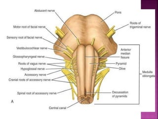
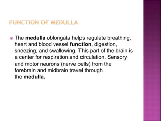
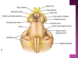
Rabbia Yousaf
(Pharm-D)
Rashid Latif College of Pharmacy
 The brainstem is made up of
 the medulla oblongata
 the pons
 the midbrain
 it occupies the posterior cranial fossa of the
skull.
 It is stalk like in shape and connects the
narrow spinal cord with the expanded
forebrain
 The brainstem has three broad functions:
 (1) it serves as a conduit for the ascending
tracts and descending tracts connecting the
spinal cord to the different parts of the
higher centers in the forebrain.
 (2) it contains important reflex centers
associated with the control of respiration the
cardiovascular system and with the control
of consciousness.
 (3) it contains the important nuclei of
cranial nerves .
 The medulla oblongata connects the pons superiorly
with the spinal cord inferiorly.
 The medulla oblongata is conical in shape, its broad
extremity being directed superiorly .
 The central canal of the spinal cord continues
upward into the lower half of the medulla; in the
upper half of the medulla, it expands as the cavity
of the fourth ventricle .
 On the anterior surface of the medulla is the
anterior median fissure, which is continuous
inferiorly with the anterior median fissure
of the spinal cord .
 On each side of the median fissure, there is a
swelling called the pyramid.
 The pyramids are composed of bundles of
nerve fibers, called corticospinal fibers,
which originate in large nerve cells in the
precentral gyrus of the cerebral cortex.
 The pyramids taper inferiorly, and it is here
that the majority of the descending fibers
cross over to the opposite side, forming the
decussation of the pyramids.
 Posterolateral to the pyramids are the
olives, which are oval elevations produced
by the underlying inferior olivary nuclei.
 The posterior surface of the superior half of the medulla
oblongata forms the lower part of the floor of the fourth
ventricle.
 The posterior surface of the inferior half of the medulla is
continuous with the posterior aspect of the spinal cord
and possesses a posterior median sulcus.
 On each side of the median sulcus, there is an
elongated swelling, the gracile tubercle, produced by
the underlying gracile nucleus.
 Lateral to the gracile tubercle is a similar swelling, the
cuneate tubercle, produced by the underlying cuneate
nucleus.
 The medulla oblongata helps regulate breathing,
heart and blood vessel function, digestion,
sneezing, and swallowing. This part of the brain is
a center for respiration and circulation. Sensory
and motor neurons (nerve cells) from the
forebrain and midbrain travel through
the medulla.
 The pons is anterior to the cerebellum and
connects the medulla oblongata to the midbrain.
 It is about 1 inch (2.5 cm) long. It is a bridge
between various parts of the nervous system,
including the cerebellum and cerebrum, There
are many important nerves that originate in
the pons.
 The anterior surface is convex from side to side
and shows many transverse fibers that converge
on each side to form the middle cerebellar
peduncle .
 There is a shallow groove in the midline, the
basilar groove, which lodges the basilar
artery.
 On the anterolateral surface of the pons,
the trigeminal nerve emerges on each side.
 Each nerve consists of a smaller, medial
part, known as the motor root, and a larger,
lateral part, known as the sensory root.
 In the groove between the pons and the
medulla oblongata, there emerge, from
medial to lateral, the abducent, facial, and
vestibulocochlear nerves
 The posterior surface of the pons is hidden
from view by the cerebellum.
 It forms the upper half of the floor of the
fourth ventricle and is triangular in shape
 The posterior surface is limited laterally by
the superior cerebellar peduncles and is
divided into symmetrical halves by a median
sulcus.
 Lateral to this sulcus is an elongated
elevation, the medial eminence, which is
bounded laterally by a sulcus, the sulcus
limitans .
 The inferior end of the medial eminence is slightly
expanded to form the facial colliculus, which is
produced by the root of the facial nerve winding
around the nucleus of the abducent nerve .
 The floor of the superior part of the sulcus limitans
is bluish-gray in color and is called the substantia
ferruginea; it owes its color to a group of deeply
pigmented nerve cells.
 Lateral to the sulcus limitans is the area vestibuli
produced by the underlying vestibular nuclei
 It is involved in the control of breathing,
communication between different parts of the
brain, and sensations such as hearing, taste, and
balance.
 The pons has also been associated with the
control of sleep cycles.
 The midbrain measures about 0.8 inch (2 cm)
in length and connects the pons and
cerebellum with the forebrain.
 The midbrain is traversed by a narrow
channel, the cerebral aqueduct, which is
filled with cerebrospinal fluid.
 On the posterior surface are four colliculi.
These are rounded eminences that are
divided into superior and inferior pairs by a
vertical and a transverse groove.
 The superior colliculi are centers for visual
reflexes, and the inferior colliculi are lower
auditory centers.
 In the midline below the inferior colliculi,
the trochlear nerves emerge.
 These are small-diameter nerves that wind
around the lateral aspect of the midbrain .
 On the lateral aspect of the midbrain, the
superior and inferior brachia ascend in an
anterolateral direction.
 The superior brachium passes from the
superior colliculus to the lateral geniculate
body and the optic tract.
 The inferior brachium connects the inferior
colliculus to the medial geniculate body.
 On the anterior aspect of the midbrain ,
there is a deep depression in the midline,
the interpeduncular fossa, which is bounded
on either side by the crus cerebri
 Many small blood vessels perforate the floor
of the interpeduncular fossa, and this region
is termed the posterior perforated
substance.
 The oculomotor nerve emerges from a
groove on the medial side of the crus
cerebri.
 The crus cerebri contains important
descending tracts and is separated from the
tegmentum by the substantia nigra .
 The corticospinal and corticonuclear fibers
occupy the middle two-thirds of the crus.
 The red nucleus is a rounded mass of gray matter
situated between the cerebral aqueduct and the
substantia nigra. Its red colour, seen in fresh
specimens, is due to its vascularity and the
presence of an iron-containing pigment in the
cytoplasm of many of its neurons.
 The substantia nigra is a large motor nucleus
situated between the tegmentum, and the crus
cerebri and is found throughout the midbrain.The
substantia nigra is concerned with muscle tone.
 The midbrain serves important functions in motor
movement, particularly movements of the eye,
and in auditory and visual processing.
 Controlling responses to sight.
 Pupil Dilation
 Regulate Muscle Movement

Brain stem

  • 1.
  • 2.
     The brainstemis made up of  the medulla oblongata  the pons  the midbrain  it occupies the posterior cranial fossa of the skull.  It is stalk like in shape and connects the narrow spinal cord with the expanded forebrain
  • 4.
     The brainstemhas three broad functions:  (1) it serves as a conduit for the ascending tracts and descending tracts connecting the spinal cord to the different parts of the higher centers in the forebrain.  (2) it contains important reflex centers associated with the control of respiration the cardiovascular system and with the control of consciousness.  (3) it contains the important nuclei of cranial nerves .
  • 6.
     The medullaoblongata connects the pons superiorly with the spinal cord inferiorly.  The medulla oblongata is conical in shape, its broad extremity being directed superiorly .  The central canal of the spinal cord continues upward into the lower half of the medulla; in the upper half of the medulla, it expands as the cavity of the fourth ventricle .
  • 7.
     On theanterior surface of the medulla is the anterior median fissure, which is continuous inferiorly with the anterior median fissure of the spinal cord .  On each side of the median fissure, there is a swelling called the pyramid.  The pyramids are composed of bundles of nerve fibers, called corticospinal fibers, which originate in large nerve cells in the precentral gyrus of the cerebral cortex.
  • 9.
     The pyramidstaper inferiorly, and it is here that the majority of the descending fibers cross over to the opposite side, forming the decussation of the pyramids.  Posterolateral to the pyramids are the olives, which are oval elevations produced by the underlying inferior olivary nuclei.
  • 10.
     The posteriorsurface of the superior half of the medulla oblongata forms the lower part of the floor of the fourth ventricle.  The posterior surface of the inferior half of the medulla is continuous with the posterior aspect of the spinal cord and possesses a posterior median sulcus.  On each side of the median sulcus, there is an elongated swelling, the gracile tubercle, produced by the underlying gracile nucleus.  Lateral to the gracile tubercle is a similar swelling, the cuneate tubercle, produced by the underlying cuneate nucleus.
  • 12.
     The medullaoblongata helps regulate breathing, heart and blood vessel function, digestion, sneezing, and swallowing. This part of the brain is a center for respiration and circulation. Sensory and motor neurons (nerve cells) from the forebrain and midbrain travel through the medulla.
  • 13.
     The ponsis anterior to the cerebellum and connects the medulla oblongata to the midbrain.  It is about 1 inch (2.5 cm) long. It is a bridge between various parts of the nervous system, including the cerebellum and cerebrum, There are many important nerves that originate in the pons.  The anterior surface is convex from side to side and shows many transverse fibers that converge on each side to form the middle cerebellar peduncle .
  • 15.
     There isa shallow groove in the midline, the basilar groove, which lodges the basilar artery.  On the anterolateral surface of the pons, the trigeminal nerve emerges on each side.  Each nerve consists of a smaller, medial part, known as the motor root, and a larger, lateral part, known as the sensory root.  In the groove between the pons and the medulla oblongata, there emerge, from medial to lateral, the abducent, facial, and vestibulocochlear nerves
  • 16.
     The posteriorsurface of the pons is hidden from view by the cerebellum.  It forms the upper half of the floor of the fourth ventricle and is triangular in shape  The posterior surface is limited laterally by the superior cerebellar peduncles and is divided into symmetrical halves by a median sulcus.  Lateral to this sulcus is an elongated elevation, the medial eminence, which is bounded laterally by a sulcus, the sulcus limitans .
  • 18.
     The inferiorend of the medial eminence is slightly expanded to form the facial colliculus, which is produced by the root of the facial nerve winding around the nucleus of the abducent nerve .  The floor of the superior part of the sulcus limitans is bluish-gray in color and is called the substantia ferruginea; it owes its color to a group of deeply pigmented nerve cells.  Lateral to the sulcus limitans is the area vestibuli produced by the underlying vestibular nuclei
  • 19.
     It isinvolved in the control of breathing, communication between different parts of the brain, and sensations such as hearing, taste, and balance.  The pons has also been associated with the control of sleep cycles.
  • 20.
     The midbrainmeasures about 0.8 inch (2 cm) in length and connects the pons and cerebellum with the forebrain.  The midbrain is traversed by a narrow channel, the cerebral aqueduct, which is filled with cerebrospinal fluid.  On the posterior surface are four colliculi. These are rounded eminences that are divided into superior and inferior pairs by a vertical and a transverse groove.
  • 22.
     The superiorcolliculi are centers for visual reflexes, and the inferior colliculi are lower auditory centers.  In the midline below the inferior colliculi, the trochlear nerves emerge.  These are small-diameter nerves that wind around the lateral aspect of the midbrain .
  • 23.
     On thelateral aspect of the midbrain, the superior and inferior brachia ascend in an anterolateral direction.  The superior brachium passes from the superior colliculus to the lateral geniculate body and the optic tract.  The inferior brachium connects the inferior colliculus to the medial geniculate body.
  • 25.
     On theanterior aspect of the midbrain , there is a deep depression in the midline, the interpeduncular fossa, which is bounded on either side by the crus cerebri  Many small blood vessels perforate the floor of the interpeduncular fossa, and this region is termed the posterior perforated substance.  The oculomotor nerve emerges from a groove on the medial side of the crus cerebri.
  • 28.
     The cruscerebri contains important descending tracts and is separated from the tegmentum by the substantia nigra .  The corticospinal and corticonuclear fibers occupy the middle two-thirds of the crus.
  • 29.
     The rednucleus is a rounded mass of gray matter situated between the cerebral aqueduct and the substantia nigra. Its red colour, seen in fresh specimens, is due to its vascularity and the presence of an iron-containing pigment in the cytoplasm of many of its neurons.  The substantia nigra is a large motor nucleus situated between the tegmentum, and the crus cerebri and is found throughout the midbrain.The substantia nigra is concerned with muscle tone.
  • 30.
     The midbrainserves important functions in motor movement, particularly movements of the eye, and in auditory and visual processing.  Controlling responses to sight.  Pupil Dilation  Regulate Muscle Movement