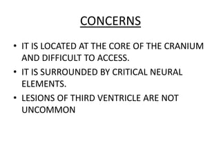
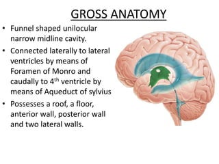
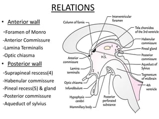
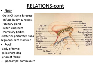
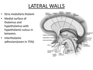
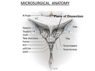
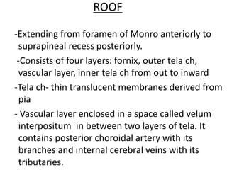
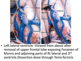
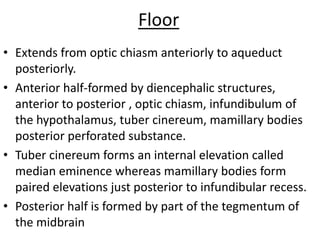
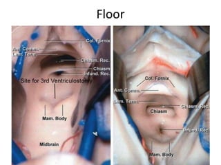
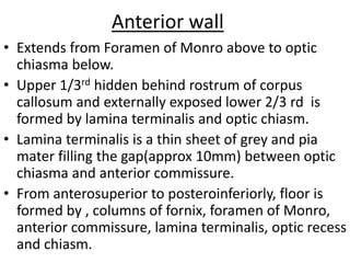
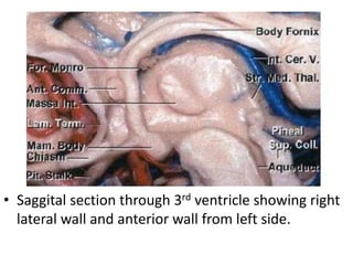
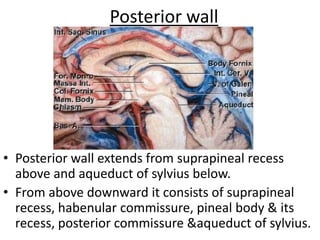
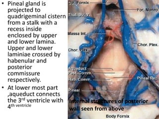
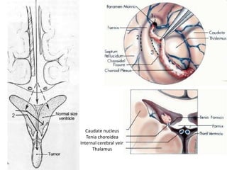
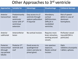
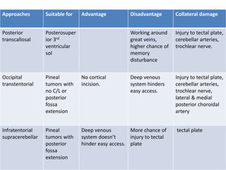
SURGICAL ANATOMY OF THIRD
VENTRICLE
DR PARTHA SARATHI MONDAL
SENIOR RESIDENT
NEUROSURGERY
CONCERNS
• IT IS LOCATED AT THE CORE OF THE CRANIUM
AND DIFFICULT TO ACCESS.
• IT IS SURROUNDED BY CRITICAL NEURAL
ELEMENTS.
• LESIONS OF THIRD VENTRICLE ARE NOT
UNCOMMON
VENTRICULAR SYSTEM
GROSS ANATOMY
• Funnel shaped unilocular
narrow midline cavity.
• Connected laterally to lateral
ventricles by means of
Foramen of Monro and
caudally to 4th ventricle by
means of Aqueduct of sylvius.
• Possesses a roof, a floor,
anterior wall, posterior wall
and two lateral walls.
RELATIONS
• Anterior wall
-Foramen of Monro
-Anterior Commissure
-Lamina Terminalis
-Optic chiasma
• Posterior wall
-Suprapineal rescess(4)
-Habenular commissure
-Pineal recess(5) & gland
-Posterior commissure
-Aqueduct of sylvius
RELATIONS-cont
• Floor
-Optic Chiasma & recess
- Infundibulum & recess
-Pituitary gland
-Tuber cinereum
-Mamillary bodies
-Posterior perforated subs
Tegmentum of midbrain
• Roof
-Body of fornix
-Tella choroidea
-Crura of fornix
- Hippocampal commissure
LATERAL WALLS
• Stria medullaris thalami
• Medial surface of
thalamus and
hypothalamus with
hypothalamic sulcus in
between.
• Interthalamic
adhesions(seen in 75%)
MICROSURGICAL ANATOMY
ROOF
-Extending from foramen of Monro anteriorly to
suprapineal recess posteriorly.
-Consists of four layers: fornix, outer tela ch,
vascular layer, inner tela ch from out to inward
-Tela ch- thin translucent membranes derived from
pia
- Vascular layer enclosed in a space called velum
interpositum in between two layers of tela. It
contains posterior choroidal artery with its
branches and internal cerebral veins with its
tributaries.
• Left lateral ventricle -Viewed from above after
removal of upper frontal lobe exposing Foramen of
Monro and adjoining parts of Rt lateral and 3rd
ventricle.Dissection done through Tenia fornicis
Floor
• Extends from optic chiasm anteriorly to aqueduct
posteriorly.
• Anterior half-formed by diencephalic structures,
anterior to posterior , optic chiasm, infundibulum of
the hypothalamus, tuber cinereum, mamillary bodies
posterior perforated substance.
• Tuber cinereum forms an internal elevation called
median eminence whereas mamillary bodies form
paired elevations just posterior to infundibular recess.
• Posterior half is formed by part of the tegmentum of
the midbrain
Floor
Anterior wall
• Extends from Foramen of Monro above to optic
chiasma below.
• Upper 1/3rd hidden behind rostrum of corpus
callosum and externally exposed lower 2/3 rd is
formed by lamina terminalis and optic chiasm.
• Lamina terminalis is a thin sheet of grey and pia
mater filling the gap(approx 10mm) between optic
chiasma and anterior commissure.
• From anterosuperior to posteroinferiorly, floor is
formed by , columns of fornix, foramen of Monro,
anterior commissure, lamina terminalis, optic recess
and chiasm.
• Saggital section through 3rd ventricle showing right
lateral wall and anterior wall from left side.
Posterior wall
• Posterior wall extends from suprapineal recess
above and aqueduct of sylvius below.
• From above downward it consists of suprapineal
recess, habenular commissure, pineal body & its
recess, posterior commissure &aqueduct of sylvius.
• Pineal gland is
projected to
quadrigeminal cistern
from a stalk with a
recess inside
enclosed by upper
and lower lamina.
Upper and lower
laminiae crossed by
habenular and
posterior
commissure
respectively.
• At lower most part
,aqueduct connects
the 3rd ventricle with
4th ventricle
Internal structures of posterior
wall seen from above
Common lesions of 3rd Ventricle
Congenital lesions Acquired lesions
Aqueductal Stenosis Anterior mass- Seller-supraseller mass
Chiasmatic-hypothalamic mass
Cavum Veli Interpositi Cyst Posterior mass-pineal sol, tectal sol, Inferior
thalamic sol
Congenital Intraventricular Cysts Inferior mass- Hypothalamic hamartoma ,
Arachnoid cyst
Mass near Foramen of Monro- Colloid cyst ,
SEGA, Subependymoma
Intraventricular SOL- Choroid plexus
neoplasm(Primary or Secondary), Congenital
intraventricular cyst, Vascular malformation of
choroid plexus
SURGICAL APPROACHES
APPROACH TO THIRD
VENTRICLE
ANTERIOR
ANTERIOR
TRANSCALLOSAL
TRANSCORTICAL
(THROUGH MIDDLE
FRONTAL GYRUS)
SUBFRONTAL
POSTERIOR
TRANSCALLOSAL TRANSCORTICAL
INFRATENTORIAL
SUPRACELEBELLAR
Anterior transcallosal approach
• Basically a transcallosal-transventricular approach.
• Indications-Anterosuperior lesion or lateral ventricular
extension.
• Advantages-access to both lateral ventricles as well, less neural
incision, possible in nondilated ventricles.
• Corridor to entry into 3rd ventricle-Transforaminal,
transforniceal, transchoroidal.
• Nearby critical structures susceptible to injury-
1)Genu of internal capsule(in transforaminal)-Hemiplegia
2)Fornix-memory disturbance
3)Thalamostriate vein-drowsiness, hemiplegia, mutism.
4)Dorsomedial Nucleus of Thalamus-emotional disturbance
Caudate nucleus
Tenia choroidea
Internal cerebral vein
Thalamus
Other Approaches to 3rd ventricle
Approaches Suitable for Advantage Disadvantage Collateral damage
Anterior
transcortical
Predominantly
lateral
ventricular
tumor with 3rd
ventricluar
extension
Easy access to c/l
ventricle through
septum pellucidum
if required
Mandatory
cortical
(MFG)incision.
Difficult in
nondialted
ventricles.
Risk of speech
deficit in case of
dominant
hemisphere.
Anterior
subfrontal
Anteroinferior
lesion
No cortical incision. Requires more
brain retraction,
working around
vessels.
Perforator vessel
injury(ACOM in
particular)
Posterior
transcortical
(through
sup parietal
lobule)
Posterior 3rd
ventricle and
roof
Good access to
quadrigeminal
cistern and also to
pineal gland.
Less spacious
exposure,
Needs cortical
incision.
Optic
radiation,visuospatia
l defect, aphasia.
Approaches Suitable for Advantage Disadvantage Collateral damage
Posterior
transcallosal
Posterosuper
ior 3rd
ventricular
sol
Working around
great veins,
higher chance of
memory
disturbance
Injury to tectal plate,
cerebellar arteries,
trochlear nerve.
Occipital
transtentorial
Pineal
tumors with
no C/L or
posterior
fossa
extension
No cortical
incision.
Deep venous
system hinders
easy access.
Injury to tectal plate,
cerebellar arteries,
trochlear nerve,
lateral & medial
posterior choroidal
artery
Infratentorial
supracerebellar
Pineal
tumors with
posterior
fossa
extension
Deep venous
system doesn’t
hinder easy access.
More chance of
injury to tectal
plate
tectal plate
Thank you

Third ventricle

  • 1.
    SURGICAL ANATOMY OFTHIRD VENTRICLE DR PARTHA SARATHI MONDAL SENIOR RESIDENT NEUROSURGERY
  • 2.
    CONCERNS • IT ISLOCATED AT THE CORE OF THE CRANIUM AND DIFFICULT TO ACCESS. • IT IS SURROUNDED BY CRITICAL NEURAL ELEMENTS. • LESIONS OF THIRD VENTRICLE ARE NOT UNCOMMON
  • 3.
  • 4.
    GROSS ANATOMY • Funnelshaped unilocular narrow midline cavity. • Connected laterally to lateral ventricles by means of Foramen of Monro and caudally to 4th ventricle by means of Aqueduct of sylvius. • Possesses a roof, a floor, anterior wall, posterior wall and two lateral walls.
  • 5.
    RELATIONS • Anterior wall -Foramenof Monro -Anterior Commissure -Lamina Terminalis -Optic chiasma • Posterior wall -Suprapineal rescess(4) -Habenular commissure -Pineal recess(5) & gland -Posterior commissure -Aqueduct of sylvius
  • 6.
    RELATIONS-cont • Floor -Optic Chiasma& recess - Infundibulum & recess -Pituitary gland -Tuber cinereum -Mamillary bodies -Posterior perforated subs Tegmentum of midbrain • Roof -Body of fornix -Tella choroidea -Crura of fornix - Hippocampal commissure
  • 7.
    LATERAL WALLS • Striamedullaris thalami • Medial surface of thalamus and hypothalamus with hypothalamic sulcus in between. • Interthalamic adhesions(seen in 75%)
  • 8.
  • 9.
    ROOF -Extending from foramenof Monro anteriorly to suprapineal recess posteriorly. -Consists of four layers: fornix, outer tela ch, vascular layer, inner tela ch from out to inward -Tela ch- thin translucent membranes derived from pia - Vascular layer enclosed in a space called velum interpositum in between two layers of tela. It contains posterior choroidal artery with its branches and internal cerebral veins with its tributaries.
  • 10.
    • Left lateralventricle -Viewed from above after removal of upper frontal lobe exposing Foramen of Monro and adjoining parts of Rt lateral and 3rd ventricle.Dissection done through Tenia fornicis
  • 11.
    Floor • Extends fromoptic chiasm anteriorly to aqueduct posteriorly. • Anterior half-formed by diencephalic structures, anterior to posterior , optic chiasm, infundibulum of the hypothalamus, tuber cinereum, mamillary bodies posterior perforated substance. • Tuber cinereum forms an internal elevation called median eminence whereas mamillary bodies form paired elevations just posterior to infundibular recess. • Posterior half is formed by part of the tegmentum of the midbrain
  • 12.
  • 13.
    Anterior wall • Extendsfrom Foramen of Monro above to optic chiasma below. • Upper 1/3rd hidden behind rostrum of corpus callosum and externally exposed lower 2/3 rd is formed by lamina terminalis and optic chiasm. • Lamina terminalis is a thin sheet of grey and pia mater filling the gap(approx 10mm) between optic chiasma and anterior commissure. • From anterosuperior to posteroinferiorly, floor is formed by , columns of fornix, foramen of Monro, anterior commissure, lamina terminalis, optic recess and chiasm.
  • 14.
    • Saggital sectionthrough 3rd ventricle showing right lateral wall and anterior wall from left side.
  • 15.
    Posterior wall • Posteriorwall extends from suprapineal recess above and aqueduct of sylvius below. • From above downward it consists of suprapineal recess, habenular commissure, pineal body & its recess, posterior commissure &aqueduct of sylvius.
  • 16.
    • Pineal glandis projected to quadrigeminal cistern from a stalk with a recess inside enclosed by upper and lower lamina. Upper and lower laminiae crossed by habenular and posterior commissure respectively. • At lower most part ,aqueduct connects the 3rd ventricle with 4th ventricle Internal structures of posterior wall seen from above
  • 17.
    Common lesions of3rd Ventricle Congenital lesions Acquired lesions Aqueductal Stenosis Anterior mass- Seller-supraseller mass Chiasmatic-hypothalamic mass Cavum Veli Interpositi Cyst Posterior mass-pineal sol, tectal sol, Inferior thalamic sol Congenital Intraventricular Cysts Inferior mass- Hypothalamic hamartoma , Arachnoid cyst Mass near Foramen of Monro- Colloid cyst , SEGA, Subependymoma Intraventricular SOL- Choroid plexus neoplasm(Primary or Secondary), Congenital intraventricular cyst, Vascular malformation of choroid plexus
  • 18.
  • 19.
    APPROACH TO THIRD VENTRICLE ANTERIOR ANTERIOR TRANSCALLOSAL TRANSCORTICAL (THROUGHMIDDLE FRONTAL GYRUS) SUBFRONTAL POSTERIOR TRANSCALLOSAL TRANSCORTICAL INFRATENTORIAL SUPRACELEBELLAR
  • 20.
    Anterior transcallosal approach •Basically a transcallosal-transventricular approach. • Indications-Anterosuperior lesion or lateral ventricular extension. • Advantages-access to both lateral ventricles as well, less neural incision, possible in nondilated ventricles. • Corridor to entry into 3rd ventricle-Transforaminal, transforniceal, transchoroidal. • Nearby critical structures susceptible to injury- 1)Genu of internal capsule(in transforaminal)-Hemiplegia 2)Fornix-memory disturbance 3)Thalamostriate vein-drowsiness, hemiplegia, mutism. 4)Dorsomedial Nucleus of Thalamus-emotional disturbance
  • 21.
  • 22.
    Other Approaches to3rd ventricle Approaches Suitable for Advantage Disadvantage Collateral damage Anterior transcortical Predominantly lateral ventricular tumor with 3rd ventricluar extension Easy access to c/l ventricle through septum pellucidum if required Mandatory cortical (MFG)incision. Difficult in nondialted ventricles. Risk of speech deficit in case of dominant hemisphere. Anterior subfrontal Anteroinferior lesion No cortical incision. Requires more brain retraction, working around vessels. Perforator vessel injury(ACOM in particular) Posterior transcortical (through sup parietal lobule) Posterior 3rd ventricle and roof Good access to quadrigeminal cistern and also to pineal gland. Less spacious exposure, Needs cortical incision. Optic radiation,visuospatia l defect, aphasia.
  • 23.
    Approaches Suitable forAdvantage Disadvantage Collateral damage Posterior transcallosal Posterosuper ior 3rd ventricular sol Working around great veins, higher chance of memory disturbance Injury to tectal plate, cerebellar arteries, trochlear nerve. Occipital transtentorial Pineal tumors with no C/L or posterior fossa extension No cortical incision. Deep venous system hinders easy access. Injury to tectal plate, cerebellar arteries, trochlear nerve, lateral & medial posterior choroidal artery Infratentorial supracerebellar Pineal tumors with posterior fossa extension Deep venous system doesn’t hinder easy access. More chance of injury to tectal plate tectal plate
  • 24.