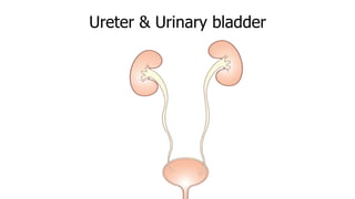
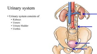
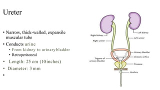
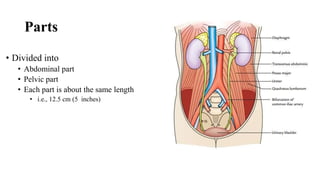
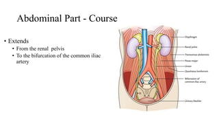
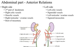
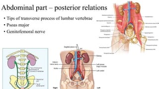
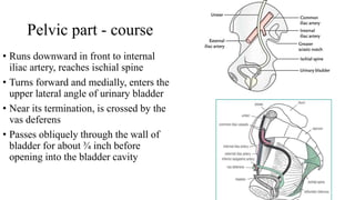
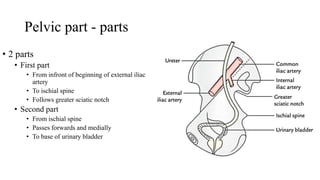
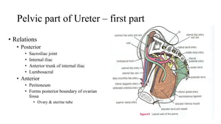
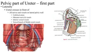
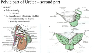
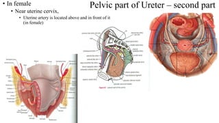
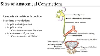
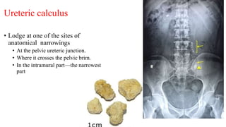
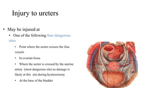
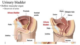
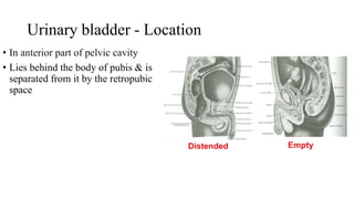
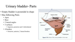
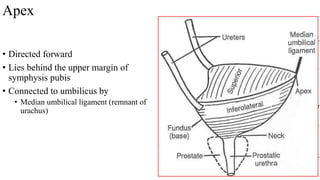
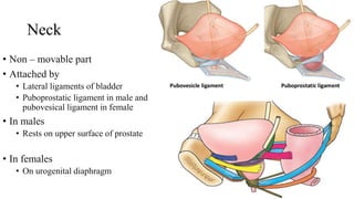
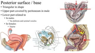
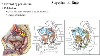
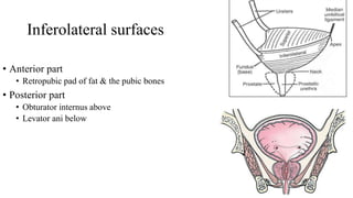
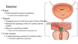
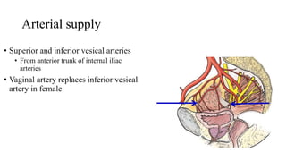
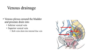
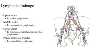
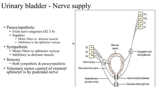
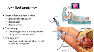
The urinary system consists of the kidneys, ureters, urinary bladder, and urethra. The ureters are thin muscular tubes that conduct urine from the kidneys to the urinary bladder. Each ureter is around 25 cm long and divided into abdominal and pelvic parts. The urinary bladder is a hollow muscular organ that acts as a reservoir for urine in the pelvis. It has a capacity of 200-300 ml in adults and is supplied by superior and inferior vesical arteries. The document provides detailed descriptions of the anatomy and relations of the ureters and urinary bladder.
1. The innervation of the wall of the abdominal cavity
a) the anterior branches of thoracic nerves
b) the lumbar plexus
2. The lumbar part of the sympathetic trunk
3. The abdominal part of the vagus (10th) nerve
4. The sacral plexus
5. The sacral part of the sympathetic trunk
6. The sacral part of the parasympathetic parts of autonomic division of CNS
anatomy of duodenum, location or position of duodenum, parts of duodenum, relations of each parts of duodenum, ligaments of treitz, visceral and peritoneal relation of duodenum, blood supply of duodenum, innervation of duodenum, clinical aspects of duodenum, duodenal ulcer, diverticulum, deodinitis, duodenal obstruction
The arterial supply to the upper limb is delivered via five main vessels (proximal to distal):
Subclavian artery
Axillary artery
Brachial artery
Radial artery
Ulnar artery
In this article, we shall look at the anatomy of the arteries of the upper limb – their anatomical course, branches and clinical correlations.
At the end of the presentation ,we should be able to describe the:
Location, shape and relations of the right and left adrenal glands.
Blood supply, lymphatic drainage and nerve supply of right and left adrenal glands
Parts of adrenal glands and function of each part.
Development of adrenal gland and common anomalies.
Anatomy of Blood vessels of abdomen pelvic cavities. Portacaval & Cavacaval A...Eneutron
1. The abdominal aorta
a. the parietal branches
b. the visceral branches
2. The common iliac arteries and veins
3. The external iliac artery and veins
4. The internal iliac artery and veins
5. The inferior vena cava
6. The portal vein
7. The cavacacal Anastomoses
8. The portacaval Anastomoses
9. The Fetal Circulation
Here is the powerpoint on relevent anatomy of multiple differentials for Inguinoscrtal swelling special for surgical diagnosis with very reliable References.
Palestine last event orientationfvgnh .pptxRaedMohamed3
An EFL lesson about the current events in Palestine. It is intended to be for intermediate students who wish to increase their listening skills through a short lesson in power point.
Model Attribute Check Company Auto PropertyCeline George
In Odoo, the multi-company feature allows you to manage multiple companies within a single Odoo database instance. Each company can have its own configurations while still sharing common resources such as products, customers, and suppliers.
Acetabularia Information For Class 9 .docxvaibhavrinwa19
Acetabularia acetabulum is a single-celled green alga that in its vegetative state is morphologically differentiated into a basal rhizoid and an axially elongated stalk, which bears whorls of branching hairs. The single diploid nucleus resides in the rhizoid.
Francesca Gottschalk - How can education support child empowerment.pptxEduSkills OECD
Francesca Gottschalk from the OECD’s Centre for Educational Research and Innovation presents at the Ask an Expert Webinar: How can education support child empowerment?
Macroeconomics- Movie Location
This will be used as part of your Personal Professional Portfolio once graded.
Objective:
Prepare a presentation or a paper using research, basic comparative analysis, data organization and application of economic information. You will make an informed assessment of an economic climate outside of the United States to accomplish an entertainment industry objective.
Honest Reviews of Tim Han LMA Course Program.pptxtimhan337
Personal development courses are widely available today, with each one promising life-changing outcomes. Tim Han’s Life Mastery Achievers (LMA) Course has drawn a lot of interest. In addition to offering my frank assessment of Success Insider’s LMA Course, this piece examines the course’s effects via a variety of Tim Han LMA course reviews and Success Insider comments.
A Strategic Approach: GenAI in EducationPeter Windle
Artificial Intelligence (AI) technologies such as Generative AI, Image Generators and Large Language Models have had a dramatic impact on teaching, learning and assessment over the past 18 months. The most immediate threat AI posed was to Academic Integrity with Higher Education Institutes (HEIs) focusing their efforts on combating the use of GenAI in assessment. Guidelines were developed for staff and students, policies put in place too. Innovative educators have forged paths in the use of Generative AI for teaching, learning and assessments leading to pockets of transformation springing up across HEIs, often with little or no top-down guidance, support or direction.
This Gasta posits a strategic approach to integrating AI into HEIs to prepare staff, students and the curriculum for an evolving world and workplace. We will highlight the advantages of working with these technologies beyond the realm of teaching, learning and assessment by considering prompt engineering skills, industry impact, curriculum changes, and the need for staff upskilling. In contrast, not engaging strategically with Generative AI poses risks, including falling behind peers, missed opportunities and failing to ensure our graduates remain employable. The rapid evolution of AI technologies necessitates a proactive and strategic approach if we are to remain relevant.
Introduction to AI for Nonprofits with Tapp NetworkTechSoup
Dive into the world of AI! Experts Jon Hill and Tareq Monaur will guide you through AI's role in enhancing nonprofit websites and basic marketing strategies, making it easy to understand and apply.
Welcome to TechSoup New Member Orientation and Q&A (May 2024).pdfTechSoup
In this webinar you will learn how your organization can access TechSoup's wide variety of product discount and donation programs. From hardware to software, we'll give you a tour of the tools available to help your nonprofit with productivity, collaboration, financial management, donor tracking, security, and more.
4. Ureter
• Narrow, thick-walled, expansile
muscular tube
• Conducts urine
• From kidney to urinary bladder
• Retroperitoneal
• Length: 25 cm (10inches)
• Diameter: 3 mm
•
5. Formation
• Formation of Renal pelvis
• Is a funnel-shaped structure
• Formed by
• Union of major calyces
• Location
• Partially outside and partly inside the kidney
• Formation of ureter
• Renal pelvis continues as ureter
• At lower pole of kidney
6. Parts
• Divided into
• Abdominal part
• Pelvic part
• Each part is about the same length
• i.e., 12.5 cm (5 inches)
7. Abdominal Part - Course
• Extends
• From the renal pelvis
• To the bifurcation of the common iliac
artery
8. Abdominal part - Anterior Relations
• Left side
• Left colic vessels
• Sigmoidal vessels
• Left testicular / ovarian vessels
• Sigmoid mesocolon
• Right side
• 2nd part of duodenum
• Right colic vessels
• Iliocolic vessels
• Right testicular / ovarian vessels
• Root of mesentery
9. • Tips of transverse process of lumbar vertebrae
• Psoas major
• Genitofemoral nerve
Abdominal part – posterior relations
10. Pelvic part - course
• Runs downward in front to internal
iliac artery, reaches ischial spine
• Turns forward and medially, enters the
upper lateral angle of urinary bladder
• Near its termination, is crossed by the
vas deferens
• Passes obliquely through the wall of
bladder for about ¾ inch before
opening into the bladder cavity
11. Pelvic part - parts
• 2 parts
• First part
• From infront of beginning of external iliac
artery
• To ischial spine
• Follows greater sciatic notch
• Second part
• From ischial spine
• Passes forwards and medially
• To base of urinary bladder
12. Pelvic part of Ureter – first part
• Relations
• Posterior
• Sacroiliac joint
• Internal iliac
• Anterior trunk of internal iliac
• Lumbosacral
• Anterior
• Peritoneum
• Forms posterior boundary of ovarian
fossa
• Ovary & uterine tube
13. Pelvic part of Ureter – first part
• Laterally
• Ureter crosses in front of
• All nerves and vessels on lateral pelvic wall
• Umbilical artery
• Obturator nerve & vessels
• Inferior vesical vessels
• Vesical branch of Middle rectal vessels
14. Pelvic part of Ureter – second part
• In male
• Inferolaterally
• Levator ani
• At lateral aspect of urinary bladder
• Crossed infront by vas deferens
• Below by seminal vesicle
15. Pelvic part of Ureter – second part
• In female
• Near uterine cervix,
• Uterine artery is located above and in front of it
(in female)
16. Sites of Anatomical Constrictions
• Lumen is not uniform throughout
• Has three constrictions
• At pelviureteric junction
• At pelvic brim
• Where it crosses common iliac artery
• At uretero-vesical junction
• Where ureter enters into bladder
17. Ureteric calculus
• Lodge at one of the sites of
anatomical narrowings
• At the pelvic ureteric junction.
• Where it crosses the pelvic brim.
• In the intramural part—the narrowest
part
18. Arterial supply
• Derives its arterial supply from the
branches of all arteries related to it
• Branches from
• Renal artery
• Gonadal artery
• Abdominal artery
• Common iliac artery
• Internal iliac
19. Clinical anatomy
• Arteries supplying ureter form an
anastomosis
• During transplantation
• Surgeons should bear in their mind that
stripping off fascia around ureter
• It will hamper the blood supply of
the ureter and may cause its necrosis
20. Nerve Supply
• Sympathetic supply
• T12–L1 spinal segments
• Through renal, aortic and hypogastric
plexuses
• Parasympathetic supply
• S2–S4 spinal segments
• Through pelvic splanchnic nerves
• Sensory fibres pass through
• Both sympathetic and parasympathetic
nerves
21. Injury to ureters
• May be injured at
• One of the following four dangerous
sites
• Point where the ureter crosses the iliac
vessels
• In ovarian fossa
• Where the ureter is crossed by the uterine
artery (most dangerous site) as damage is
likely at this site during hysterectomy
• At the base of the bladder
23. Urinary bladder - Location
• In anterior part of pelvic cavity
• Lies behind the body of pubis & is
separated from it by the retropubic
space
24. Urinary bladder - Capacity
• Normal capacity in adult
• 200-300 ml
• Desire to micturate
• Beyond 220 cc
• Painful
• Up to 500 cc is tolerated
• Beyond this becomes painful
25. Urinary bladder- Parts
• Empty bladder is pyramidal in shape
• Has following Parts
• Apex
• Base
• 2 lateral angles
• 3 surfaces
• Superior, posterior and 2 inferolateral
• 4 borders
• Posterior, anterior, 2 lateral borders
26. Apex
• Directed forward
• Lies behind the upper margin of
symphysis pubis
• Connected to umbilicus by
• Median umbilical ligament (remnant of
urachus)
27. Neck
• Non – movable part
• Attached by
• Lateral ligaments of bladder
• Puboprostatic ligament in male and
pubovesical ligament in female
• In males
• Rests on upper surface of prostate
• In females
• On urogenital diaphragm
28. Posterior surface / base
• Triangular in shape
• Upper part covered by peritoneum in male
• Lower part related to
• In males
• Vas deferens and seminal vesicles
• In females
• Vagina
29. Superior surface
• Covered by peritoneum
• Related to
• Coils of ileum or sigmoid colon in males
• Uterus in females
30. Inferolateral surfaces
• Anterior part
• Retropubic pad of fat & the pubic bones
• Posterior part
• Obturator internus above
• Levator ani below
31. Interior
• Rugae
• Folds formed by mucous membrane
• Except in the triangular region
• Trigone
• Triangular area over the lower part of base of bladder
• Between the openings of the two ureters and the
urethra
• Mucosa is smooth (no folds)
• Due to its firm attachment to muscular coat
• Uvula vesicae
• A slight projection posterior to urethral orifice
• Formed by middle lobe of prostate
32. Arterial supply
• Superior and inferior vesical arteries
• From anterior trunk of internal iliac
arteries
• Vaginal artery replaces inferior vesical
artery in female
33. Venous drainage
• Venous plexus around the bladder
and prostate drain into
• Inferior vesical vein
• Superior vesical vein
• Both veins drain into internal iliac vein
34. Lymphatic drainage
• Upper ureter
• To lumbar lymph nodes
• Middle ureter
• To common iliac lymph nodes
• Lower ureter
• To common , external and internal iliac
lymph nodes
• Pelvic ureter and bladder
• To internal iliac lymph nodes
35. Urinary bladder - Nerve supply
• Parasympathetic
• From nervi erigentes (S2 3 4)
• Supplies
• Motor fibers to detrusor muscle
• Inhibitory to the sphincter vesicae
• Sympathetic
• Motor fibers to sphincter vesicae
• Inhibitory to detrusor muscle
• Sensory
• Both sympathetic & parasympathetic
• Voluntary motor control of external
sphincter is by pudendal nerve
Anatomy
36. Applied anatomy
• Obstruction in urine outflow
• Hypertrophy of bladder
• Hydroureter
• Hydronephrosis
• Cystoscopy
• Examining interior of urinary bladder
• By a hollow tube with a lens
• Pyelography
• Examining ureter and renal pelvis and
calyces by radiograph