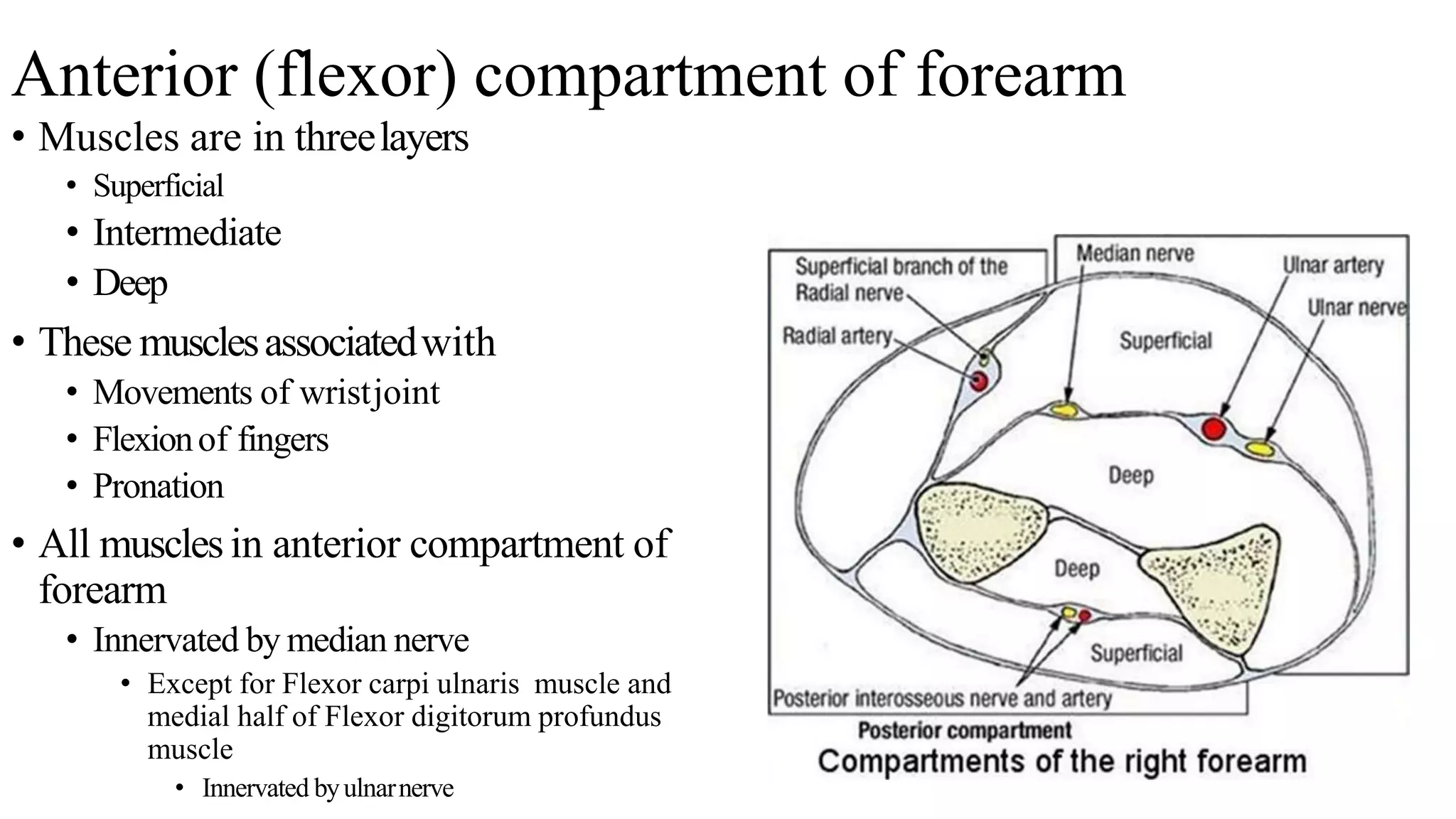
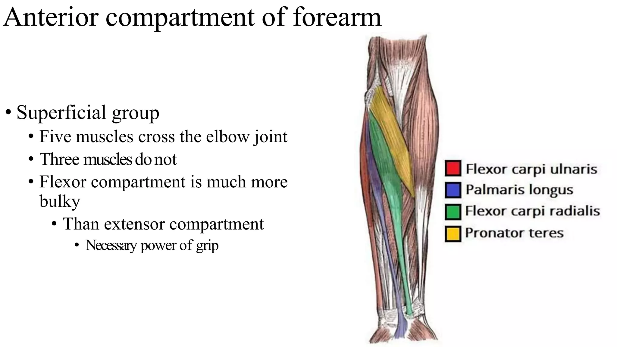
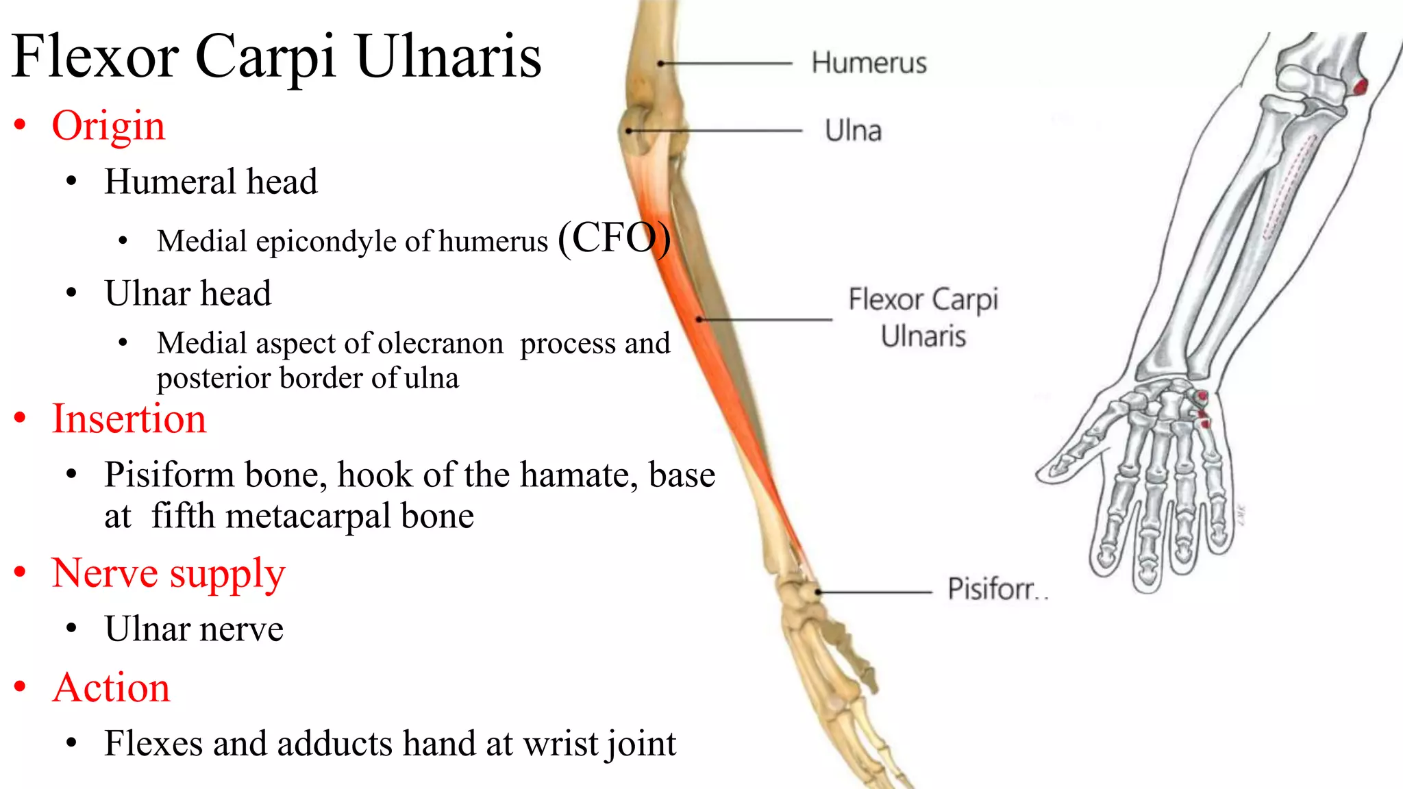
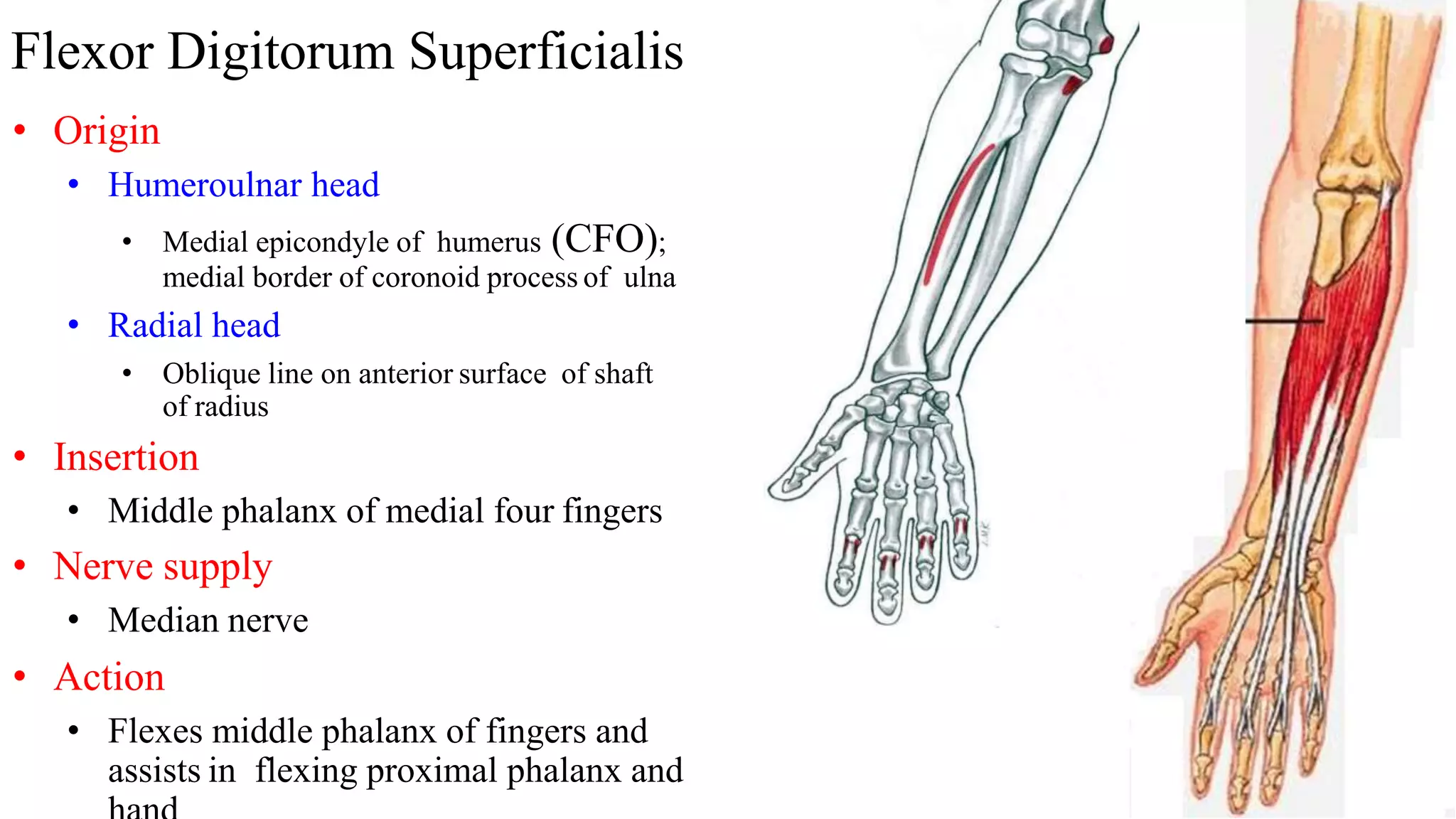
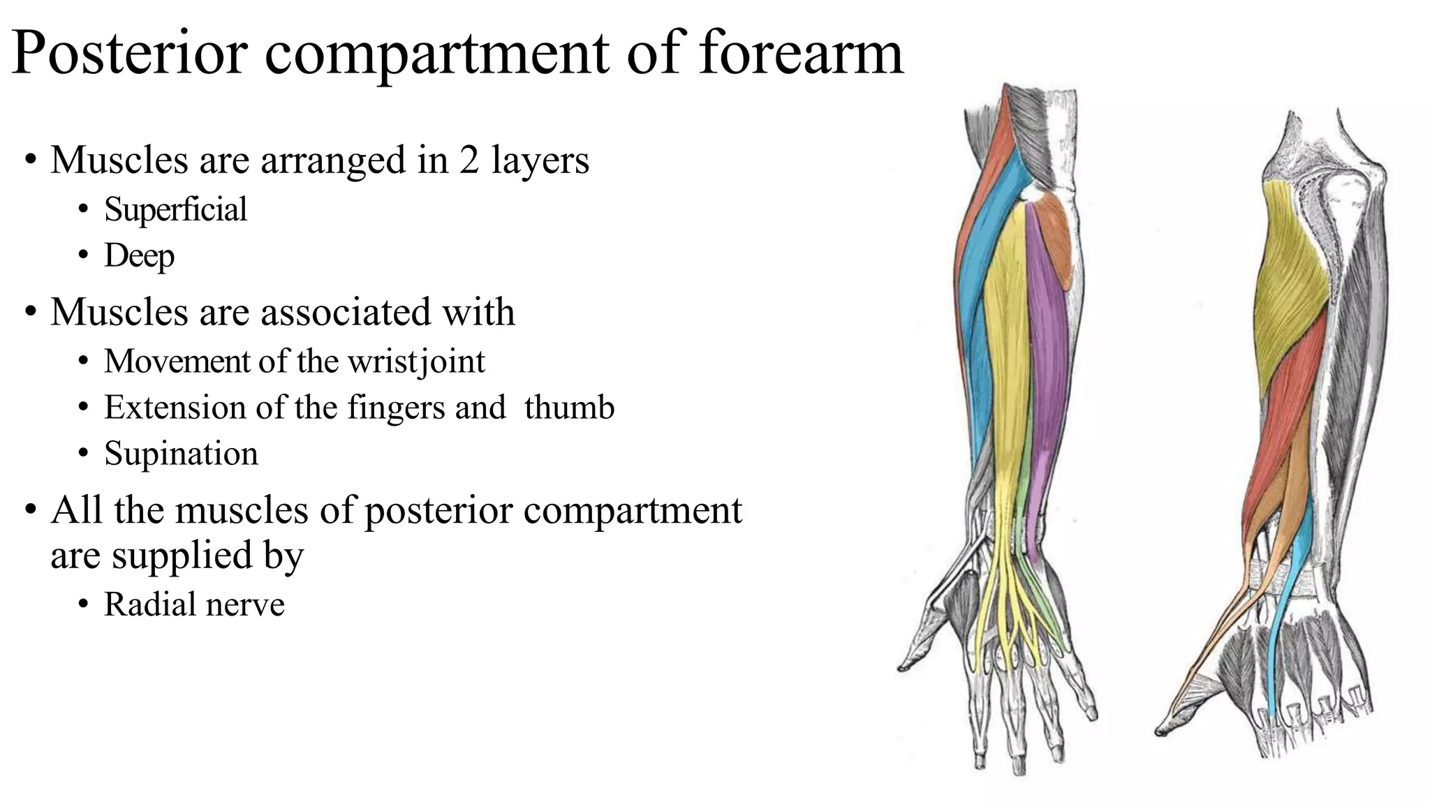
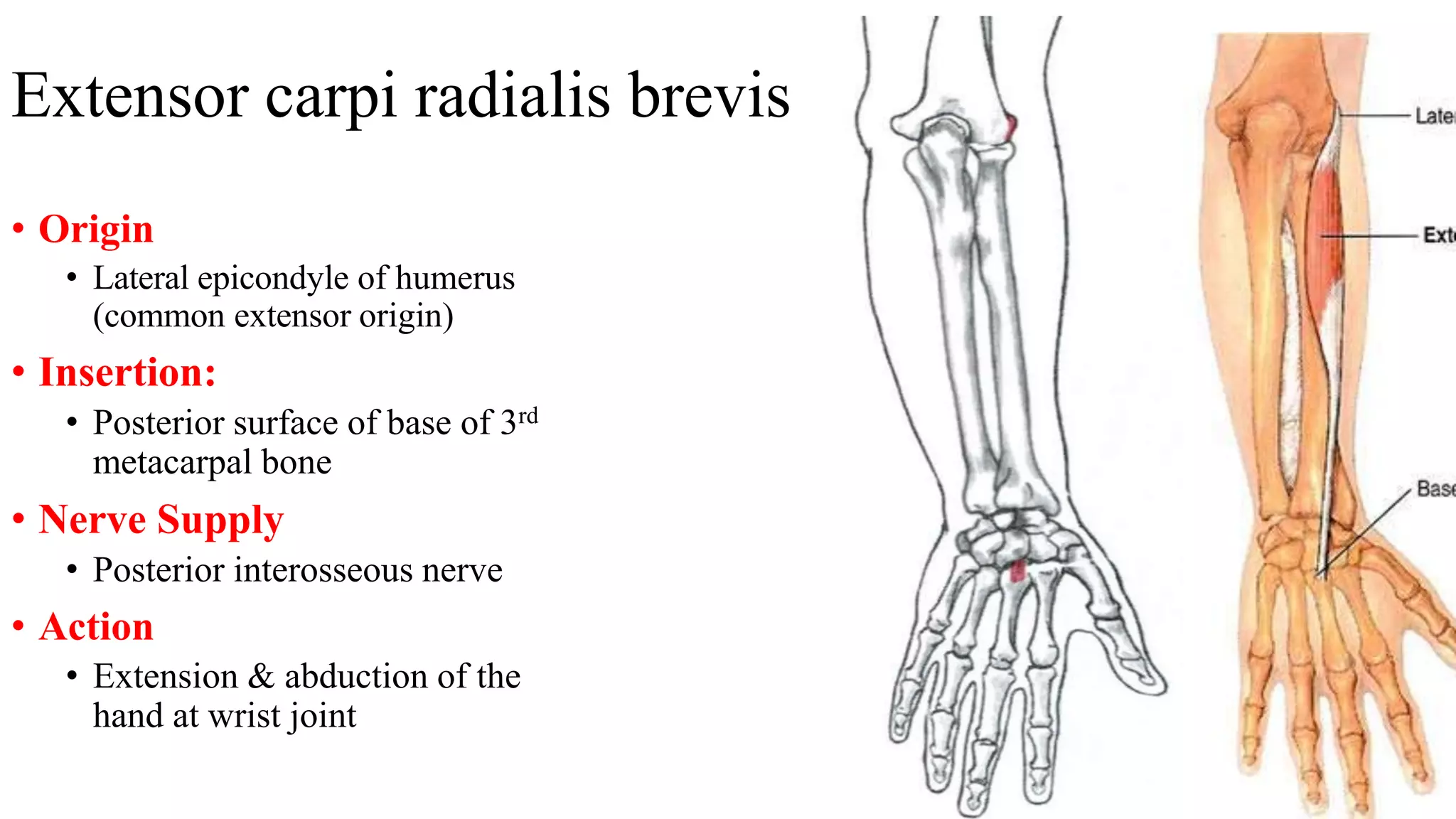
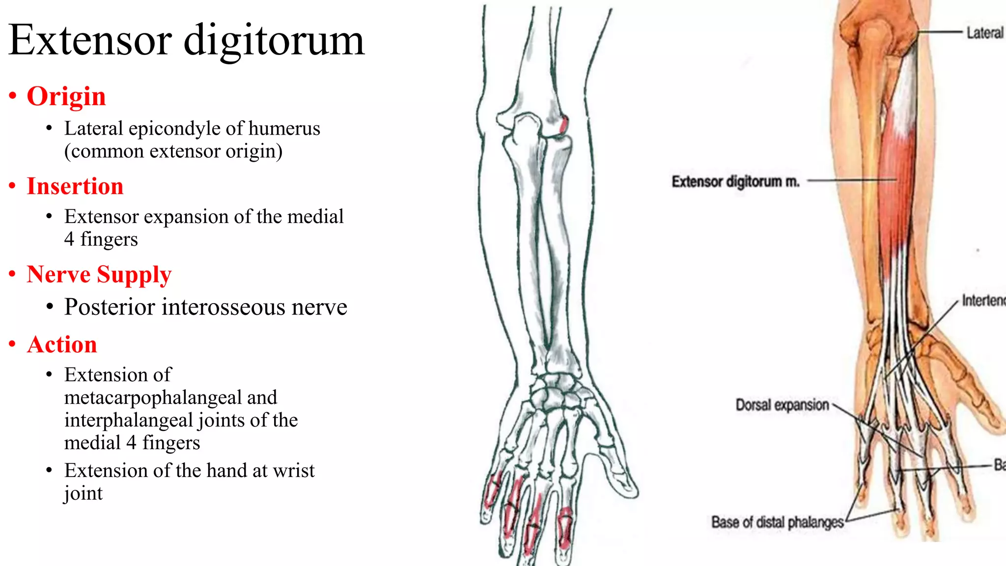
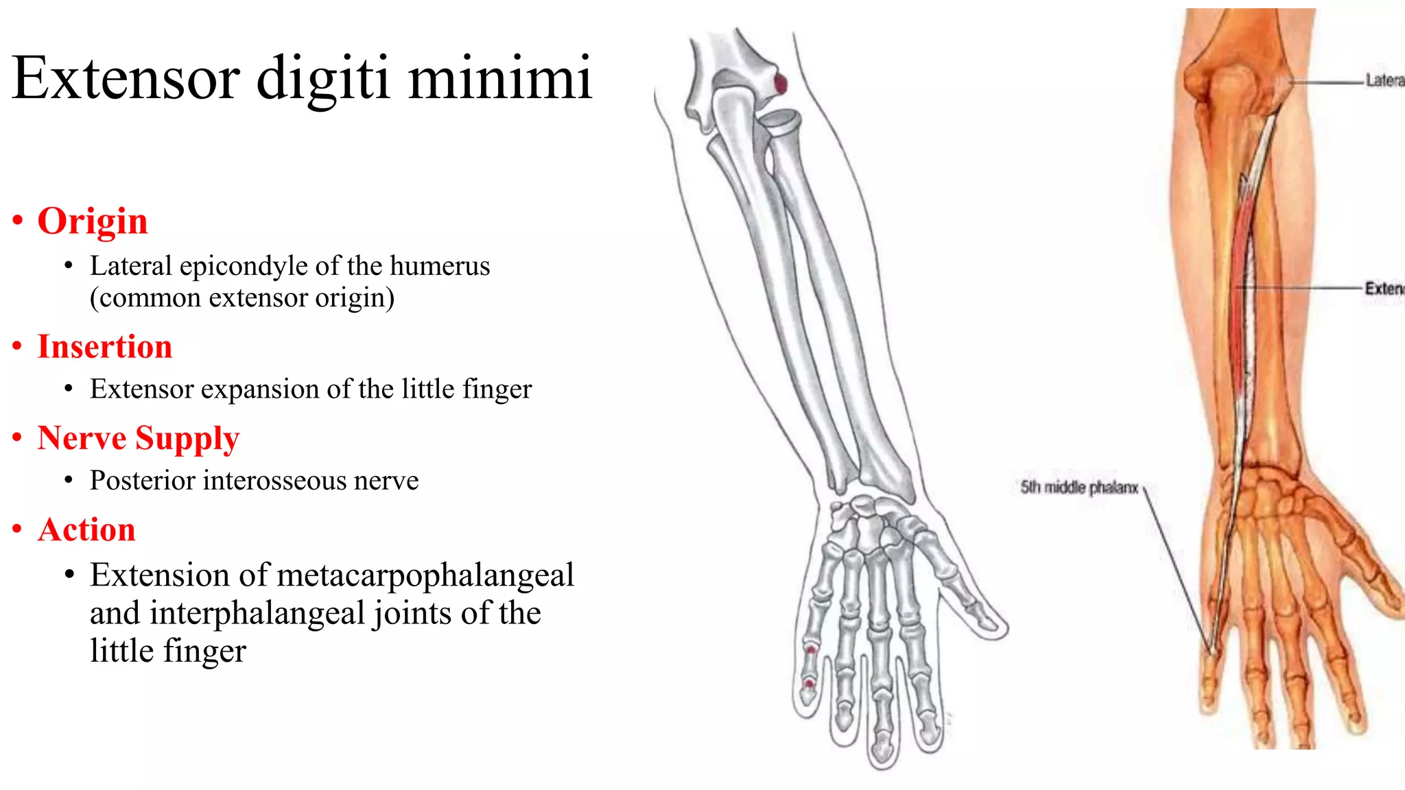
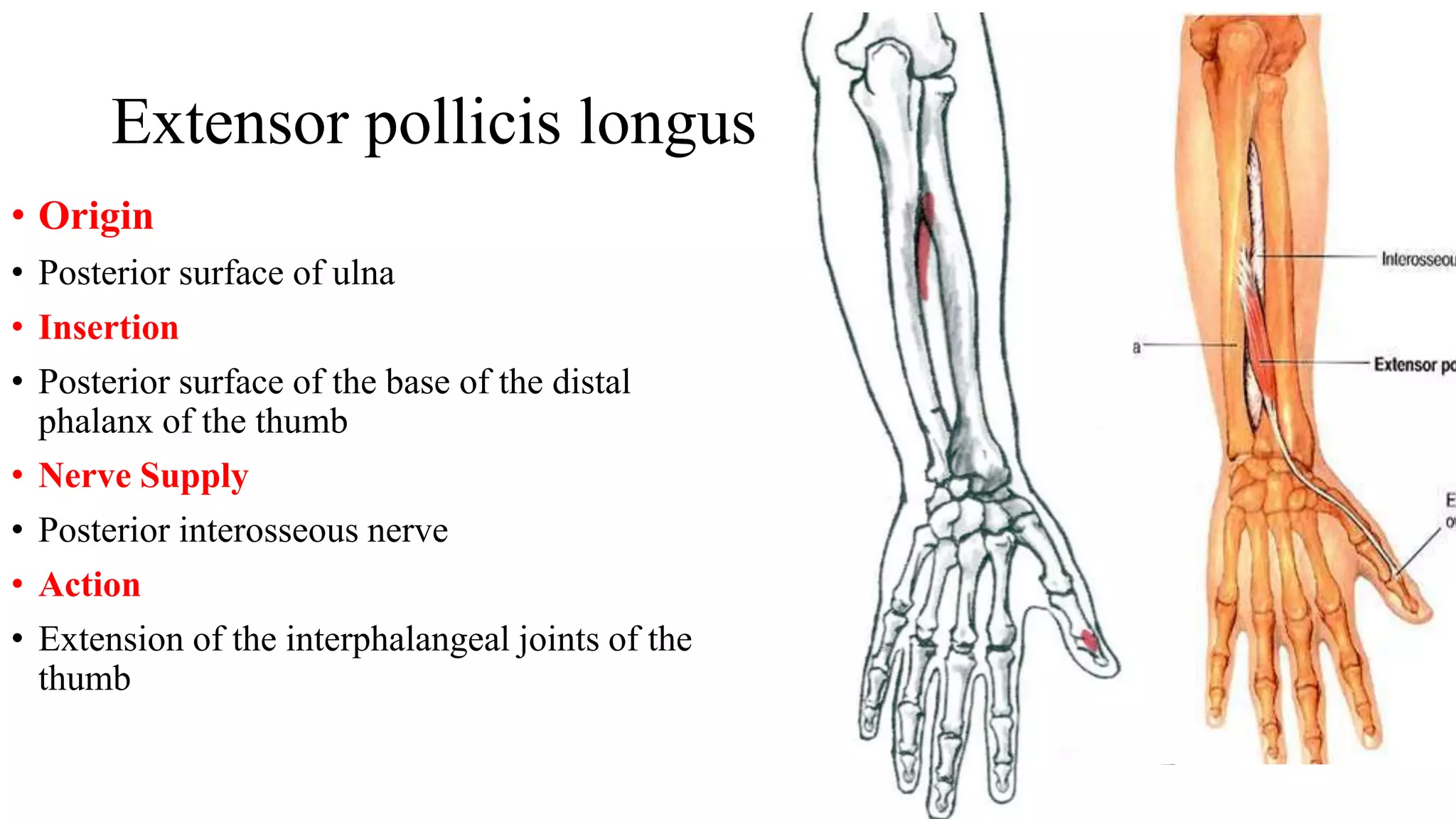
Forearm muscles
Compartments
• Forearm is divided into
• Anterior and posterior compartments by
• Lateral intermuscularseptum
• Aninterosseousmembrane
• Attachment of deepfasciaalongtheposterior border of the
ulna
Anterior (flexor) compartment of forearm
• Muscles are in threelayers
• Superficial
• Intermediate
• Deep
• These musclesassociatedwith
• Movements of wristjoint
• Flexionof fingers
• Pronation
• All muscles in anterior compartment of
forearm
• Innervated by median nerve
• Except for Flexor carpi ulnaris muscle and
medial half of Flexor digitorum profundus
muscle
• Innervated byulnarnerve
Anterior compartment of forearm
• Superficial group
• Five muscles cross the elbow joint
• Three musclesdonot
• Flexor compartment is much more
bulky
• Than extensor compartment
• Necessary power of grip
Pronator Teres
• Origin
• Humeral head
• Medial epicondyle of humerus (CFO)
• Ulnar head
• Medial border of coronoid process of ulna
• Action
• Middle of lateral aspect of shaft of radius
• Nerve supply
• Median nerve
• Action
• Pronation and flexion of elbow
Flexor carpi radialis
• Origin
• Medial epicondyle of humerus (CFO)
• Insertion
• Bases of second and third metacarpal bones
• Nerve supply
• Median nerve
• Action
• Flexes elbow & wrist
• Abducts hand at wrist joint
Palmaris longus
• Origin
• Medial epicondyle of humerus (CFO)
• Insertion
• Flexor retinaculum and palmar aponeurosis
• Nerve supply
• Median nerve
• Action
• Flexes elbow & wrist
Flexor Carpi Ulnaris
• Origin
• Humeral head
• Medial epicondyle of humerus (CFO)
• Ulnar head
• Medial aspect of olecranon process and
posterior border of ulna
• Insertion
• Pisiform bone, hook of the hamate, base
at fifth metacarpal bone
• Nerve supply
• Ulnar nerve
• Action
• Flexes and adducts hand at wrist joint
Flexor Digitorum Superficialis
• Origin
• Humeroulnar head
• Medial epicondyle of humerus (CFO);
medial border of coronoid process of ulna
• Radial head
• Oblique line on anterior surface of shaft
of radius
• Insertion
• Middle phalanx of medial four fingers
• Nerve supply
• Median nerve
• Action
• Flexes middle phalanx of fingers and
assists in flexing proximal phalanx and
Deep group
• 3 muscles
• Flexor digitorum profundus
• Flexor pollicis longus
• Pronator quadratus
Flexor digitorum profundus
• Origin
• Anteromedial surface of shaft of
ulna
• Insertion
• Distal phalanges of medial four
fingers
• Action
• Flexion of middle and proximal
phalanges and wrist
• Nerve supply
• Ulnar (medial half)
Flexor pollicis longus
• Origin
• Anterior surface of shaft of
radius
• Insertion
• Distal phalanx of thumb
• Action
• Flexes distal phalanx of
thumb
• Nerve supply
• Anterior interosseous branch
of median nerve
Pronator quadratus
• Origin
• Lower ¼ of anterior surface of
shaft of ulna
• Insertion
• Lower ¼ of anterior surface of
shaft of radius
• Action
• Pronates radioulnar joint
• Nerve Supply
• Anterior interosseous
• Branch of median nerve
Space of Parona
• Space in front of pronator quadratus
• Deep to long flexor tendons of fingers and their synovial
sheaths
• Limited proximally by
• Oblique origin of flexor digitorum superficialis
• Space involved in proximal extensions of synovial
sheath infections
• Can be drained through radial and ulnar incisions to the
side of the flexor tendons
Posterior compartment of forearm
• Muscles are arranged in 2 layers
• Superficial
• Deep
• Muscles are associated with
• Movement of the wristjoint
• Extension of the fingers and thumb
• Supination
• All the muscles of posterior compartment
are supplied by
• Radial nerve
Arrangement of posterior compartment muscles
• Upper part
• Anconeus (superficial)
• Supinator (deep)
• Lateral part of humerus
• Pass along the radial side
• Brachioradialis
• Extensors carpi radialis longus
• Extensors carpi radialis brevis
• Passing along posterior surface of the
forearm
• Extensor digitorum
• Extensor digiti minimi and
• Extensor carpi ulnaris
• At lower end of forearm
• These two groups are separated by
three muscles
• They emerge from deeply in between
them and go to the thumb
• Abductor pollicis longus
• Extensors pollicis longus and
• Extensors pollicis longus brevis
• One muscle for the forefinger runs
deeply to reach the back of the hand
• Extensor indicis
Superficial muscles of the back of forearm
• There are 7 muscles
• Common extensor origin
• Four of the superficialmuscles
• Arisefromthetipoflateralepicondyleof thehumerus
• Extensor carpi radialis brevis
• Extensor digitorum
• Extensor digiti minimi, and
• Extensor carpi ulnaris
• All seven muscles cross elbow joint
Superficial muscles of back of forearm
• Categorized into two groups
• Lateral and posterior
• Each group consists of three muscles
• Lateral group
• Brachioradialis
• Extensor carpi radialis longus
• Extensor carpi radialis brevis
• Posterior group
• Extensor digitorum
• Extensor digiti minimi
• Extensor carpi ulnaris
• In the upper end
• Anconeus
Brachioradialis
• Origin
• Upper 2/3rd of lateral supracondylar ridge of
humerus
• Insertion
• Into base of styloid process of radius
• Nerve Supply
• Radial nerve
• Action
• Flexion of the forearm (specially in midprone
position)
Extensor carpi radialis longus
• Origin
• From the lower 1/3rd of the lateral
supracondylar ridge of the humerus
• Insertion
• Into posterior surface of base of 2nd
metacarpal bone
• Nerve Supply
• Radial nerve
• Action
• Extension & Abduction of wrist
joint
• Extension of elbow joint
Extensor carpi radialis brevis
• Origin
• Lateral epicondyle of humerus
(common extensor origin)
• Insertion:
• Posterior surface of base of 3rd
metacarpal bone
• Nerve Supply
• Posterior interosseous nerve
• Action
• Extension & abduction of the
hand at wrist joint
Extensor digitorum
• Origin
• Lateral epicondyle of humerus
(common extensor origin)
• Insertion
• Extensor expansion of the medial
4 fingers
• Nerve Supply
• Posterior interosseous nerve
• Action
• Extension of
metacarpophalangeal and
interphalangeal joints of the
medial 4 fingers
• Extension of the hand at wrist
joint
Extensor digiti minimi
• Origin
• Lateral epicondyle of the humerus
(common extensor origin)
• Insertion
• Extensor expansion of the little finger
• Nerve Supply
• Posterior interosseous nerve
• Action
• Extension of metacarpophalangeal
and interphalangeal joints of the
little finger
Extensor carpi ulnaris
• Origin
• Lateral epicondyle of the humerus
(common extensor origin)
• Insertion
• Posterior surface of the base of the 5th
metacarpal bone
• Nerve Supply
• Posterior interosseous nerve
• Action
• Extension & Adduction of the
hand at wrist joint
Anconeus
• Origin
• Posterior aspect of the lateral
epicondyle of the humerus
• Insertion
• Lateral surface of olecranon
process of ulna
• Nerve Supply
• Posterior interosseous nerve
• Action
• Helps the triceps in extension of
the elbow joint
Deep Layer
• There are five deep muscles
• Three deep extensorsof the forearm
• Function on thumb
• Abductor pollicis longus(APL)
• Extensor pollicis brevis (EPB)
• Extensor pollicis longus (EPL)
• To acquire insertion on the three bones of
thumb ‘crop out’
• From furrow in lateralelementof forearmbetweenlateral
andposterior groupsof superficialextensor
• One muscle
• Functiononindexfinger
• Extensor indicis
• In the upper part
• Supinator
Supinator
• Origin
• Lateral epicondyle of the humerus
• Annular ligament of the superior radio-ulnar joint
• Supinator fossa and crest of the ulna
• Insertion
• Upper 1/3rd of the lateral surface of shaft of radius
• Nerve Supply
• Posterior interosseous nerve
• Action
• Supination of the forearm
• At radio-ulnar joint
Abductor pollicis longus
• Origin
• Middle of the posterior surface of radius and ulna
• Interosseus membrane
• Insertion
• Posterior surface of the base of the 1st metacarpal
bone
• Nerve Supply
• Posterior interosseous nerve
• Action
• Abduction and extension of the thumb at
the carpo-metacarpal joint
Extensor pollicis brevis
• Origin
• Posterior surface of the shaft of radius
• Insertion
• Posterior surface of the base of the proximal
phalanx of the thumb
• Nerve Supply
• Posterior interosseous nerve
• Action
• Extension of the metacarpophalangeal joint of
the thumb
Extensor pollicis longus
• Origin
• Posterior surface of ulna
• Insertion
• Posterior surface of the base of the distal
phalanx of the thumb
• Nerve Supply
• Posterior interosseous nerve
• Action
• Extension of the interphalangeal joints of the
thumb
Extensor indicis
• Origin
• Posterior surface of ulna (below extensor
pollicis longus)
• Insertion
• Extensor expansion of the index finger.
• Nerve Supply
• Posterior interosseous nerve
• Action
• Extension of the interphalangeal joints of
the thumb
Wrist Drop
• Sign of radial nerve injury
• There are two common characteristic sites
of damage
• Axilla
• Injured viahumeral dislocationsor fractures of the
proximalhumerus
• Radial groove of the humerus
• Injured via a humeral shaft fracture
• Radial nerve innervates
• All muscles in extensor compartment of
forearm
• In radial nerve lesion
• These muscles are paralysed
• Muscles that flex the wrist
• Innervated by median nerve
• Flexor muscles produces unopposed flexion at
the wrist joint – wrist drop

Forearm muscles.pptx

  • 1.
  • 2.
    Compartments • Forearm isdivided into • Anterior and posterior compartments by • Lateral intermuscularseptum • Aninterosseousmembrane • Attachment of deepfasciaalongtheposterior border of the ulna
  • 3.
    Anterior (flexor) compartmentof forearm • Muscles are in threelayers • Superficial • Intermediate • Deep • These musclesassociatedwith • Movements of wristjoint • Flexionof fingers • Pronation • All muscles in anterior compartment of forearm • Innervated by median nerve • Except for Flexor carpi ulnaris muscle and medial half of Flexor digitorum profundus muscle • Innervated byulnarnerve
  • 4.
    Anterior compartment offorearm • Superficial group • Five muscles cross the elbow joint • Three musclesdonot • Flexor compartment is much more bulky • Than extensor compartment • Necessary power of grip
  • 5.
    Pronator Teres • Origin •Humeral head • Medial epicondyle of humerus (CFO) • Ulnar head • Medial border of coronoid process of ulna • Action • Middle of lateral aspect of shaft of radius • Nerve supply • Median nerve • Action • Pronation and flexion of elbow
  • 6.
    Flexor carpi radialis •Origin • Medial epicondyle of humerus (CFO) • Insertion • Bases of second and third metacarpal bones • Nerve supply • Median nerve • Action • Flexes elbow & wrist • Abducts hand at wrist joint
  • 7.
    Palmaris longus • Origin •Medial epicondyle of humerus (CFO) • Insertion • Flexor retinaculum and palmar aponeurosis • Nerve supply • Median nerve • Action • Flexes elbow & wrist
  • 8.
    Flexor Carpi Ulnaris •Origin • Humeral head • Medial epicondyle of humerus (CFO) • Ulnar head • Medial aspect of olecranon process and posterior border of ulna • Insertion • Pisiform bone, hook of the hamate, base at fifth metacarpal bone • Nerve supply • Ulnar nerve • Action • Flexes and adducts hand at wrist joint
  • 9.
    Flexor Digitorum Superficialis •Origin • Humeroulnar head • Medial epicondyle of humerus (CFO); medial border of coronoid process of ulna • Radial head • Oblique line on anterior surface of shaft of radius • Insertion • Middle phalanx of medial four fingers • Nerve supply • Median nerve • Action • Flexes middle phalanx of fingers and assists in flexing proximal phalanx and
  • 10.
    Deep group • 3muscles • Flexor digitorum profundus • Flexor pollicis longus • Pronator quadratus
  • 11.
    Flexor digitorum profundus •Origin • Anteromedial surface of shaft of ulna • Insertion • Distal phalanges of medial four fingers • Action • Flexion of middle and proximal phalanges and wrist • Nerve supply • Ulnar (medial half)
  • 12.
    Flexor pollicis longus •Origin • Anterior surface of shaft of radius • Insertion • Distal phalanx of thumb • Action • Flexes distal phalanx of thumb • Nerve supply • Anterior interosseous branch of median nerve
  • 13.
    Pronator quadratus • Origin •Lower ¼ of anterior surface of shaft of ulna • Insertion • Lower ¼ of anterior surface of shaft of radius • Action • Pronates radioulnar joint • Nerve Supply • Anterior interosseous • Branch of median nerve
  • 14.
    Space of Parona •Space in front of pronator quadratus • Deep to long flexor tendons of fingers and their synovial sheaths • Limited proximally by • Oblique origin of flexor digitorum superficialis • Space involved in proximal extensions of synovial sheath infections • Can be drained through radial and ulnar incisions to the side of the flexor tendons
  • 15.
    Posterior compartment offorearm • Muscles are arranged in 2 layers • Superficial • Deep • Muscles are associated with • Movement of the wristjoint • Extension of the fingers and thumb • Supination • All the muscles of posterior compartment are supplied by • Radial nerve
  • 16.
    Arrangement of posteriorcompartment muscles • Upper part • Anconeus (superficial) • Supinator (deep) • Lateral part of humerus • Pass along the radial side • Brachioradialis • Extensors carpi radialis longus • Extensors carpi radialis brevis • Passing along posterior surface of the forearm • Extensor digitorum • Extensor digiti minimi and • Extensor carpi ulnaris • At lower end of forearm • These two groups are separated by three muscles • They emerge from deeply in between them and go to the thumb • Abductor pollicis longus • Extensors pollicis longus and • Extensors pollicis longus brevis • One muscle for the forefinger runs deeply to reach the back of the hand • Extensor indicis
  • 17.
    Superficial muscles ofthe back of forearm • There are 7 muscles • Common extensor origin • Four of the superficialmuscles • Arisefromthetipoflateralepicondyleof thehumerus • Extensor carpi radialis brevis • Extensor digitorum • Extensor digiti minimi, and • Extensor carpi ulnaris • All seven muscles cross elbow joint
  • 18.
    Superficial muscles ofback of forearm • Categorized into two groups • Lateral and posterior • Each group consists of three muscles • Lateral group • Brachioradialis • Extensor carpi radialis longus • Extensor carpi radialis brevis • Posterior group • Extensor digitorum • Extensor digiti minimi • Extensor carpi ulnaris • In the upper end • Anconeus
  • 19.
    Brachioradialis • Origin • Upper2/3rd of lateral supracondylar ridge of humerus • Insertion • Into base of styloid process of radius • Nerve Supply • Radial nerve • Action • Flexion of the forearm (specially in midprone position)
  • 20.
    Extensor carpi radialislongus • Origin • From the lower 1/3rd of the lateral supracondylar ridge of the humerus • Insertion • Into posterior surface of base of 2nd metacarpal bone • Nerve Supply • Radial nerve • Action • Extension & Abduction of wrist joint • Extension of elbow joint
  • 21.
    Extensor carpi radialisbrevis • Origin • Lateral epicondyle of humerus (common extensor origin) • Insertion: • Posterior surface of base of 3rd metacarpal bone • Nerve Supply • Posterior interosseous nerve • Action • Extension & abduction of the hand at wrist joint
  • 22.
    Extensor digitorum • Origin •Lateral epicondyle of humerus (common extensor origin) • Insertion • Extensor expansion of the medial 4 fingers • Nerve Supply • Posterior interosseous nerve • Action • Extension of metacarpophalangeal and interphalangeal joints of the medial 4 fingers • Extension of the hand at wrist joint
  • 23.
    Extensor digiti minimi •Origin • Lateral epicondyle of the humerus (common extensor origin) • Insertion • Extensor expansion of the little finger • Nerve Supply • Posterior interosseous nerve • Action • Extension of metacarpophalangeal and interphalangeal joints of the little finger
  • 24.
    Extensor carpi ulnaris •Origin • Lateral epicondyle of the humerus (common extensor origin) • Insertion • Posterior surface of the base of the 5th metacarpal bone • Nerve Supply • Posterior interosseous nerve • Action • Extension & Adduction of the hand at wrist joint
  • 25.
    Anconeus • Origin • Posterioraspect of the lateral epicondyle of the humerus • Insertion • Lateral surface of olecranon process of ulna • Nerve Supply • Posterior interosseous nerve • Action • Helps the triceps in extension of the elbow joint
  • 26.
    Deep Layer • Thereare five deep muscles • Three deep extensorsof the forearm • Function on thumb • Abductor pollicis longus(APL) • Extensor pollicis brevis (EPB) • Extensor pollicis longus (EPL) • To acquire insertion on the three bones of thumb ‘crop out’ • From furrow in lateralelementof forearmbetweenlateral andposterior groupsof superficialextensor • One muscle • Functiononindexfinger • Extensor indicis • In the upper part • Supinator
  • 27.
    Supinator • Origin • Lateralepicondyle of the humerus • Annular ligament of the superior radio-ulnar joint • Supinator fossa and crest of the ulna • Insertion • Upper 1/3rd of the lateral surface of shaft of radius • Nerve Supply • Posterior interosseous nerve • Action • Supination of the forearm • At radio-ulnar joint
  • 28.
    Abductor pollicis longus •Origin • Middle of the posterior surface of radius and ulna • Interosseus membrane • Insertion • Posterior surface of the base of the 1st metacarpal bone • Nerve Supply • Posterior interosseous nerve • Action • Abduction and extension of the thumb at the carpo-metacarpal joint
  • 29.
    Extensor pollicis brevis •Origin • Posterior surface of the shaft of radius • Insertion • Posterior surface of the base of the proximal phalanx of the thumb • Nerve Supply • Posterior interosseous nerve • Action • Extension of the metacarpophalangeal joint of the thumb
  • 30.
    Extensor pollicis longus •Origin • Posterior surface of ulna • Insertion • Posterior surface of the base of the distal phalanx of the thumb • Nerve Supply • Posterior interosseous nerve • Action • Extension of the interphalangeal joints of the thumb
  • 31.
    Extensor indicis • Origin •Posterior surface of ulna (below extensor pollicis longus) • Insertion • Extensor expansion of the index finger. • Nerve Supply • Posterior interosseous nerve • Action • Extension of the interphalangeal joints of the thumb
  • 32.
    Wrist Drop • Signof radial nerve injury • There are two common characteristic sites of damage • Axilla • Injured viahumeral dislocationsor fractures of the proximalhumerus • Radial groove of the humerus • Injured via a humeral shaft fracture • Radial nerve innervates • All muscles in extensor compartment of forearm • In radial nerve lesion • These muscles are paralysed • Muscles that flex the wrist • Innervated by median nerve • Flexor muscles produces unopposed flexion at the wrist joint – wrist drop