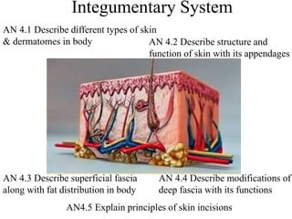
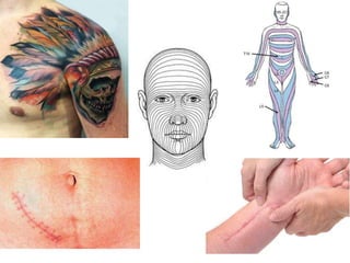
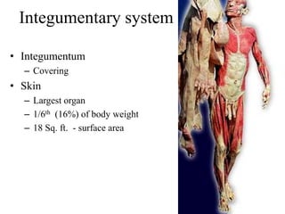
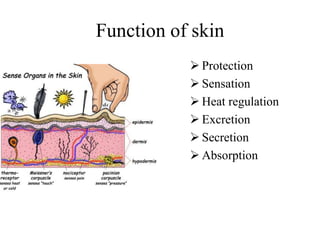
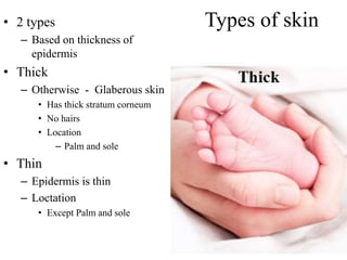
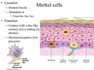
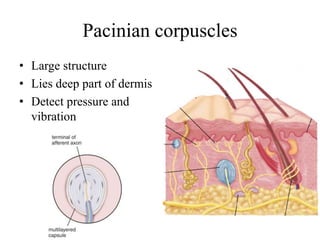
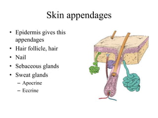
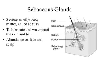
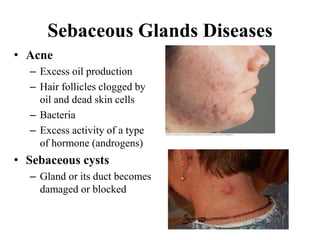
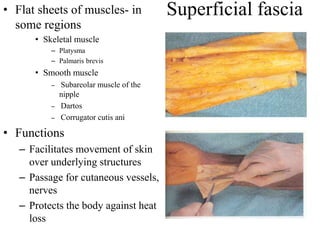
Integumentary System
AN 4.1 Describe different types of skin
& dermatomes in body AN 4.2 Describe structure and
function of skin with its appendages
AN 4.3 Describe superficial fascia
along with fat distribution in body
AN 4.4 Describe modifications of
deep fascia with its functions
AN4.5 Explain principles of skin incisions
Integumentary system
• Integumentum
– Covering
• Skin
– Largest organ
– 1/6th (16%) of body weight
– 18 Sq. ft. - surface area
Function of skin
 Protection
 Sensation
 Heat regulation
 Excretion
 Secretion
 Absorption
Types of skin
• 2 types
– Based on thickness of
epidermis
• Thick
– Otherwise - Glaberous skin
• Has thick stratum corneum
• No hairs
• Location
– Palm and sole
• Thin
– Epidermis is thin
– Loctation
• Except Palm and sole
Structure
• Formed by 2 layers
• Epidermis
• Dermis
Epidermis
• Superficial layer
• Avascular
• Stratified squamous epithelium
• Gives rise to
– Hair, nail sweat and sebaceous
glands
Cells of epidermis (4)
• 2 types of cells
• Keratinocytes (85-90%)
• Non-keratinocytes
– Melanocytes (8%)
– Dendritic cell of Langerhans
– Cells of Merkel
Keratinocytes
• 90 % of epidermis cells
• Environmental barrier
– via keratinization
• Regenerative (25-45 days)
• Protective
– Pathogens / Fungi /
parasites / Viruses
– Heat / UV radiation
– Water loss
Melanocyte
Merkel cells
• Location
– Stratum basale
– Abundant in
• Fingertips, lips, face
• Function
– Contact with a disc like
sensory nerve ending (in
dermis)
– Mechanoreceptors (low
pressure)
Dendritic cells of Langerhans
• Specialized Monocytes
• Location
– Stratum spinosum
• Origin
– Mesoderm (bone marrow)
• Function
– Antigen presenting cells
– Phagocytic
• Easily damaged by
– UV light
Layers of epidermis
Stratum basale
• Deepest layer of epidermis
• Consists of
– Single layer of cuboidal
cells
• These divide and give
keratinocytes (cells of
skin)
• So this layer is known as
– Stratum germinativum
Stratum spinosum
• Several layers of cells
• Polygonal cells
• Cells have the capacity
of mitosis
Stratum granulosum
• Consists of
– 1 – 3 cell thick layer of
keratinocytes
• Contains
– Granules of keratohyalin
protein
Stratum lucidum
(Clear)
• Present only in thick skin
Stratum corneum
• Superficial layer
• Cells are nonnucleated,
flat, scale like cells
• Cells are filled with keratin
Dermis
 Lies deep to Epidermis
 Contains:
 Blood vessels
 Sensory nerve endings
 Sudoriferous glands -
sweat
 Sebaceous glands - oil
 Hair follicles
 Arrector pili muscles
 Two layers
 Reticular layer(deeper
layer)
 Papillary layer (superfial)
Papillary layer
• Superficial layer of dermis
• Composed of loose areolar
connective tissue
• Has fingerlike projections
called papillae
• Provide the dermis a
"bumpy" surface that
interdigitates with the
epidermis
– Strengthening the connection
between the two layers of
skin
– Has thin collagen fibres
• Inner, thicker layer
• Composed of
– Dense irregular connective tissue
– Collagen / Reticular / Elastic fibres
– Langer lines
– Orientation of collagen fibres
– Surgery incisions made parallel
Reticular layer
Langer's lines /
Tension lines /
Cleavage lines
• Correspond to the
natural orientation
of collagen fibers in
the dermis
Stretch marks in skin
• Damage to the collagen
fibers in dermis due to
over stretching as in
pregnancy or abdominal
enlargement
– Striae gravidarum
Flexure lines
• Found in the vicinity of
synovial joints
• Here the skin is attached
strongly to underlying deep
fascia
• Prominent on the flexor
surfaces of palms, soles and
digits
• Skin lines don't necessarily
coincide with the underlying
joint line
Wrinkles
• If fibres loose their
tense wrinkles formed
Meissners corpuscles
• Rapidly adapting
Mechanoreceptor
• Lie in dermal papilla
• To detect light touch
Pacinian corpuscles
• Large structure
• Lies deep part of dermis
• Detect pressure and
vibration
Free nerve ending
• Lies in stratum granulosum
• Detect hot, cold
Dermatome
• Area of skin supplied by
a single nerve root
Skin appendages
• Epidermis gives this
appendages
• Hair follicle, hair
• Nail
• Sebaceous glands
• Sweat glands
– Apocrine
– Eccrine
Hair
• Shaft-portion that
project above skin
surface
• Root-embedded in skin,
penetrates into dermis
or subQ
• Hair Follicle- surrounds
the root
• Bulb-base of hair
follicle
Hair root
• Surrounded by hair
follicle
Sebaceous Glands
• Secrete an oily/waxy
matter, called sebum
• To lubricate and waterproof
the skin and hair
• Abundance on face and
scalp
Sebaceous Glands Diseases
• Acne
– Excess oil production
– Hair follicles clogged by
oil and dead skin cells
– Bacteria
– Excess activity of a type
of hormone (androgens)
• Sebaceous cysts
– Gland or its duct becomes
damaged or blocked
Sweat glands
• Produce sweat
• Helps to cool the body
• Two types
• Eccrine (sudoriferous)
– Opens on the surface of skin
through a duct
– Most numerous (3-4 million)
– Numerous on palms, sole of feet
and forehead
• Apocrine
– Empty into hair follicles
– Lies in armpits and genital areas
Sweat Glands Diseases
• Hyperhidrosis -
Excessive sweating
• Hypohidrosis -
diminished or absent
perspiration
Superficial fascia
• Mixture of adipose and loose
areolar tissues
• Unites the skin to the
underlying structures
• Most distinct
– Lower part of abdomen
– Perineum and limbs
• Very thin
– Eyelids, auricle, scrotum
(devoid of adipose tissue)
• Very dense
– Scalp, palm & sole
• Function
– Facilitate movements of skin
– Bad conductor of heat
Superficial fascia
• Flat sheets of muscles- in
some regions
• Skeletal muscle
– Platysma
– Palmaris brevis
• Smooth muscle
– Subareolar muscle of the
nipple
– Dartos
– Corrugator cutis ani
• Functions
– Facilitates movement of skin
over underlying structures
– Passage for cutaneous vessels,
nerves
– Protects the body against heat
loss
Deep fascia
• Beneath superficial
fascia
• Devoid of fat
• Inelastic & tough
• Either invests or bind
structures
Modifications of deep fascia
• Intermuscular septa
• Retiinaculum
• Epimysium, Perimysium
and endomysium
• Epineurium, perineurium
and endoneurium
• Sheath
Intradermal Injections
• Injection of the substance
into the dermis
• Indications
– Identify skin allergy to any
drugs
• Tuberculin injection
• Allergy sensitivity tests
• Local anesthesia
• BCG vaccine
• Angle- 10-15 degree
• Sites
– Forearm
Subcutaneous injection
• Cyanotic (cyan = blue)
– Who has stopped breathing
• Because the hemoglobin is
depleted of oxygen
• Jaundice (jaund = yellow)
– Build up of bilirubin (yellow
pigment) in the blood
• Erythema (ery = red)
– Engorgement of capillaries in
the dermis
• Skin injury, infection, heat
exposure, inflammation,
allergies, emotional state,
hypertension
• Pallor - paleness
– Emotional state, anemia, low
blood pressure
Skin color as diagnostic clues
for medical conditions
Integumentary  system -mbbs.pptx

Integumentary system -mbbs.pptx

  • 1.
    Integumentary System AN 4.1Describe different types of skin & dermatomes in body AN 4.2 Describe structure and function of skin with its appendages AN 4.3 Describe superficial fascia along with fat distribution in body AN 4.4 Describe modifications of deep fascia with its functions AN4.5 Explain principles of skin incisions
  • 3.
    Integumentary system • Integumentum –Covering • Skin – Largest organ – 1/6th (16%) of body weight – 18 Sq. ft. - surface area
  • 4.
    Function of skin Protection  Sensation  Heat regulation  Excretion  Secretion  Absorption
  • 5.
    Types of skin •2 types – Based on thickness of epidermis • Thick – Otherwise - Glaberous skin • Has thick stratum corneum • No hairs • Location – Palm and sole • Thin – Epidermis is thin – Loctation • Except Palm and sole
  • 6.
    Structure • Formed by2 layers • Epidermis • Dermis
  • 7.
    Epidermis • Superficial layer •Avascular • Stratified squamous epithelium • Gives rise to – Hair, nail sweat and sebaceous glands
  • 8.
    Cells of epidermis(4) • 2 types of cells • Keratinocytes (85-90%) • Non-keratinocytes – Melanocytes (8%) – Dendritic cell of Langerhans – Cells of Merkel
  • 9.
    Keratinocytes • 90 %of epidermis cells • Environmental barrier – via keratinization • Regenerative (25-45 days) • Protective – Pathogens / Fungi / parasites / Viruses – Heat / UV radiation – Water loss
  • 10.
  • 12.
    Merkel cells • Location –Stratum basale – Abundant in • Fingertips, lips, face • Function – Contact with a disc like sensory nerve ending (in dermis) – Mechanoreceptors (low pressure)
  • 13.
    Dendritic cells ofLangerhans • Specialized Monocytes • Location – Stratum spinosum • Origin – Mesoderm (bone marrow) • Function – Antigen presenting cells – Phagocytic • Easily damaged by – UV light
  • 14.
  • 15.
    Stratum basale • Deepestlayer of epidermis • Consists of – Single layer of cuboidal cells • These divide and give keratinocytes (cells of skin) • So this layer is known as – Stratum germinativum
  • 16.
    Stratum spinosum • Severallayers of cells • Polygonal cells • Cells have the capacity of mitosis
  • 17.
    Stratum granulosum • Consistsof – 1 – 3 cell thick layer of keratinocytes • Contains – Granules of keratohyalin protein
  • 18.
  • 19.
    Stratum corneum • Superficiallayer • Cells are nonnucleated, flat, scale like cells • Cells are filled with keratin
  • 20.
    Dermis  Lies deepto Epidermis  Contains:  Blood vessels  Sensory nerve endings  Sudoriferous glands - sweat  Sebaceous glands - oil  Hair follicles  Arrector pili muscles  Two layers  Reticular layer(deeper layer)  Papillary layer (superfial)
  • 21.
    Papillary layer • Superficiallayer of dermis • Composed of loose areolar connective tissue • Has fingerlike projections called papillae • Provide the dermis a "bumpy" surface that interdigitates with the epidermis – Strengthening the connection between the two layers of skin – Has thin collagen fibres
  • 22.
    • Inner, thickerlayer • Composed of – Dense irregular connective tissue – Collagen / Reticular / Elastic fibres – Langer lines – Orientation of collagen fibres – Surgery incisions made parallel Reticular layer
  • 23.
    Langer's lines / Tensionlines / Cleavage lines • Correspond to the natural orientation of collagen fibers in the dermis
  • 24.
    Stretch marks inskin • Damage to the collagen fibers in dermis due to over stretching as in pregnancy or abdominal enlargement – Striae gravidarum
  • 25.
    Flexure lines • Foundin the vicinity of synovial joints • Here the skin is attached strongly to underlying deep fascia • Prominent on the flexor surfaces of palms, soles and digits • Skin lines don't necessarily coincide with the underlying joint line
  • 26.
    Wrinkles • If fibresloose their tense wrinkles formed
  • 27.
    Meissners corpuscles • Rapidlyadapting Mechanoreceptor • Lie in dermal papilla • To detect light touch
  • 28.
    Pacinian corpuscles • Largestructure • Lies deep part of dermis • Detect pressure and vibration
  • 29.
    Free nerve ending •Lies in stratum granulosum • Detect hot, cold
  • 30.
    Dermatome • Area ofskin supplied by a single nerve root
  • 31.
    Skin appendages • Epidermisgives this appendages • Hair follicle, hair • Nail • Sebaceous glands • Sweat glands – Apocrine – Eccrine
  • 32.
    Hair • Shaft-portion that projectabove skin surface • Root-embedded in skin, penetrates into dermis or subQ • Hair Follicle- surrounds the root • Bulb-base of hair follicle
  • 33.
    Hair root • Surroundedby hair follicle
  • 34.
    Sebaceous Glands • Secretean oily/waxy matter, called sebum • To lubricate and waterproof the skin and hair • Abundance on face and scalp
  • 35.
    Sebaceous Glands Diseases •Acne – Excess oil production – Hair follicles clogged by oil and dead skin cells – Bacteria – Excess activity of a type of hormone (androgens) • Sebaceous cysts – Gland or its duct becomes damaged or blocked
  • 36.
    Sweat glands • Producesweat • Helps to cool the body • Two types • Eccrine (sudoriferous) – Opens on the surface of skin through a duct – Most numerous (3-4 million) – Numerous on palms, sole of feet and forehead • Apocrine – Empty into hair follicles – Lies in armpits and genital areas
  • 37.
    Sweat Glands Diseases •Hyperhidrosis - Excessive sweating • Hypohidrosis - diminished or absent perspiration
  • 38.
    Superficial fascia • Mixtureof adipose and loose areolar tissues • Unites the skin to the underlying structures • Most distinct – Lower part of abdomen – Perineum and limbs • Very thin – Eyelids, auricle, scrotum (devoid of adipose tissue) • Very dense – Scalp, palm & sole • Function – Facilitate movements of skin – Bad conductor of heat
  • 39.
    Superficial fascia • Flatsheets of muscles- in some regions • Skeletal muscle – Platysma – Palmaris brevis • Smooth muscle – Subareolar muscle of the nipple – Dartos – Corrugator cutis ani • Functions – Facilitates movement of skin over underlying structures – Passage for cutaneous vessels, nerves – Protects the body against heat loss
  • 40.
    Deep fascia • Beneathsuperficial fascia • Devoid of fat • Inelastic & tough • Either invests or bind structures
  • 41.
    Modifications of deepfascia • Intermuscular septa • Retiinaculum • Epimysium, Perimysium and endomysium • Epineurium, perineurium and endoneurium • Sheath
  • 42.
    Intradermal Injections • Injectionof the substance into the dermis • Indications – Identify skin allergy to any drugs • Tuberculin injection • Allergy sensitivity tests • Local anesthesia • BCG vaccine • Angle- 10-15 degree • Sites – Forearm
  • 43.
  • 44.
    • Cyanotic (cyan= blue) – Who has stopped breathing • Because the hemoglobin is depleted of oxygen • Jaundice (jaund = yellow) – Build up of bilirubin (yellow pigment) in the blood • Erythema (ery = red) – Engorgement of capillaries in the dermis • Skin injury, infection, heat exposure, inflammation, allergies, emotional state, hypertension • Pallor - paleness – Emotional state, anemia, low blood pressure Skin color as diagnostic clues for medical conditions