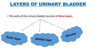
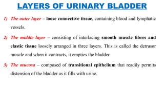
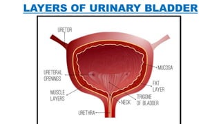
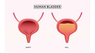
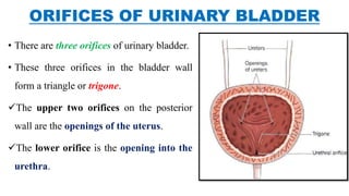
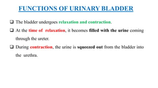
2. Urinary bladder is a TEMPORARY RESERVOIR OF URINE.
It is an inverted triangle shape.
Its shape and size vary from individual to individual and also depends on the
urine volume is stores.
It receives urine from the kidneys via the Ureters - stores it within , and expels it
during urination via Urethra.
An empty bladder is about the size and shape of a pear, becomes more oval as it
fills with urine.
The normal capacity of the bladder is 400-600ml.
URINARY BLADDER
3. • It is a hollow muscular, and distensible (or elastic) organ,
• LOCATION – Pelvic cavity. (it is situated in the pelvic cavity behind the
pubis, in front of the rectum in the male and in front of the anterior wall
of the vagina in females). When distended, the bladder rises into the
abdominal cavity.
• SIZE and POSITION – vary, depending on the volume of urine it contains.
STRUCTURE OF URINARY BLADDER
4. Filling beyond 220 ml causes micturition.
Filling up to 500 ml may be tolerated, but beyond this it
becomes painful.
5. • The external features of the bladder are:
1) Apex – located superiorly.
2) Body – main part of the bladder, located between the apex and the fundus
3) Fundus (or base) – located posteriorly. It is triangular-shaped.
4) Neck – the lowest and most fixed part of the bladder. It is situated where the
inferolateral and the posterior surfaces of the bladder meet. It is continuous
with the urethra.
6.
7. LAYERS OF URINARY BLADDER
• The walls of the urinary bladder consists of three layers.
8. 1) The outer layer – loose connective tissue, containing blood and lymphatic
vessels.
2) The middle layer – consisting of interlacing smooth muscle fibres and
elastic tissue loosely arranged in three layers. This is called the detrusor
muscle and when it contracts, it empties the bladder.
3) The mucosa – composed of transitional epithelium that readily permits
distension of the bladder as it fills with urine.
LAYERS OF URINARY BLADDER
11. ORIFICES OF URINARY BLADDER
• There are three orifices of urinary bladder.
• These three orifices in the bladder wall
form a triangle or trigone.
The upper two orifices on the posterior
wall are the openings of the uterus.
The lower orifice is the opening into the
urethra.
12.
13. BLOOD SUPPLY OF URINARY BLADDER
The superior vesical artery supplies blood to the upper part of the bladder.
The lower part of the bladder is supplied by the inferior vesical artery.
(Vesical artery - The superior vesical artery is a branch of the umbilical artery. The
inferior vesical artery is a pelvic branch of the internal iliac artery in men; and in
women it branches from the vaginal artery). (iliac artery – large artery of the
abdomen) (umbilical artery – paired artery found in abdominal and pelvic regions)
In females, the uterine and vaginal arteries provide additional blood supply.
14. • Bladder receives blood supply from both the autonomic (sympathetic and
parasympathetic) and somatic nervous system:
1) Sympathetic – hypogastric nerve (T12 – L2). It causes relaxation of the
detrusor muscle, promoting urine retention.
2) Parasympathetic – pelvic nerve (S2-S4). Increased signals from this
nerve causes contraction of the detrusor muscle, stimulating micturition.
3) Somatic – pudendal nerve (S2-4). It stimulates the external urethral
sphincter, providing voluntary control over micturition.
NERVE SUPPLY OF URINARY BLADDER
15. FUNCTIONS OF URINARY BLADDER
The bladder undergoes relaxation and contraction.
At the time of relaxation, it becomes filled with the urine coming
through the ureter.
During contraction, the urine is squeezed out from the bladder into
the urethra.