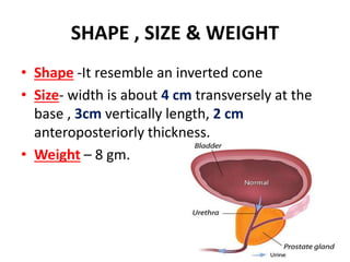
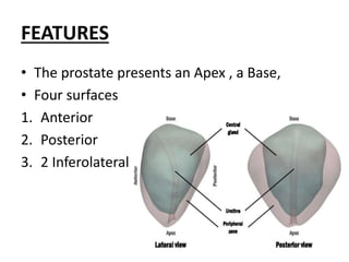
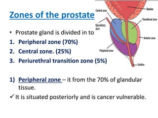
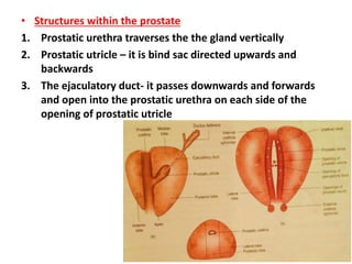
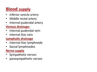
PROSTATE GLAND
BY
DR.ANKUSH D. KHEDKAR
ASST. PROF. IN RACHANA SHARIR
INTRODUCTION
• The Prostate is an accessory gland of the male
reproductive system.
• The Prostate is firm in consistency because of
fibromuscular stroma in which the glandular
elements are embedded.
• In the female the prostate id represented by
the paraurethral gland of skene.
SITUATION
• The prostate lies in the lesser pelvis,
• below the neck of the urinary bladder,
• Behind the lower part of pubic symphysis and the upper
part of pubic arch
• Lies in front of the ampulla of the rectum.
SHAPE , SIZE & WEIGHT
• Shape -It resemble an inverted cone
• Size- width is about 4 cm transversely at the
base , 3cm vertically length, 2 cm
anteroposteriorly thickness.
• Weight – 8 gm.
FEATURES
• The prostate presents an Apex , a Base,
• Four surfaces
1. Anterior
2. Posterior
3. 2 Inferolateral
• APEX-
The apex is directed downwards
Surrounds the junction of prostatic and
membranous parts of posterior urethra
It is separated from the anal canal by the
perineal body.
• BASE
The base is directed upwards, and structurally
continuous with the neck of the bladder.
Surface
1. Anterior surface-
Narrow & convex from side to side.
It lies 2cm behind the pubic symphysis.
Its upper part is connected to the pubic bone
by the Puboprostatic ligament.
The lower end of this surface is pierced by the
Urethra.
2) Posterior surface
This surface triangular in shape
It is flattened from side to side and convex
from above downwards
It is separated from the rectum by the fascia
of Denonvilliers
This surface lies 4 cm from the anus, and can
easily palpated on digital examination through
the rectum.
3 & 4 ) 2 Inferolateral surface-
This surface is related the side walls of pelvis.
Zones of the prostate
• Prostate gland is divided in to
1. Peripheral zone (70%)
2. Central zone. (25%)
3. Periurethral transition zone (5%)
1) Peripheral zone – it from the 70% of glandular
tissue.
It is situated posteriorly and is cancer vulnerable.
2) Central zone –
• It constitute 25% of glandular tissue situated posterior to the
urethral lumen and above the ejaculatory duct
• This central zone not involved in any diseases.
3) Periurethral transition zone-
• It constitute 5% of glandular tissue.
• From this zone Benign prostatic hyperplasia arises.
Sphincture related to prostate
1. Internal urethral sphincture – it subserves sexual function of
closing during ejaculation.
• If this sphincture gets resected , retrograde ejaculation occurs
2. External urethral sphincture- it is seen at the junction of prostatic
and membranous parts of the urethra
• It is horse shoe shaped with most of the bulk lying anteriorly
Lobes
The prostate gland was described earlier as
having five lobes initially
• One anterior, one posterior, one median, 2
lateral
As of now, the glandular tissue is divided into
3 lobes
• 2 lateral and One median
Capsules and ligaments of prostate
1. True capsule- it is formed by the condensation of the peripheral
part of gland.
• It is fibromuscular in structure and is continuous with stroma of
the gland.
2. False capsule- it lies outside the true capsule
• Anteriorly it is continuous with the puboprostatic ligamnent , on
each side prostatic venous plexus , posteriorly it is avascular , and
it is formed by the rectovesical fascia of denonvilliers.
• Structures within the prostate
1. Prostatic urethra traverses the the gland vertically
2. Prostatic utricle – it is bind sac directed upwards and
backwards
3. The ejaculatory duct- it passes downwards and forwards
and open into the prostatic urethra on each side of the
opening of prostatic utricle
Blood supply
• Inferior vesicle artery
• Middle rectal artery
• Internal pudendal artery
Venous drainage-
• Internal pudendal vein
• Internal iliac vein
Lymphatic drainage
• Internal iliac lymphnode
• Sacral lymphnodes
Nerve supply
• Sympathetic nerves
• parasympathetic nerves
Applied aspect
• Senile enlargement of prostate – after 50 years of age
the prostate is often enlarged due to benign
hypertrophy
• Digital examination of prostate from the anus , in the
case of enlarged prostate.
• Prostatectomy – removal of prostate
• Prostatitis- inflamation of the prostate
• Benign prostatic hyperplasia (BPH)- occure in
periurethral zone
• Carcinoma of prostate- it occurs in peripheral zone, it
usually occurs at the age of 50 -55 years.
THANK YOU

Prostate gland

  • 1.
    PROSTATE GLAND BY DR.ANKUSH D.KHEDKAR ASST. PROF. IN RACHANA SHARIR
  • 2.
    INTRODUCTION • The Prostateis an accessory gland of the male reproductive system. • The Prostate is firm in consistency because of fibromuscular stroma in which the glandular elements are embedded. • In the female the prostate id represented by the paraurethral gland of skene.
  • 3.
    SITUATION • The prostatelies in the lesser pelvis, • below the neck of the urinary bladder, • Behind the lower part of pubic symphysis and the upper part of pubic arch • Lies in front of the ampulla of the rectum.
  • 4.
    SHAPE , SIZE& WEIGHT • Shape -It resemble an inverted cone • Size- width is about 4 cm transversely at the base , 3cm vertically length, 2 cm anteroposteriorly thickness. • Weight – 8 gm.
  • 5.
    FEATURES • The prostatepresents an Apex , a Base, • Four surfaces 1. Anterior 2. Posterior 3. 2 Inferolateral
  • 6.
    • APEX- The apexis directed downwards Surrounds the junction of prostatic and membranous parts of posterior urethra It is separated from the anal canal by the perineal body. • BASE The base is directed upwards, and structurally continuous with the neck of the bladder.
  • 7.
    Surface 1. Anterior surface- Narrow& convex from side to side. It lies 2cm behind the pubic symphysis. Its upper part is connected to the pubic bone by the Puboprostatic ligament. The lower end of this surface is pierced by the Urethra.
  • 8.
    2) Posterior surface Thissurface triangular in shape It is flattened from side to side and convex from above downwards It is separated from the rectum by the fascia of Denonvilliers This surface lies 4 cm from the anus, and can easily palpated on digital examination through the rectum. 3 & 4 ) 2 Inferolateral surface- This surface is related the side walls of pelvis.
  • 9.
    Zones of theprostate • Prostate gland is divided in to 1. Peripheral zone (70%) 2. Central zone. (25%) 3. Periurethral transition zone (5%) 1) Peripheral zone – it from the 70% of glandular tissue. It is situated posteriorly and is cancer vulnerable.
  • 10.
    2) Central zone– • It constitute 25% of glandular tissue situated posterior to the urethral lumen and above the ejaculatory duct • This central zone not involved in any diseases. 3) Periurethral transition zone- • It constitute 5% of glandular tissue. • From this zone Benign prostatic hyperplasia arises.
  • 11.
    Sphincture related toprostate 1. Internal urethral sphincture – it subserves sexual function of closing during ejaculation. • If this sphincture gets resected , retrograde ejaculation occurs 2. External urethral sphincture- it is seen at the junction of prostatic and membranous parts of the urethra • It is horse shoe shaped with most of the bulk lying anteriorly
  • 12.
    Lobes The prostate glandwas described earlier as having five lobes initially • One anterior, one posterior, one median, 2 lateral As of now, the glandular tissue is divided into 3 lobes • 2 lateral and One median
  • 13.
    Capsules and ligamentsof prostate 1. True capsule- it is formed by the condensation of the peripheral part of gland. • It is fibromuscular in structure and is continuous with stroma of the gland. 2. False capsule- it lies outside the true capsule • Anteriorly it is continuous with the puboprostatic ligamnent , on each side prostatic venous plexus , posteriorly it is avascular , and it is formed by the rectovesical fascia of denonvilliers.
  • 14.
    • Structures withinthe prostate 1. Prostatic urethra traverses the the gland vertically 2. Prostatic utricle – it is bind sac directed upwards and backwards 3. The ejaculatory duct- it passes downwards and forwards and open into the prostatic urethra on each side of the opening of prostatic utricle
  • 15.
    Blood supply • Inferiorvesicle artery • Middle rectal artery • Internal pudendal artery Venous drainage- • Internal pudendal vein • Internal iliac vein Lymphatic drainage • Internal iliac lymphnode • Sacral lymphnodes Nerve supply • Sympathetic nerves • parasympathetic nerves
  • 16.
    Applied aspect • Senileenlargement of prostate – after 50 years of age the prostate is often enlarged due to benign hypertrophy • Digital examination of prostate from the anus , in the case of enlarged prostate. • Prostatectomy – removal of prostate • Prostatitis- inflamation of the prostate • Benign prostatic hyperplasia (BPH)- occure in periurethral zone • Carcinoma of prostate- it occurs in peripheral zone, it usually occurs at the age of 50 -55 years.
  • 17.