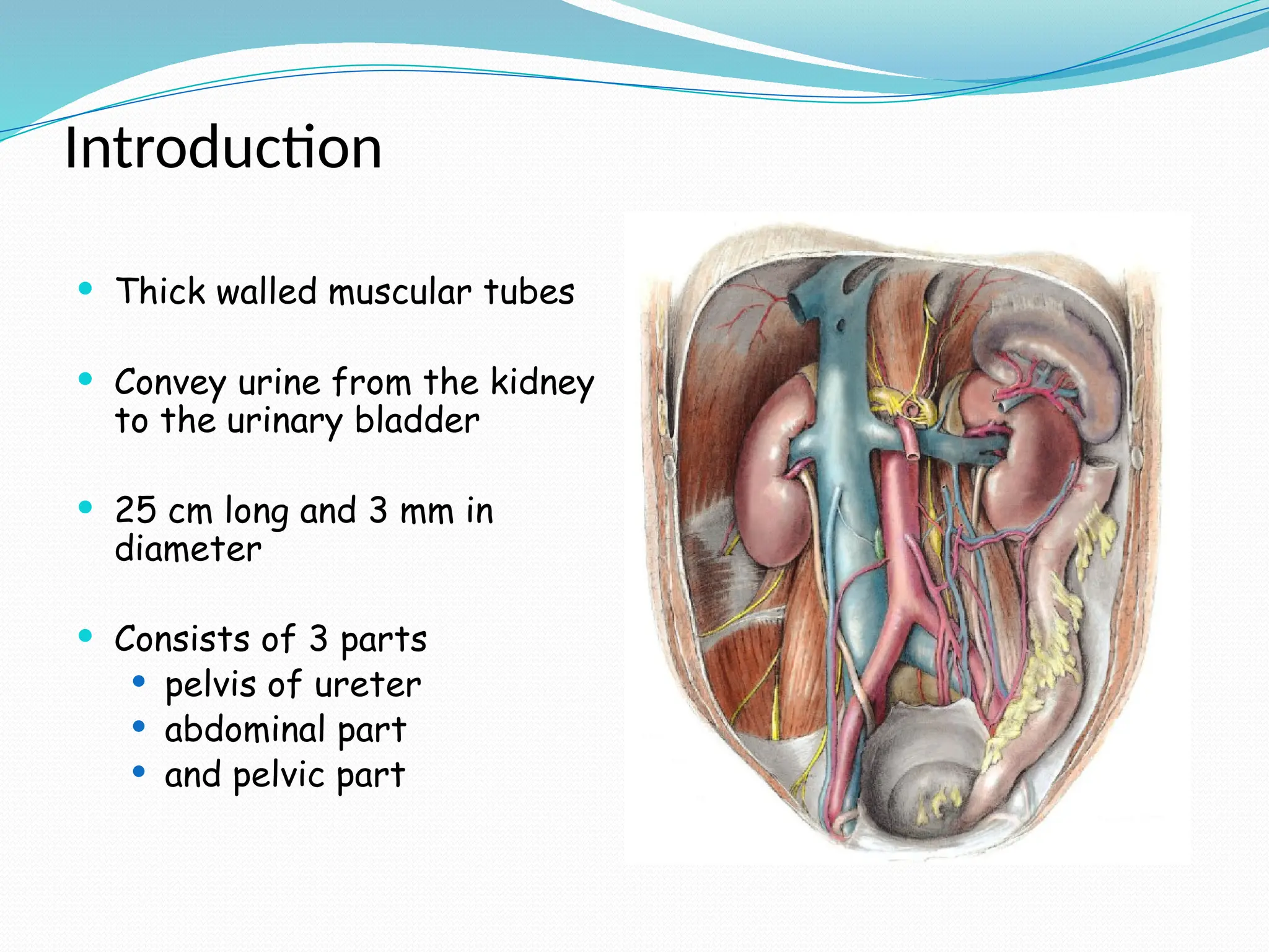
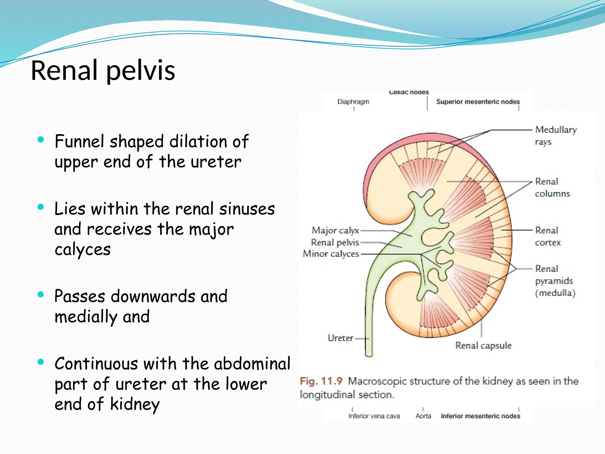
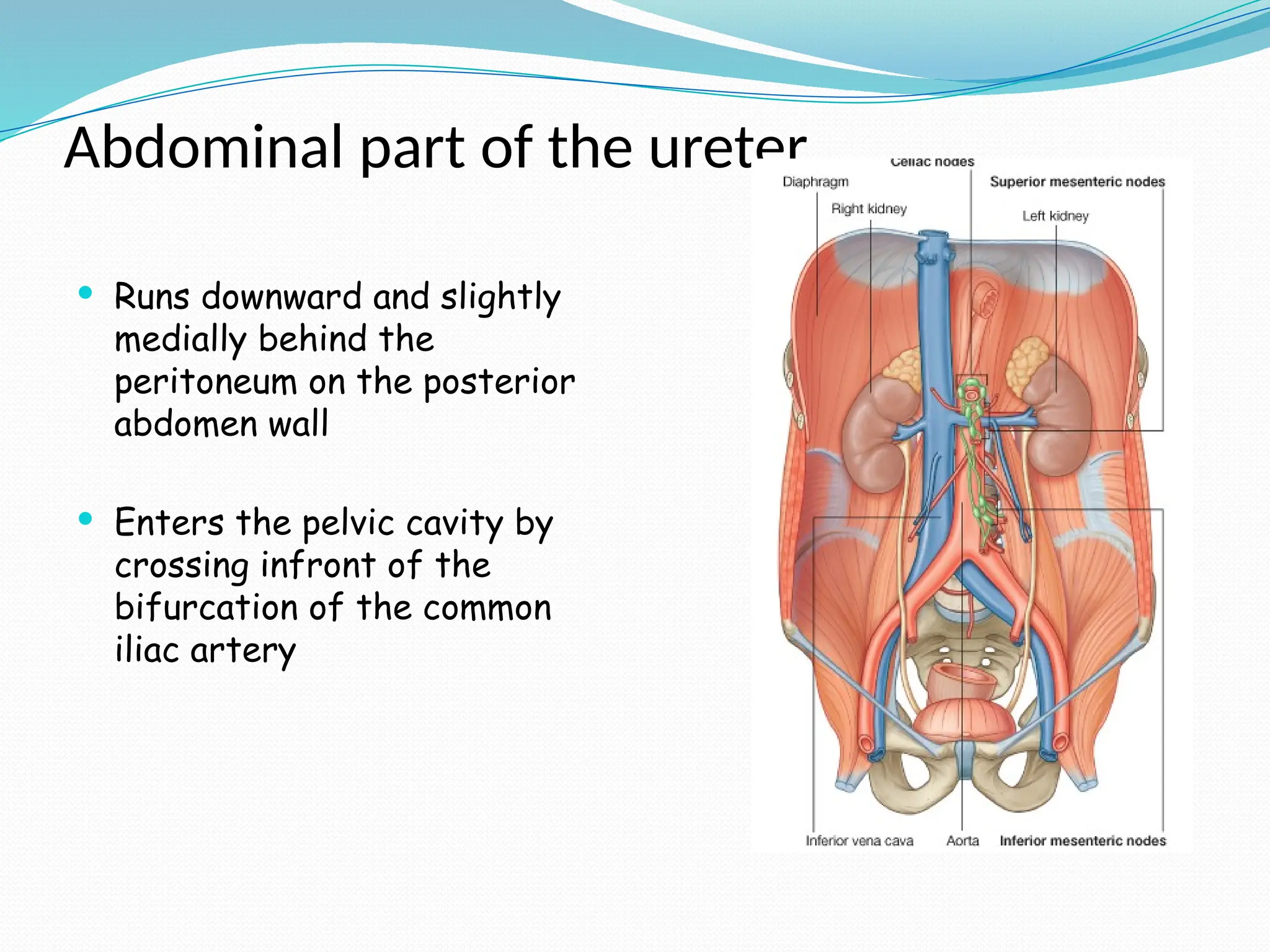
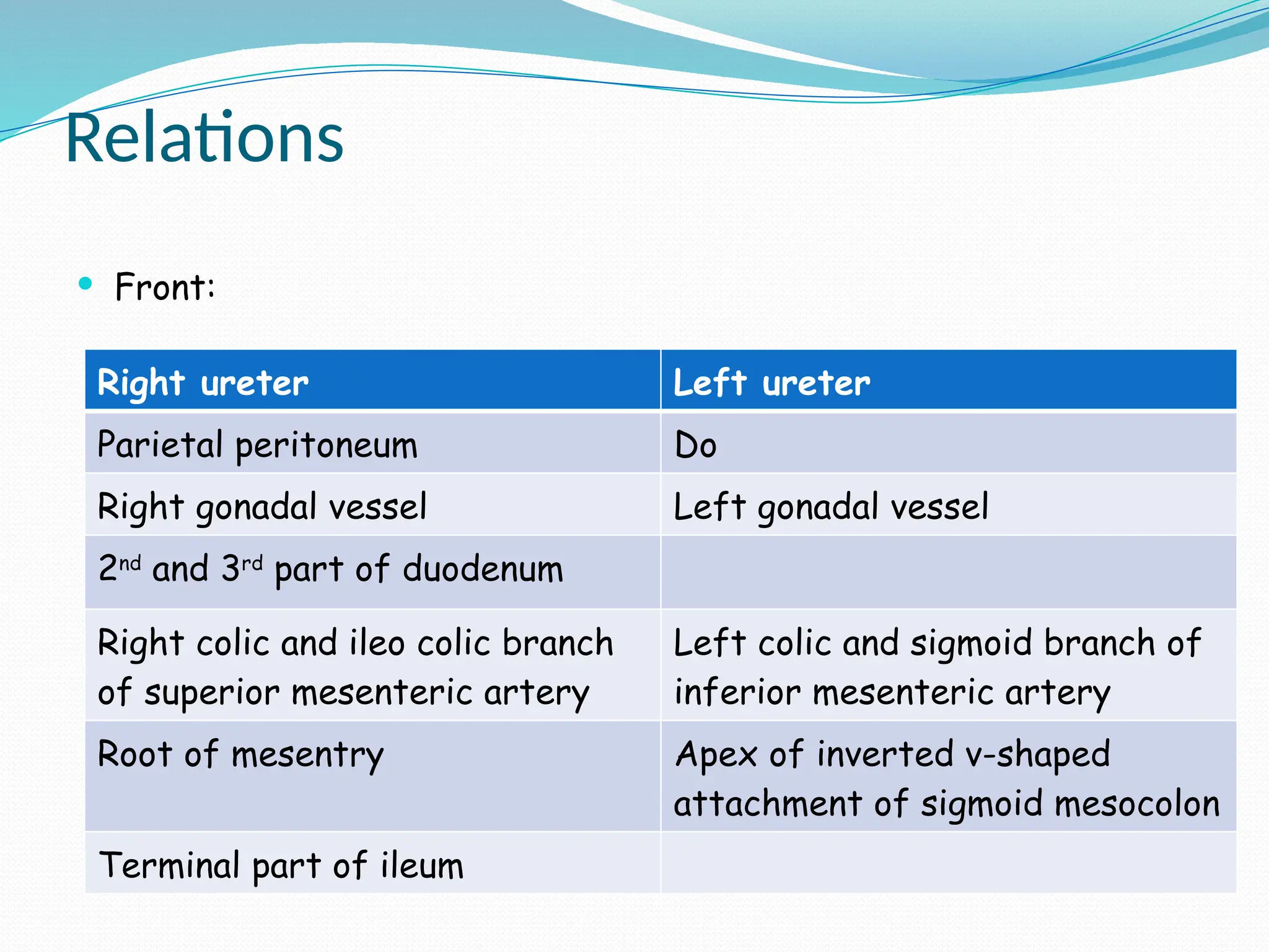
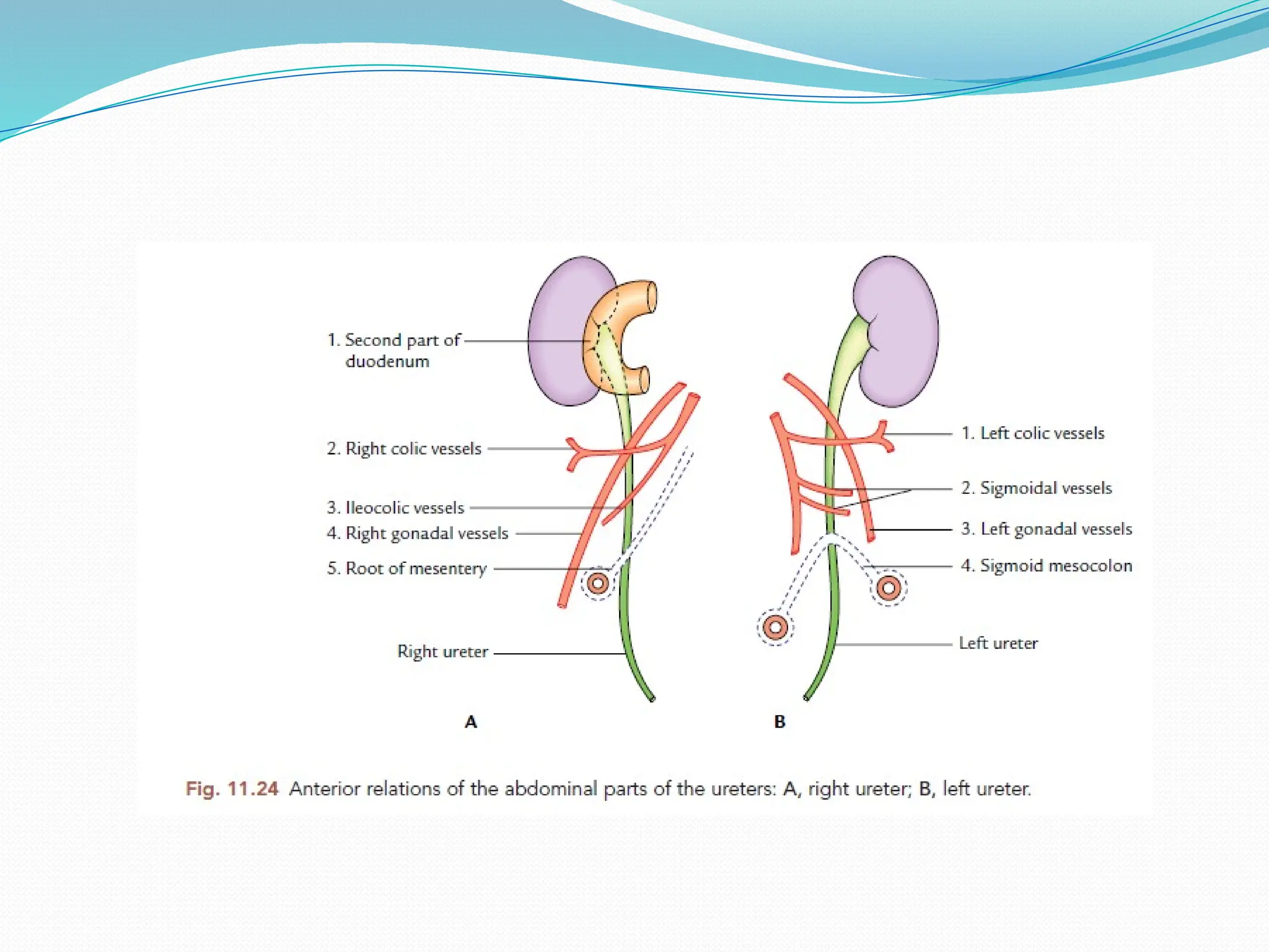
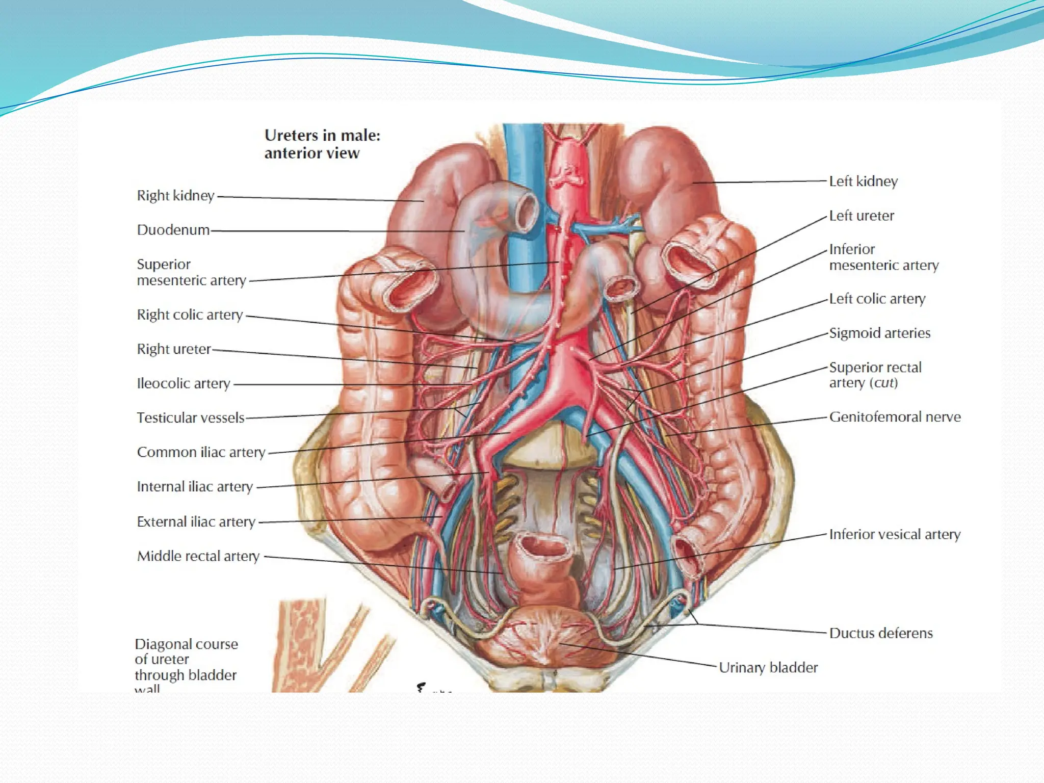
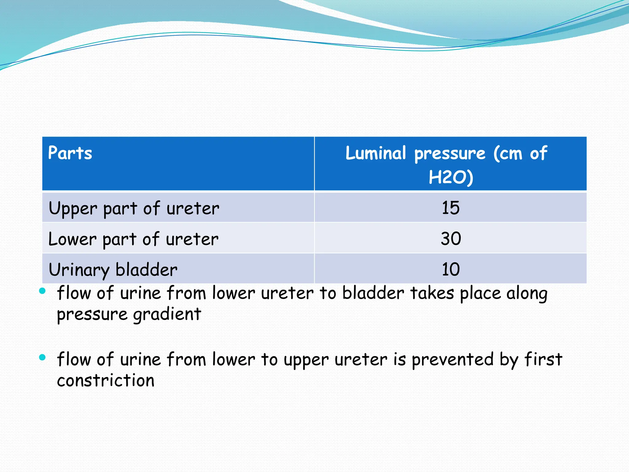
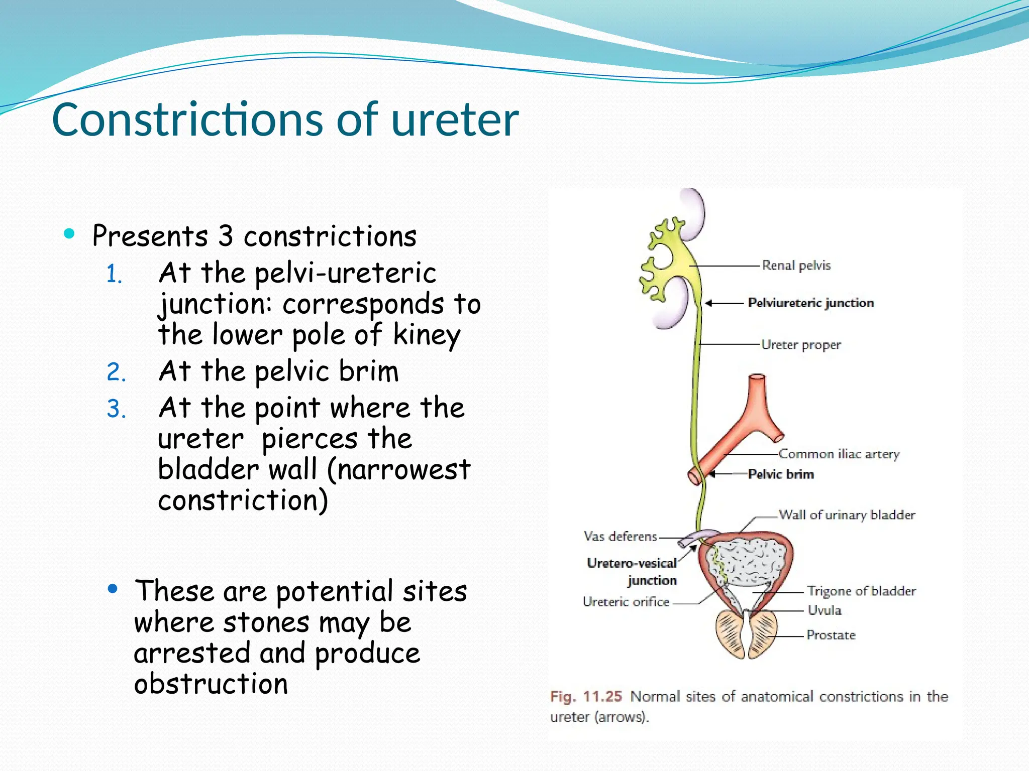
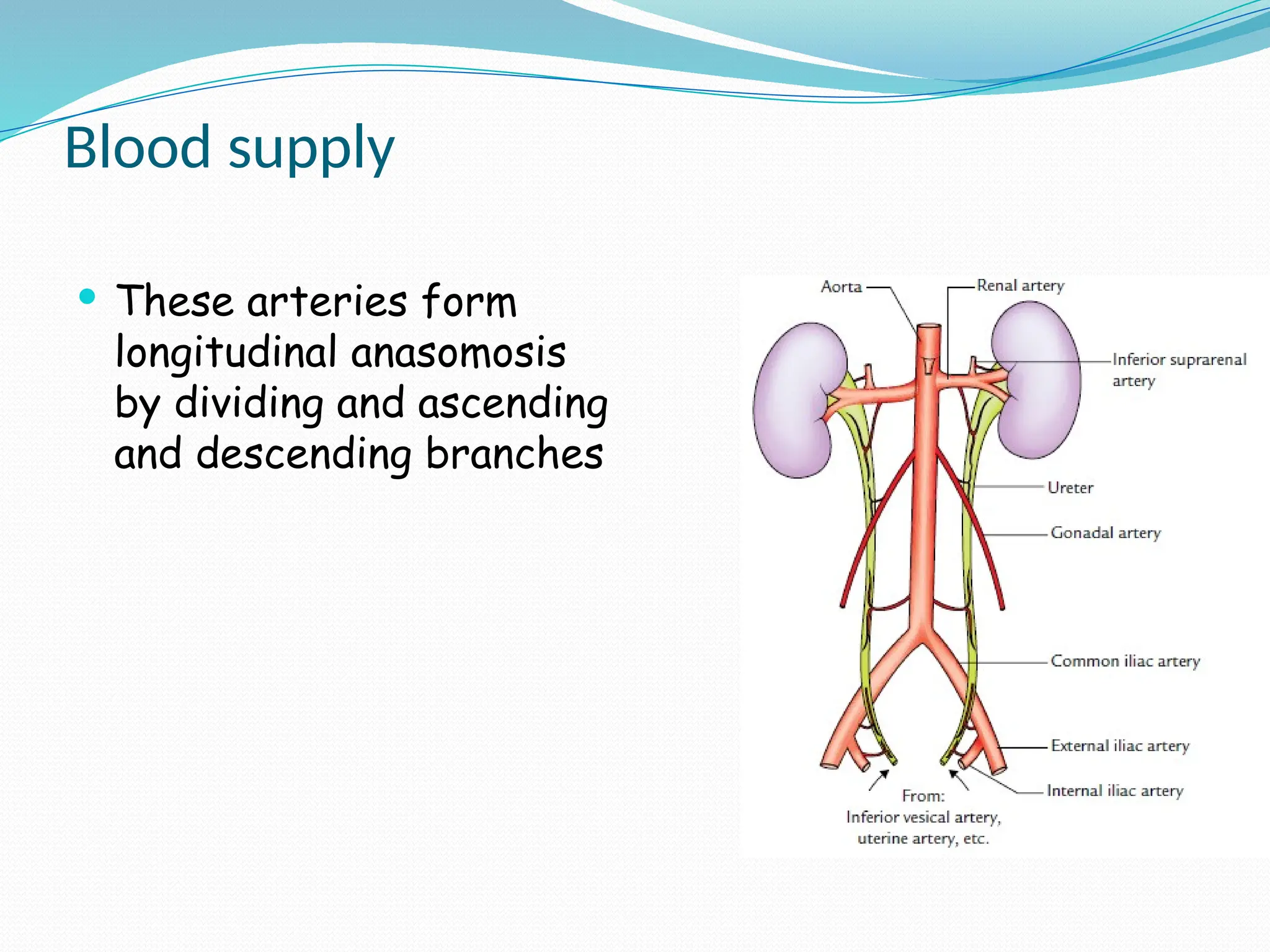
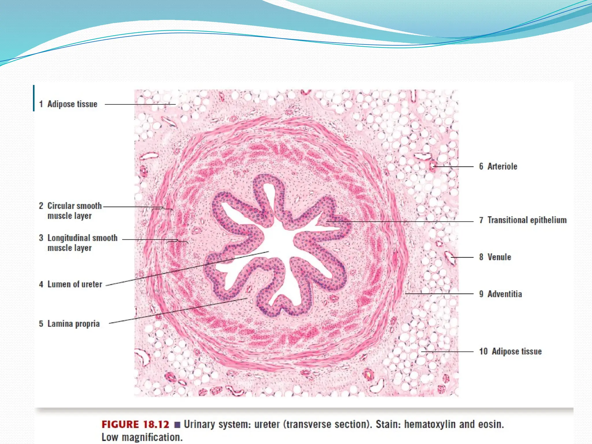
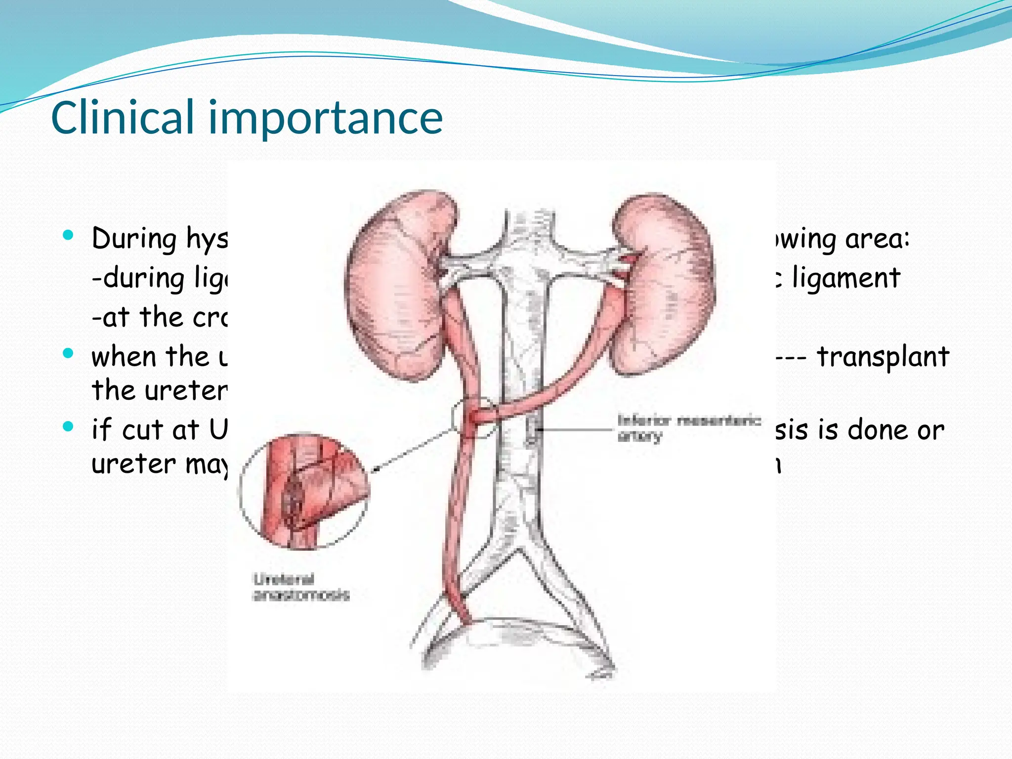
THE URETER
Introduction
 Thick walled muscular tubes
 Convey urine from the kidney
to the urinary bladder
 25 cm long and 3 mm in
diameter
 Consists of 3 parts
 pelvis of ureter
 abdominal part
 and pelvic part
Renal pelvis
 Funnel shaped dilation of
upper end of the ureter
 Lies within the renal sinuses
and receives the major
calyces
 Passes downwards and
medially and
 Continuous with the abdominal
part of ureter at the lower
end of kidney
Abdominal part of the ureter
 Runs downward and slightly
medially behind the
peritoneum on the posterior
abdomen wall
 Enters the pelvic cavity by
crossing infront of the
bifurcation of the common
iliac artery
Relations
 Front:
Right ureter Left ureter
Parietal peritoneum Do
Right gonadal vessel Left gonadal vessel
2nd
and 3rd
part of duodenum
Right colic and ileo colic branch
of superior mesenteric artery
Left colic and sigmoid branch of
inferior mesenteric artery
Root of mesentry Apex of inverted v-shaped
attachment of sigmoid mesocolon
Terminal part of ileum
 Behind:
 Psoas major and its fascia
 Genitofemoral nerve
 Tips of transverse process
of all lumbar vertebra
Pelvic part of the ureter
 In lesser or true pelvis
 Runs downwards beneath peritoneum of lateral pelvic wall
following the anterior margin of the greater sciatic notch and
reaches the ischial spine - first/vertical part
 From the ischial spine it turns forwards and medially to reach
the base of the urinary bladder – second/oblique part
 Finally pierces superolateral angle of base of urinary bladder
and undergoes oblique course within the wall of bladder and
opens at the lateral angle of its trigone- third/intravesical
part
Relations
1st
part:
 Medially:
 Covered with peritoneum
 Behind:
 Internal iliac vessels
 Lumbosacral trunk
 Sacroiliac joint
 Laterally:
 Obliterated umblical artery
 Obturater nerve and blood vessels
 Inferior vesical artery
 Middle rectal artery
 Uterine and ovarian artery in female
2nd
part:
In male
 Below and laterally: levator
ani muscles
 Above and medially: covered
with peritoneum
At lateral angle of base of
bladder,
 Above and medially: crossed
by vas deferens
 Below: upper end of seminal
vesicles
In female,
 Above:
 lower attached margin of broad ligament
 Uterine artery
 Below:
 levator ani muscle
 mackenrodt’s ligament
 lateral fornix of vagina
 Medially:
 supra vaginal part of cervix uteri
 anterior vaginal wall
 Laterally: uterine artery
3rd
part:
 Course is represented by ureteric fold of mucous membrane
extending upward and laterally from ureteric opening in the
interior of bladder
Anatomic factors preventing reflux of urine from bladder to ureter
 Oblique direction of intra- vesical part of ureter: obliquity is
maintained by contraction of trigonal muscle acting from
posterior wall of proximal urethra
 posterior wall of ureter is firmly anchored to detrusor muscle of
bladder and fascia of waldeyer, anterior wall is flexible
 increased intra- vesical pressure--- anterior wall is compressed
against posterior wall--- acts as flap valve--- prevents reflux
 flow of urine from lower ureter to bladder takes place along
pressure gradient
 flow of urine from lower to upper ureter is prevented by first
constriction
Parts Luminal pressure (cm of
H2O)
Upper part of ureter 15
Lower part of ureter 30
Urinary bladder 10
Constrictions of ureter
 Presents 3 constrictions
1. At the pelvi-ureteric
junction: corresponds to
the lower pole of kiney
2. At the pelvic brim
3. At the point where the
ureter pierces the
bladder wall (narrowest
constriction)
 These are potential sites
where stones may be
arrested and produce
obstruction
Blood supply
 These arteries form
longitudinal anasomosis
by dividing and ascending
and descending branches
Nerve supply
 Sympathetic from T10 - L1
 This explains why referred pain in renal colic from loin to the
infra-umbilical part of abdominal wall including the groin along
the aforeside spinal nerves
 There is increased peristalsis of urethral muscles during colic
pain--- irritates genitofemoral nerve behind the ureter----
contraction of cremasteric muscles--- upward retraction of
testes to superficial inguinal ring
 Parasympathetic: vagus and pelvic splanchnic nerves (S2 – S4)
Lymphatic drainage
 Upper part: para-aortic lymph node
 Middle part: common iliac lymph node
 Lower part: internal and external iliac lymph nodes
Microscopic structure
 Mucosa
 Muscular layer
 Adventitia
Clinical importance
 During hysterectomy, ureter may be involved in following area:
-during ligation of ovarian vessel in infundibulo pelvic ligament
-at the crossing of ureter and uterine artery
 when the ureter is accidentally cut at LOWER level --- transplant
the ureter to bladder
 if cut at UPPER level --- uretero- ureteric anastomosis is done or
ureter may be transplanted to ileum or sigmoid colon

the ureter (2).pptxllllllllllllllllllllll

  • 1.
  • 2.
    Introduction  Thick walledmuscular tubes  Convey urine from the kidney to the urinary bladder  25 cm long and 3 mm in diameter  Consists of 3 parts  pelvis of ureter  abdominal part  and pelvic part
  • 3.
    Renal pelvis  Funnelshaped dilation of upper end of the ureter  Lies within the renal sinuses and receives the major calyces  Passes downwards and medially and  Continuous with the abdominal part of ureter at the lower end of kidney
  • 4.
    Abdominal part ofthe ureter  Runs downward and slightly medially behind the peritoneum on the posterior abdomen wall  Enters the pelvic cavity by crossing infront of the bifurcation of the common iliac artery
  • 5.
    Relations  Front: Right ureterLeft ureter Parietal peritoneum Do Right gonadal vessel Left gonadal vessel 2nd and 3rd part of duodenum Right colic and ileo colic branch of superior mesenteric artery Left colic and sigmoid branch of inferior mesenteric artery Root of mesentry Apex of inverted v-shaped attachment of sigmoid mesocolon Terminal part of ileum
  • 7.
     Behind:  Psoasmajor and its fascia  Genitofemoral nerve  Tips of transverse process of all lumbar vertebra
  • 8.
    Pelvic part ofthe ureter  In lesser or true pelvis  Runs downwards beneath peritoneum of lateral pelvic wall following the anterior margin of the greater sciatic notch and reaches the ischial spine - first/vertical part  From the ischial spine it turns forwards and medially to reach the base of the urinary bladder – second/oblique part  Finally pierces superolateral angle of base of urinary bladder and undergoes oblique course within the wall of bladder and opens at the lateral angle of its trigone- third/intravesical part
  • 10.
    Relations 1st part:  Medially:  Coveredwith peritoneum  Behind:  Internal iliac vessels  Lumbosacral trunk  Sacroiliac joint  Laterally:  Obliterated umblical artery  Obturater nerve and blood vessels  Inferior vesical artery  Middle rectal artery  Uterine and ovarian artery in female
  • 13.
    2nd part: In male  Belowand laterally: levator ani muscles  Above and medially: covered with peritoneum At lateral angle of base of bladder,  Above and medially: crossed by vas deferens  Below: upper end of seminal vesicles
  • 15.
    In female,  Above: lower attached margin of broad ligament  Uterine artery  Below:  levator ani muscle  mackenrodt’s ligament  lateral fornix of vagina  Medially:  supra vaginal part of cervix uteri  anterior vaginal wall  Laterally: uterine artery
  • 16.
    3rd part:  Course isrepresented by ureteric fold of mucous membrane extending upward and laterally from ureteric opening in the interior of bladder
  • 17.
    Anatomic factors preventingreflux of urine from bladder to ureter  Oblique direction of intra- vesical part of ureter: obliquity is maintained by contraction of trigonal muscle acting from posterior wall of proximal urethra  posterior wall of ureter is firmly anchored to detrusor muscle of bladder and fascia of waldeyer, anterior wall is flexible  increased intra- vesical pressure--- anterior wall is compressed against posterior wall--- acts as flap valve--- prevents reflux
  • 18.
     flow ofurine from lower ureter to bladder takes place along pressure gradient  flow of urine from lower to upper ureter is prevented by first constriction Parts Luminal pressure (cm of H2O) Upper part of ureter 15 Lower part of ureter 30 Urinary bladder 10
  • 19.
    Constrictions of ureter Presents 3 constrictions 1. At the pelvi-ureteric junction: corresponds to the lower pole of kiney 2. At the pelvic brim 3. At the point where the ureter pierces the bladder wall (narrowest constriction)  These are potential sites where stones may be arrested and produce obstruction
  • 20.
    Blood supply  Thesearteries form longitudinal anasomosis by dividing and ascending and descending branches
  • 21.
    Nerve supply  Sympatheticfrom T10 - L1  This explains why referred pain in renal colic from loin to the infra-umbilical part of abdominal wall including the groin along the aforeside spinal nerves  There is increased peristalsis of urethral muscles during colic pain--- irritates genitofemoral nerve behind the ureter---- contraction of cremasteric muscles--- upward retraction of testes to superficial inguinal ring  Parasympathetic: vagus and pelvic splanchnic nerves (S2 – S4)
  • 22.
    Lymphatic drainage  Upperpart: para-aortic lymph node  Middle part: common iliac lymph node  Lower part: internal and external iliac lymph nodes
  • 23.
    Microscopic structure  Mucosa Muscular layer  Adventitia
  • 24.
    Clinical importance  Duringhysterectomy, ureter may be involved in following area: -during ligation of ovarian vessel in infundibulo pelvic ligament -at the crossing of ureter and uterine artery  when the ureter is accidentally cut at LOWER level --- transplant the ureter to bladder  if cut at UPPER level --- uretero- ureteric anastomosis is done or ureter may be transplanted to ileum or sigmoid colon