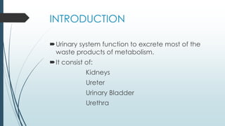
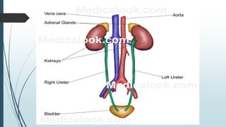
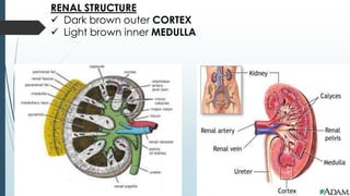
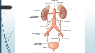
URINARY SYSTEM
INTRODUCTION
๏‚ดUrinary system function to excrete most of the
waste products of metabolism.
๏‚ดIt consist of:
Kidneys
Ureter
Urinary Bladder
Urethra
KIDNEYS
๏‚ดKidneys play a major role in controlling the water
and electrolyte balance within the body and in
maintaining the acid base balance of the
blood.
๏‚ดThey are reddish brown and lie behind the
peritoneum high up on the posterior abdominal
wall on either side of vertebral column; they are
largely under cover of the costal margin.
๏ƒผ On the medial concave border of each kidney is a
vertical slit that is bounded by thick lips of renal substance
and is called the HILUM.
๏ƒผ The hilum extends into a large cavity called the RENAL
SINUS.
๏ƒผ The hilum transmits from front backward, the renal vein,
two renal arteries, ureter and third renal artery (VAUA).
COVERINGS
๏ƒผ Fibrous capsule
๏ƒผ Perirenal fat
๏ƒผ Renal fascia
๏ƒผ Pararenal fat
RENAL STRUCTURE
๏ƒผ Dark brown outer CORTEX
๏ƒผ Light brown inner MEDULLA
RELATIONS
ANTERIORLY
POSTERIORLY
Diaphragm, costodiaphragmatic recess of
pleura, 12th rib, psoas, quadratus lumborum and transversus
abdominus muscles. The subcostal, iliohypogastric and
ilioinguinal nerves.
BLOOD SUPPLY
ARTERIES
Renal artery 5 Segmental arteries
Lobar arteries 2-3 Interlobar arteries
Arcuate arteries Interlobular arteries
Afferent Glomerular Arterioles
VEINS
Renal vein emerges from hilum in front
of renal artery and drains into Inferior Vena
Cava
LYMPH DRAINAGE
Lateral Aortic lymph nodes
NERVE SUPPLY
Renal Sympathetic Plexus
URETER
๏‚ดThe two ureters are muscular tubes that extend from
the kidneys to the posterior surface of the urinary
bladder.
๏‚ดIt is 10 inch long.
๏‚ดIt lies within hilum and receives major calyces.
๏‚ดIt has 3 constrictions along its course:
where renal pelvis joins ureter
where it crosses pelvic brim
where it pierces bladder wall
RELATIONS
RIGHT URETER
ANTERIORLY: duodenum, terminal part of ileum,
right colic and Ileocolic vessels, right testicular or ovarian
vessels, and the root of mesentery of small intestine.
POSTERIORLY: right psoas muscle, which separates it
from lumbar transverse processes, and the bifurcation of
right common iliac artery.
LEFT URETER
ANTERIORLY: sigmoid colon and sigmoid
mesocolon, left colic vessels, left testicular or ovarian vessels.
POSTERIORLY: left psoas muscle, which separates it
from lumbar transverse processes, and the bifurcation of left
common iliac artery.
BLOOD SUPPLY
ARTERIES
Upper end: renal
artery
Middle portion:
testicular or ovarian artery
Lower end:
superior vesical artery
VEINS
Corresponding
veins
LYMPH DRAINAGE
Lateral Aortic Nodes
Iliac nodes
NERVE SUPPLY
Renal nerve
Testicular or Ovarian nerve
Hypogastric Plexus (pelvis)
Afferent fibers travel with the sympathetic nerves and
enter spinal cord in the first and second lumbar segments.
URINARY BLADDER
๏‚ด It is situated immediately behind the pubic bones within
the pelvis.
๏‚ด It stores urine and has the maximum capacity of about
500 ml.
๏‚ด It has a strong muscular wall.
๏‚ด The empty bladder is pyramidal having an apex, a base
and a superior and two inferolateral surfaces, it also has
a neck.
๏ƒผ The APEX of bladder points anteriorly and lies behind the
upper margin of the symphysis pubis. it is connected to
the umbilicus by the MEDIAN UMBILICAL LIGAMENT.
๏ƒผ The BASE or POSTERIOR SURFACE of the bladder, faces
posteriorly and is triangular. The superolateral angles are
joined by ureters and the inferior angle gives rise to the
urethra.
๏ƒผ The upper part of the posterior surface of bladder is
covered by peritoneum, which forms the anterior wall of
rectovesical pouch.
๏ƒผ The lower part of the posterior surface is separated from
the rectum by the vasa differentia, the seminal vesicles
and the rectovesical fascia.
๏ƒผ The SUPERIOR SURFACE of the bladder is covered with
peritoneum and is related to coils of ileum or sigmoid
colon.
๏ƒผ As the bladder fills, it becomes ovoid and the superior
surface bulges upward into the abdominal cavity.
๏ƒผ The INFEROLATERAL SURFACES are related in front to
the RETROPUBIC PAD OF FAT and the pubic bones.
๏ƒผ The NECK of the bladder lies inferiorly and rests on the
upper surface of the prostate. The neck is held in
position by the PUBOPROSTATIC LIGAMENTS in male;
these are called PUBOVESICAL LIGAMENTS in female.
๏ƒผ The MUCOUS MEMBRANE of the greater part of empty
bladder is thrown into folds that disappear when the
bladder is full. The area of mucous membrane
covering the internal surface of the base of bladder is
called TRIGONE.
๏ƒผ The MUSCULAR COAT OF BLADDER is composed of
smooth muscles and is arranged as three layers of
interlacing bundles known as DETRUSOR MUSCLE.
๏ƒผ In females the base or posterior surface is separated by
the vagina from rectum.
๏ƒผ The superior surface is related to the uterovesical pouch
of peritoneum and to the body of the uterus.
๏ƒผ The neck of bladder rests on upper surface of urogenital
diaphragm.
BLOOD SUPPLY
ARTERIES
Superior and Inferior Vesical arteries (branches of
internal iliac arteries)
VEINS
From Vesical Venous plexus drains into Internal
Iliac vein
LYMPHATIC DRAINAGE
Internal and External Iliac nodes
NERVE SUPPLY
Pelvis Splanchnic nerves from second, third and
fourth Sacral nerves and pass through Inferior Hypogastric
plexus
Sympathetic nerves inhibit contraction of the
detrusor muscle of the bladder wall and stimulate closure of
the sphincter
URETHRA
Male Urethra
๏‚ดThe male urethra is about 8 in. (20 cm) long and extends
from the neck of the bladder to the external meatus on the
glans penis. It is divided into three parts: prostatic,
membranous, and penile.
๏‚ดThe prostatic urethra is about 1.25 in. (3 cm) long and passes
through the prostate from the base to the apex. It is the
widest and most dilatable portion of the urethra.
The membranous urethra is about 0.5 in. (1.25 cm)
long and lies within the urogenital diaphragm, surrounded
by the sphincter urethrae muscle. It is the least dilatable
portion of the urethra.
The penile urethra is about 6 in. (15.75 cm) long and
is enclosed in the bulb and the corpus spongiosum of the
penis. The external meatus is the narrowest part of the
entire urethra. The part of the urethra that lies within the
glans penis is dilated to form the fossa terminalis (navicular
fossa). The bulbourethral glands open into the penile
urethra below the urogenital diaphragm.
Paraurethral Glands
The paraurethral glands, which correspond to the prostate
in the male, open into the vestibule by small ducts on either
side of the urethral orifice.
Female Urethra
The female urethra is about 1.5 in. (3.8 cm) long. It extends
from the neck of the bladder to the external meatus, where
it opens into the vestibule about 1 in. (2.5 cm) below the
clitoris. It traverses the sphincter urethrae and lies
immediately in front of the vagina. At the sides of the
external urethral meatus are the small openings of the
ducts of the paraurethral glands. The urethra can be
dilated relatively easily.
URINARY SYSTEM & Details about urine & it's components

URINARY SYSTEM & Details about urine & it's components

  • 1.
  • 2.
    INTRODUCTION ๏‚ดUrinary system functionto excrete most of the waste products of metabolism. ๏‚ดIt consist of: Kidneys Ureter Urinary Bladder Urethra
  • 4.
    KIDNEYS ๏‚ดKidneys play amajor role in controlling the water and electrolyte balance within the body and in maintaining the acid base balance of the blood. ๏‚ดThey are reddish brown and lie behind the peritoneum high up on the posterior abdominal wall on either side of vertebral column; they are largely under cover of the costal margin.
  • 5.
    ๏ƒผ On themedial concave border of each kidney is a vertical slit that is bounded by thick lips of renal substance and is called the HILUM. ๏ƒผ The hilum extends into a large cavity called the RENAL SINUS. ๏ƒผ The hilum transmits from front backward, the renal vein, two renal arteries, ureter and third renal artery (VAUA). COVERINGS ๏ƒผ Fibrous capsule ๏ƒผ Perirenal fat ๏ƒผ Renal fascia ๏ƒผ Pararenal fat
  • 6.
    RENAL STRUCTURE ๏ƒผ Darkbrown outer CORTEX ๏ƒผ Light brown inner MEDULLA
  • 7.
    RELATIONS ANTERIORLY POSTERIORLY Diaphragm, costodiaphragmatic recessof pleura, 12th rib, psoas, quadratus lumborum and transversus abdominus muscles. The subcostal, iliohypogastric and ilioinguinal nerves.
  • 8.
    BLOOD SUPPLY ARTERIES Renal artery5 Segmental arteries Lobar arteries 2-3 Interlobar arteries Arcuate arteries Interlobular arteries Afferent Glomerular Arterioles VEINS Renal vein emerges from hilum in front of renal artery and drains into Inferior Vena Cava
  • 9.
    LYMPH DRAINAGE Lateral Aorticlymph nodes NERVE SUPPLY Renal Sympathetic Plexus
  • 11.
    URETER ๏‚ดThe two uretersare muscular tubes that extend from the kidneys to the posterior surface of the urinary bladder. ๏‚ดIt is 10 inch long. ๏‚ดIt lies within hilum and receives major calyces. ๏‚ดIt has 3 constrictions along its course: where renal pelvis joins ureter where it crosses pelvic brim where it pierces bladder wall
  • 13.
    RELATIONS RIGHT URETER ANTERIORLY: duodenum,terminal part of ileum, right colic and Ileocolic vessels, right testicular or ovarian vessels, and the root of mesentery of small intestine. POSTERIORLY: right psoas muscle, which separates it from lumbar transverse processes, and the bifurcation of right common iliac artery. LEFT URETER ANTERIORLY: sigmoid colon and sigmoid mesocolon, left colic vessels, left testicular or ovarian vessels. POSTERIORLY: left psoas muscle, which separates it from lumbar transverse processes, and the bifurcation of left common iliac artery.
  • 14.
    BLOOD SUPPLY ARTERIES Upper end:renal artery Middle portion: testicular or ovarian artery Lower end: superior vesical artery VEINS Corresponding veins
  • 15.
    LYMPH DRAINAGE Lateral AorticNodes Iliac nodes NERVE SUPPLY Renal nerve Testicular or Ovarian nerve Hypogastric Plexus (pelvis) Afferent fibers travel with the sympathetic nerves and enter spinal cord in the first and second lumbar segments.
  • 16.
    URINARY BLADDER ๏‚ด Itis situated immediately behind the pubic bones within the pelvis. ๏‚ด It stores urine and has the maximum capacity of about 500 ml. ๏‚ด It has a strong muscular wall. ๏‚ด The empty bladder is pyramidal having an apex, a base and a superior and two inferolateral surfaces, it also has a neck.
  • 17.
    ๏ƒผ The APEXof bladder points anteriorly and lies behind the upper margin of the symphysis pubis. it is connected to the umbilicus by the MEDIAN UMBILICAL LIGAMENT. ๏ƒผ The BASE or POSTERIOR SURFACE of the bladder, faces posteriorly and is triangular. The superolateral angles are joined by ureters and the inferior angle gives rise to the urethra. ๏ƒผ The upper part of the posterior surface of bladder is covered by peritoneum, which forms the anterior wall of rectovesical pouch. ๏ƒผ The lower part of the posterior surface is separated from the rectum by the vasa differentia, the seminal vesicles and the rectovesical fascia. ๏ƒผ The SUPERIOR SURFACE of the bladder is covered with peritoneum and is related to coils of ileum or sigmoid colon.
  • 19.
    ๏ƒผ As thebladder fills, it becomes ovoid and the superior surface bulges upward into the abdominal cavity. ๏ƒผ The INFEROLATERAL SURFACES are related in front to the RETROPUBIC PAD OF FAT and the pubic bones. ๏ƒผ The NECK of the bladder lies inferiorly and rests on the upper surface of the prostate. The neck is held in position by the PUBOPROSTATIC LIGAMENTS in male; these are called PUBOVESICAL LIGAMENTS in female. ๏ƒผ The MUCOUS MEMBRANE of the greater part of empty bladder is thrown into folds that disappear when the bladder is full. The area of mucous membrane covering the internal surface of the base of bladder is called TRIGONE. ๏ƒผ The MUSCULAR COAT OF BLADDER is composed of smooth muscles and is arranged as three layers of interlacing bundles known as DETRUSOR MUSCLE.
  • 21.
    ๏ƒผ In femalesthe base or posterior surface is separated by the vagina from rectum. ๏ƒผ The superior surface is related to the uterovesical pouch of peritoneum and to the body of the uterus. ๏ƒผ The neck of bladder rests on upper surface of urogenital diaphragm.
  • 22.
    BLOOD SUPPLY ARTERIES Superior andInferior Vesical arteries (branches of internal iliac arteries) VEINS From Vesical Venous plexus drains into Internal Iliac vein
  • 23.
    LYMPHATIC DRAINAGE Internal andExternal Iliac nodes NERVE SUPPLY Pelvis Splanchnic nerves from second, third and fourth Sacral nerves and pass through Inferior Hypogastric plexus Sympathetic nerves inhibit contraction of the detrusor muscle of the bladder wall and stimulate closure of the sphincter
  • 25.
    URETHRA Male Urethra ๏‚ดThe maleurethra is about 8 in. (20 cm) long and extends from the neck of the bladder to the external meatus on the glans penis. It is divided into three parts: prostatic, membranous, and penile. ๏‚ดThe prostatic urethra is about 1.25 in. (3 cm) long and passes through the prostate from the base to the apex. It is the widest and most dilatable portion of the urethra.
  • 26.
    The membranous urethrais about 0.5 in. (1.25 cm) long and lies within the urogenital diaphragm, surrounded by the sphincter urethrae muscle. It is the least dilatable portion of the urethra. The penile urethra is about 6 in. (15.75 cm) long and is enclosed in the bulb and the corpus spongiosum of the penis. The external meatus is the narrowest part of the entire urethra. The part of the urethra that lies within the glans penis is dilated to form the fossa terminalis (navicular fossa). The bulbourethral glands open into the penile urethra below the urogenital diaphragm.
  • 28.
    Paraurethral Glands The paraurethralglands, which correspond to the prostate in the male, open into the vestibule by small ducts on either side of the urethral orifice. Female Urethra The female urethra is about 1.5 in. (3.8 cm) long. It extends from the neck of the bladder to the external meatus, where it opens into the vestibule about 1 in. (2.5 cm) below the clitoris. It traverses the sphincter urethrae and lies immediately in front of the vagina. At the sides of the external urethral meatus are the small openings of the ducts of the paraurethral glands. The urethra can be dilated relatively easily.