2. OBJECTIVES
At the end of the lecture, students should be able
to:
Describe the course of ureter & identify the sites
of ureteric constriction
Describe the important relations & identify certain
areas (trigone, uvula vesicae) in the base of
urinary bladder.
List the blood supply, lymphatic drainage & nerve
supply of urinary bladder
Differentiate between male & female urethra
regarding length, structure, course & function.
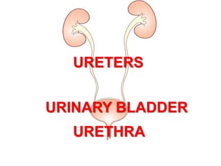
3. Ureter
A 25 – 30 cm long
muscular tube transporting
urine from kidney to urinary
bladder.
Begins as a continuation of
renal pelvis.
COURSE IN ABDOMEN:
It descends anterior to psoas major muscle (opposite the
tips of lumbar transverse processes).
It crosses the end (bifurcation) of common iliac artery to
enter the pelvis.
4. COURSE IN PELVIS &
TERMINATION:
Runs downward in front to internal
iliac artery, reaches ischial spine
Turns forward and medially ,
enters the upper lateral angle of
urinary bladder
Near its termination, is crossed by
the vas deferens
Passes obliquely through the wall
of bladder for about ¾ inch before
opening into the bladder cavity.
Bladder muscle contraction
mechanically closes off ureteral
orifice which prevents a reverse flow
of urine toward the kidney
5. Ureteric Constrictions
The normal ureter is not of
uniform caliber and
shows constrictions at
three points (possible
sites of obstruction and
stone impaction)
1. At the ureteropelvic
junction
2. At the crossing of
external/common iliac
artery
3. At site of entrance to
bladder (uretrovesical
junction; narrowest part)
1
2
3
6. Arterial Supply
Ureter is supplied by
multiple arteries
throughout its course
From above downward,
these are::
1. Renal artery
2. Gonadal artery
3. Common iliac artery
4. Internal iliac artery
2
4
3
1
7. Urinary Bladder
Located immediately behind
the pubic symphysis
Shape and relations vary
according to the amount of
urine it contains
An empty bladder:
In adults, is entirely a
pelvic organ; as it fills,
rises up into hypogastric
region of the abdomen.
In young children, it is
an abdominal organ and
projects above the pelvic
inlet
8. An empty bladder is
pyramidal in shape
having:
An apex
A base (posterior
surface)
Three surfaces:
• A superior surface
• Two infrolateral
surfaces
A neck
9. Apex:
Directed forward
Lies behind the upper
margin of the symphysis
pubis
Is connected to umbilicus
by the median umbilical
ligament (contains
urachus, the remnant of
allantoise)
10. Base (Posterior surface):
Triangular in shape
Upper part covered by
peritoneum
Lower part related to:
In males: vas deferentia
and seminal vesicles.
In females: vagina.
11. Superior surface:
Completely covered by peritoneum.
Related to:
In males: coils of ileum or sigmoid colon
In females: uterus
Male pelvis Female pelvis
12. Infrolateral surfaces:
Related:
In front to the retropubic
pad of fat & the pubic
bones
Posteriorly lie in contact
with obturator internus
above and levator ani
below
Retropubic fat
Accomodates distention of bladder
Continuous with fat in the anterior abdominal wall.
Rupture of bladder results in escape of urine to anterior
abdominal wall
13. Neck:
Lies inferiorly, and is the
most fixed part of the
bladder
Is related to lower border
of symphysis pubis
In male, rests on the
upper surface of
prostate. Here, the
smooth muscle fibers of
the bladder are
continuous with those of
the prostate.
The circular muscle
fibers thickened to form
the sphincter vesicae
14. Interior of the Urinary Bladder
Mucous membrane thrown
into folds except in the
triangular region in the base
of bladder, between the
openings of the two ureters
and the urethra. This region is
called the ‘trigone’. Here The
mucous membrane is always
smooth even when the
bladder is empty
Uvula vesicae, a small
elevation in the mucous
membrane, located just
behind the internal urethral
orifice. It is produced by the
median lobe of prostate.
15. Arterial supply: from internal iliac artery
Venous drainage: into internal iliac vein
Lymphatics: into internal iliac lymph nodes
The nerves form the vesical nerve
plexus that contains:
Sympathetic fibers derived mainly
from L1,2
Parasympathetic fibers derived from
pelvic splanchnic nerves S2,3,4
Sensory fibers from the bladder are
visceral and transmit pain sensation
resulting from overdistention
Blood & Nerve Supply
16. The normal capacity of
bladder is about 300-
500ml.
As bladder fills, it
becomes circular in
shape & its superior
surface bulges upward
into abdominal cavity.
The peritoneal lining is
peeled off the lower part
of anterior abdominal
wall and the bladder
comes into direct contact
with the anterior
abdominal wall
17. Male Urethra
About 8 inches (20cm)
long
Extends from the neck
of bladder to the
external urinary meatus
on the tip of the glans
penis
Divided into three
parts:
Prostatic
Membranous
Penile
18. Prostatic urethra
Length= 3 cm
Widest & most dilatable
Extends from neck of bladder
inside prostate gland
Structures openings into
prostatic urethra:
Ejaculatory ducts
Ducts of prostate gland
Membranous urethra
Length=1 cm
Surrounded by external
urethral sphincter
Penile (spongy) urethra
Length=16 cm
Narrowest part of whole urethra
Extends throughout penis &
opens externally through external
urethral orifice
19. Female Urethra
Length= 4 cm
Extends from neck
of urinary bladder
to open externally
through the external
urethral orifice
(anterior to the
vaginal opening)
Has only urinary
function
Trigone