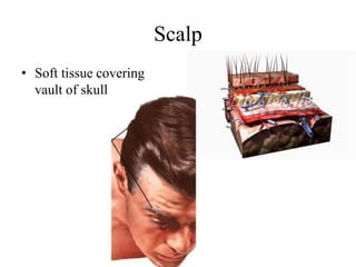
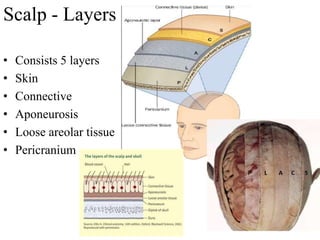
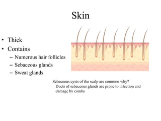
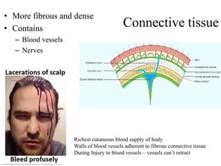
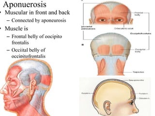
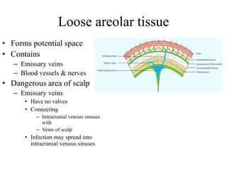
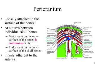
Scalp
• Soft tissue covering
vault of skull
Scalp - Extent
• Anteriorly
– Supraorbital
margin
• Posteriorly
– External occipital
protuberance &
superior nuchal
line
• On each side
– Zygomatic arch
and temporal lines
Scalp - Layers
• Consists 5 layers
• Skin
• Connective
• Aponeurosis
• Loose areolar tissue
• Pericranium
Skin
• Thick
• Contains
– Numerous hair follicles
– Sebaceous glands
– Sweat glands
Sebaceous cysts of the scalp are common why?
Ducts of sebaceous glands are prone to infection and
damage by combs
Connective tissue
• More fibrous and dense
• Contains
– Blood vessels
– Nerves
Richest cutaneous blood supply of body
Walls of blood vessels adherent to fibrous connective tissue
During Injury to blood vessels – vessels can’t retract
Aponuerosis
• Muscular in front and back
– Connected by aponeurosis
• Muscle is
– Frontal belly of oocipito
frontalis
– Occiital belly of
occipitofrontalis
• Frontal belly
– O: Skin of the forehead (no
bony attachment)
– Ins: Epicranial aponeurois
• Occipital belly:
– O: Lateral part of highest
nuchal line
– Ins: Epicranial aponeurosis
– Nerve supply: Posterior
auricular branch of facial nerve
• Nerve Supply
– Frontal belly
• Temporal branch of facial nerve
– Occiptal belly
• Posterior Auricular of Facial
nerve
Loose areolar tissue
• Forms potential space
• Contains
– Emissary veins
– Blood vessels & nerves
• Dangerous area of scalp
– Emissary veins
• Have no valves
• Connecting
– Intracranial venous sinuses
with
– Veins of scalp
• Infection may spread into
intracranial venous sinuses
Black eye
• Collection of fluid can
spreads only into the eye
lids and the root of the nose
• Because of the attachment
of frontalis into the skin
• Posteriorly
– Attachment of occipitalis
muscle to the occipital and
temporal bones
• Laterally
– Attachment of aponeurosis to
the temporal fascia
Pericranium
• Loosely attached to the
surface of the bones
• At sutures between
individual skull bones
– Periosteum on the outer
surface of the bones is
continuous with
– Endosteum on the inner
surface of the skull bones
• Firmly adherent to the
sutures
Arteries
• In front of auricle
– Supratrochlear
– Supraorbital
– Superficial
temporal
• Behind the auricle
– Posterior auricular
– Occipital
Veins
• Superficial temporal
– Joins with maxillary
• In parotid gland
– Forms retromandibular
– Divides into
• Anterior division of
retromandibular
• Posterior division of
retromandibular
Veins
• Supra trochlear & supra
orbital
– Joins at medial angle of eye
– Forms angular vein
– Continues as facial
• Facial vein meets
– Anterior division of
retromandibular
– Forms common facial vein
• Drains into IJV
Veins
• Posterior auricular
– Unites with
• Posterior division of
retromandibular
– Forms external jugular vein
• Occipital vein
– Drains into
• Suboccipital venous plexus
Nerves
• In front of auricle
– 4 sensory
• Supratrochlear
• Supraorbital
• Zygomatico temporal
• Auriculotemporal
– One motor
• Temporal branch of facial
Nerves
• Behind auricle
– 4 sensory
• Great auricular (C1&2)
• Lesser occipital (C2)
• Greater occipital (C2 DPR)
• Third occipital (C3 DPR)
– One motor
• Posterior auricular
– Facial

Scalp

  • 1.
    Scalp • Soft tissuecovering vault of skull
  • 2.
    Scalp - Extent •Anteriorly – Supraorbital margin • Posteriorly – External occipital protuberance & superior nuchal line • On each side – Zygomatic arch and temporal lines
  • 3.
    Scalp - Layers •Consists 5 layers • Skin • Connective • Aponeurosis • Loose areolar tissue • Pericranium
  • 4.
    Skin • Thick • Contains –Numerous hair follicles – Sebaceous glands – Sweat glands Sebaceous cysts of the scalp are common why? Ducts of sebaceous glands are prone to infection and damage by combs
  • 5.
    Connective tissue • Morefibrous and dense • Contains – Blood vessels – Nerves Richest cutaneous blood supply of body Walls of blood vessels adherent to fibrous connective tissue During Injury to blood vessels – vessels can’t retract
  • 6.
    Aponuerosis • Muscular infront and back – Connected by aponeurosis • Muscle is – Frontal belly of oocipito frontalis – Occiital belly of occipitofrontalis
  • 7.
    • Frontal belly –O: Skin of the forehead (no bony attachment) – Ins: Epicranial aponeurois • Occipital belly: – O: Lateral part of highest nuchal line – Ins: Epicranial aponeurosis – Nerve supply: Posterior auricular branch of facial nerve • Nerve Supply – Frontal belly • Temporal branch of facial nerve – Occiptal belly • Posterior Auricular of Facial nerve
  • 9.
    Loose areolar tissue •Forms potential space • Contains – Emissary veins – Blood vessels & nerves • Dangerous area of scalp – Emissary veins • Have no valves • Connecting – Intracranial venous sinuses with – Veins of scalp • Infection may spread into intracranial venous sinuses
  • 10.
    Black eye • Collectionof fluid can spreads only into the eye lids and the root of the nose • Because of the attachment of frontalis into the skin • Posteriorly – Attachment of occipitalis muscle to the occipital and temporal bones • Laterally – Attachment of aponeurosis to the temporal fascia
  • 11.
    Pericranium • Loosely attachedto the surface of the bones • At sutures between individual skull bones – Periosteum on the outer surface of the bones is continuous with – Endosteum on the inner surface of the skull bones • Firmly adherent to the sutures
  • 12.
    Arteries • In frontof auricle – Supratrochlear – Supraorbital – Superficial temporal • Behind the auricle – Posterior auricular – Occipital
  • 13.
    Veins • Superficial temporal –Joins with maxillary • In parotid gland – Forms retromandibular – Divides into • Anterior division of retromandibular • Posterior division of retromandibular
  • 14.
    Veins • Supra trochlear& supra orbital – Joins at medial angle of eye – Forms angular vein – Continues as facial • Facial vein meets – Anterior division of retromandibular – Forms common facial vein • Drains into IJV
  • 15.
    Veins • Posterior auricular –Unites with • Posterior division of retromandibular – Forms external jugular vein • Occipital vein – Drains into • Suboccipital venous plexus
  • 16.
    Nerves • In frontof auricle – 4 sensory • Supratrochlear • Supraorbital • Zygomatico temporal • Auriculotemporal – One motor • Temporal branch of facial
  • 17.
    Nerves • Behind auricle –4 sensory • Great auricular (C1&2) • Lesser occipital (C2) • Greater occipital (C2 DPR) • Third occipital (C3 DPR) – One motor • Posterior auricular – Facial