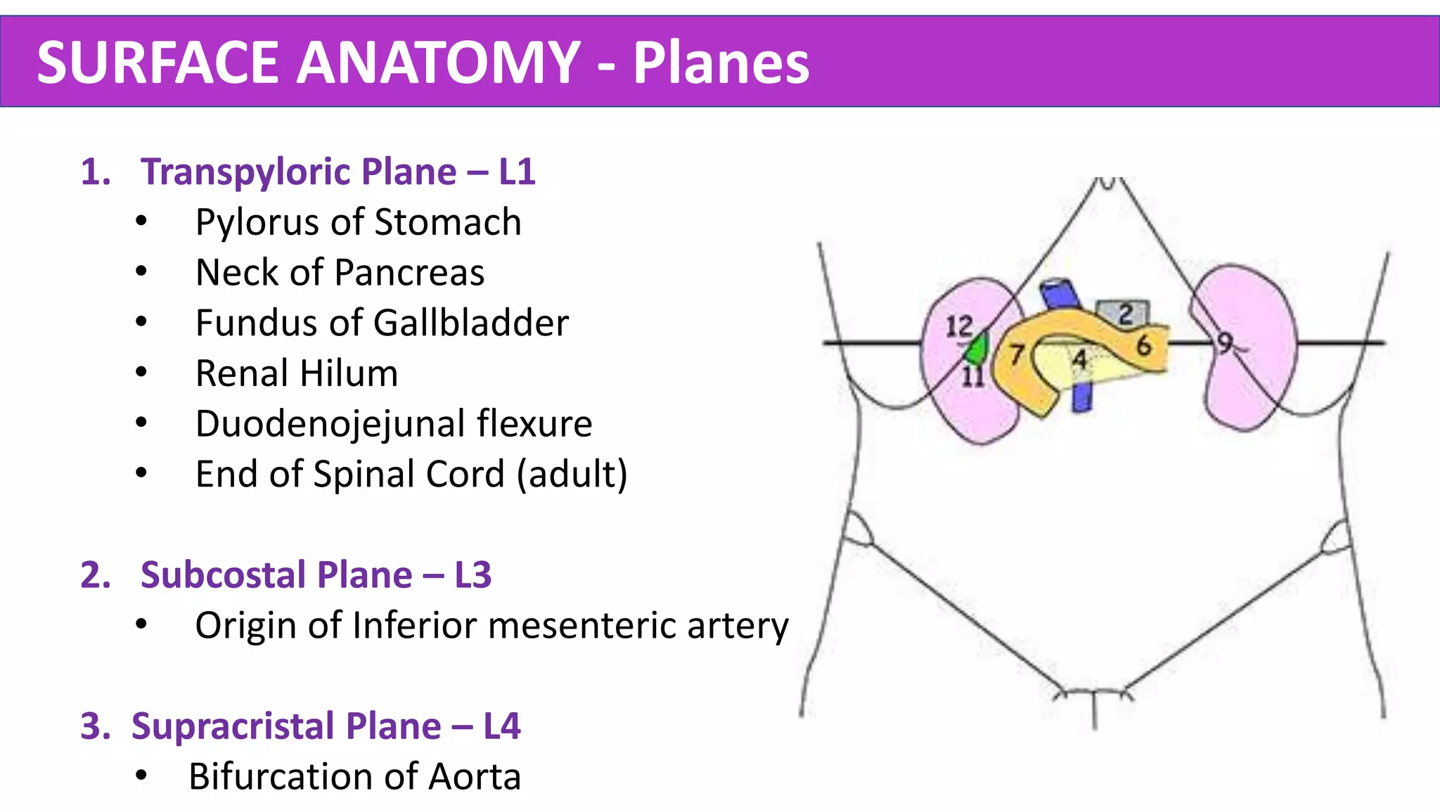
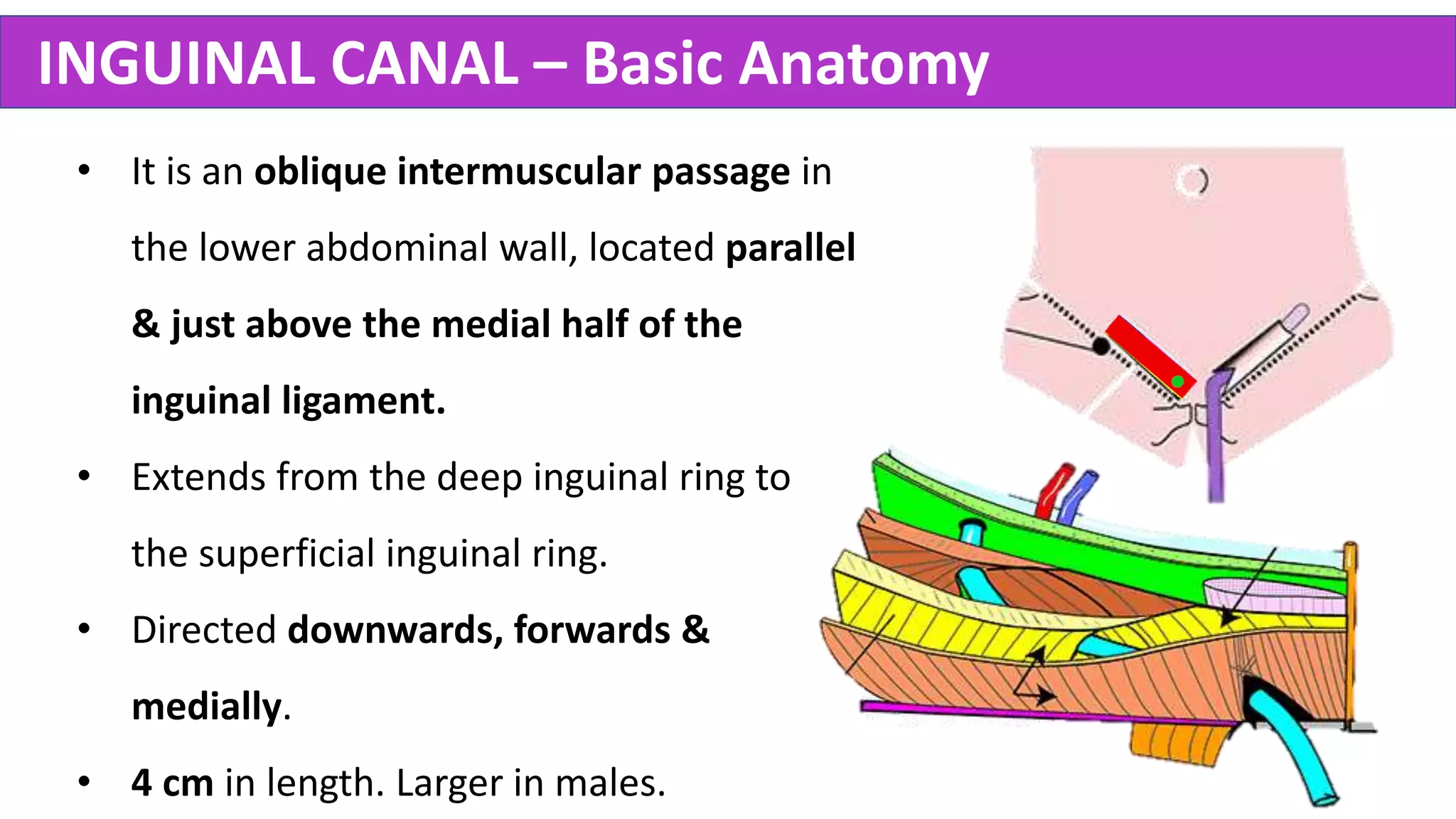
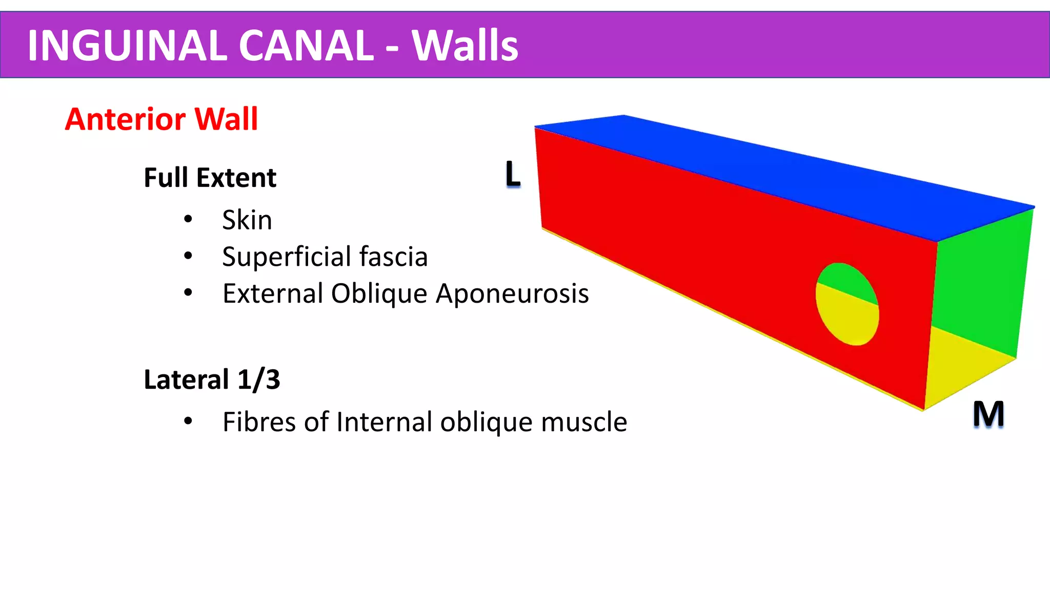
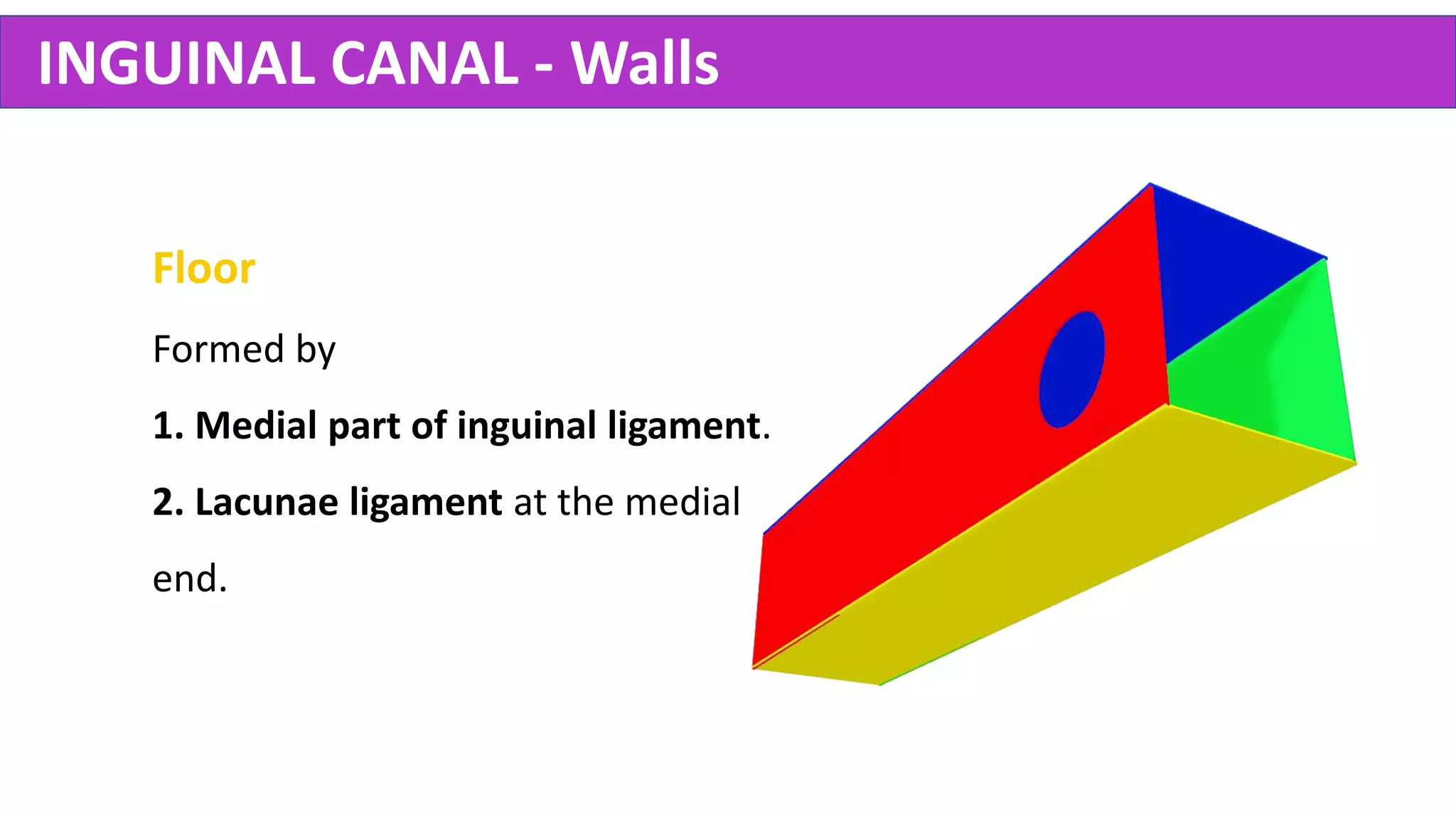
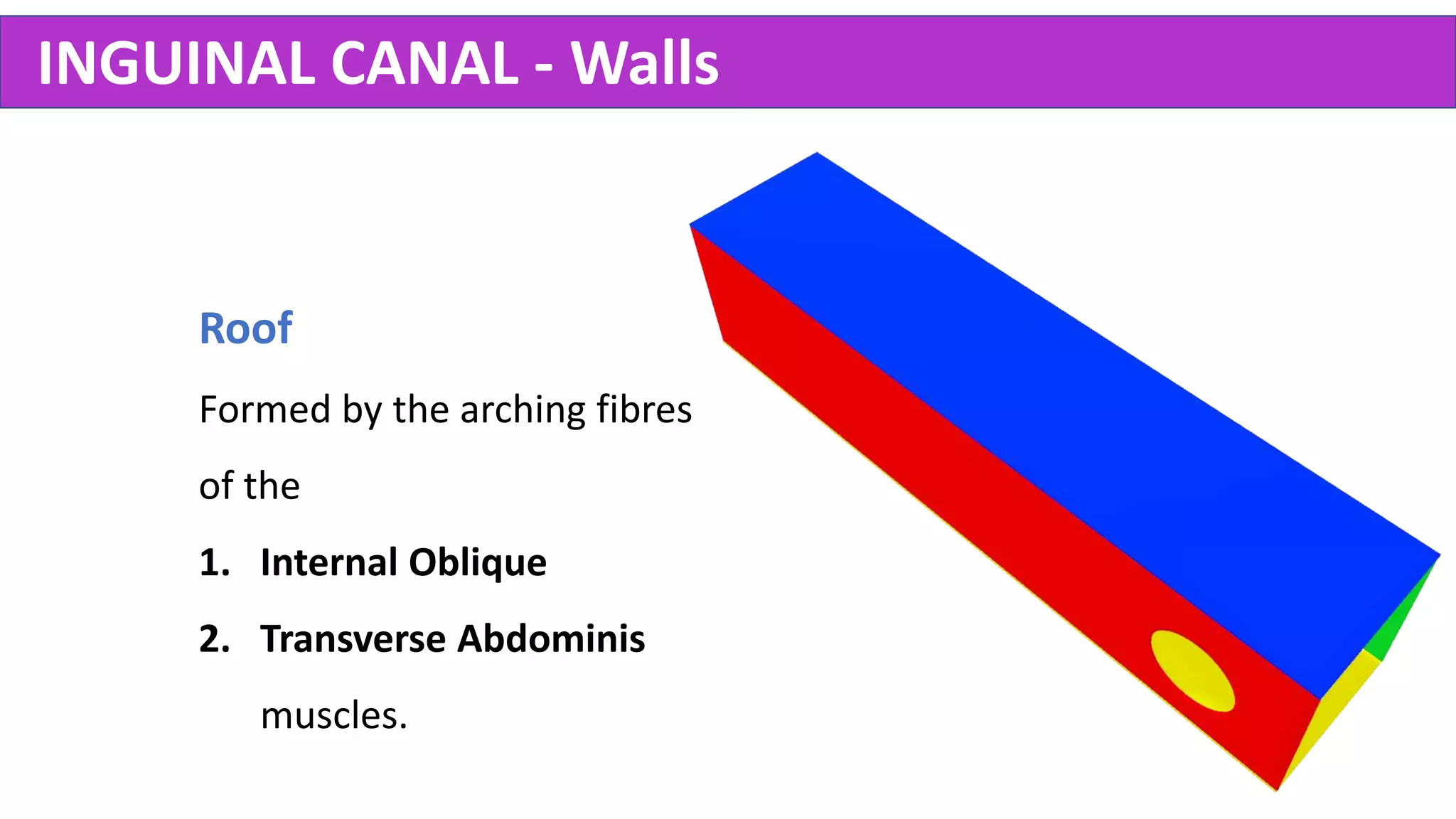
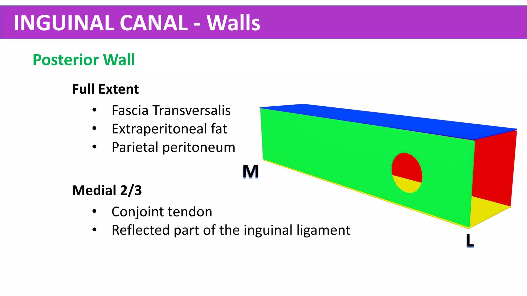
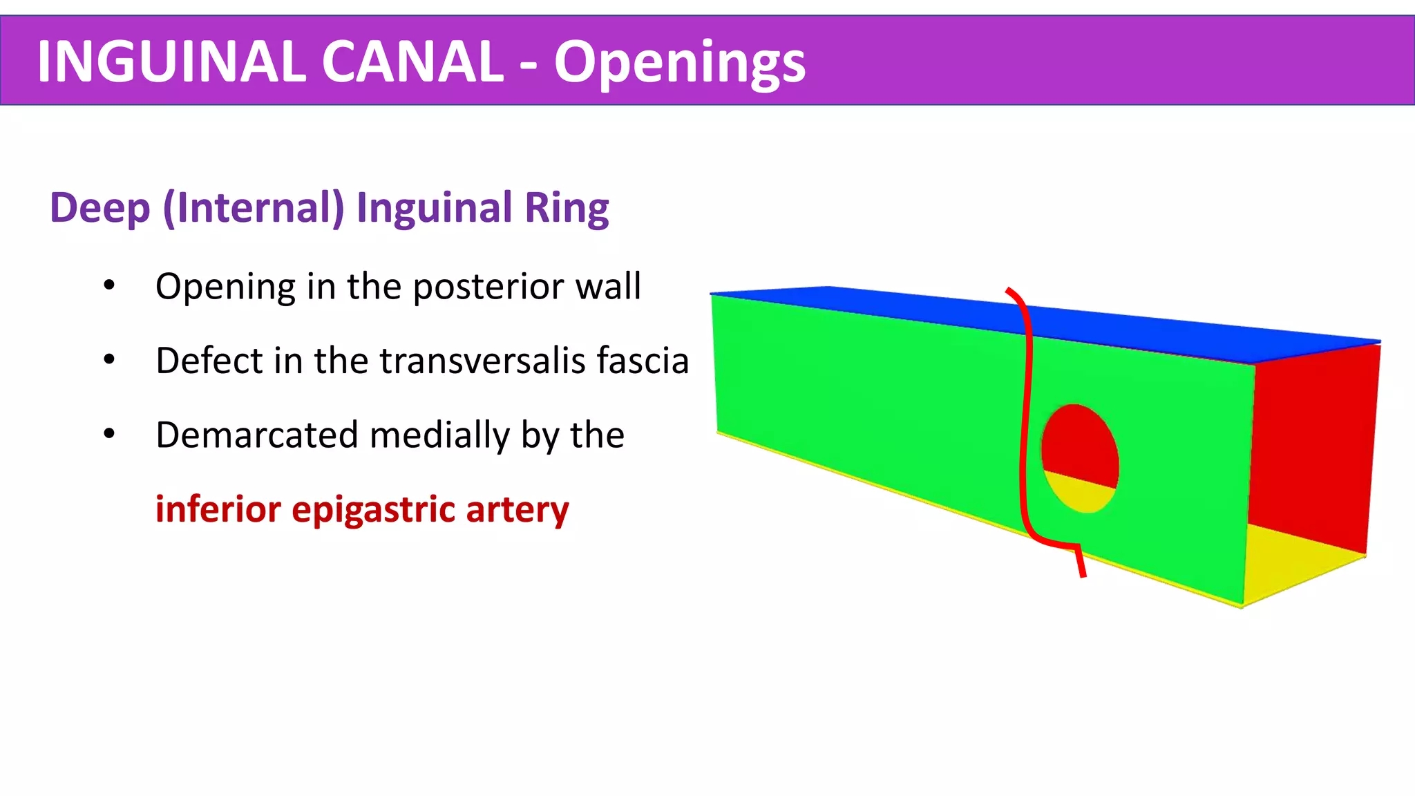
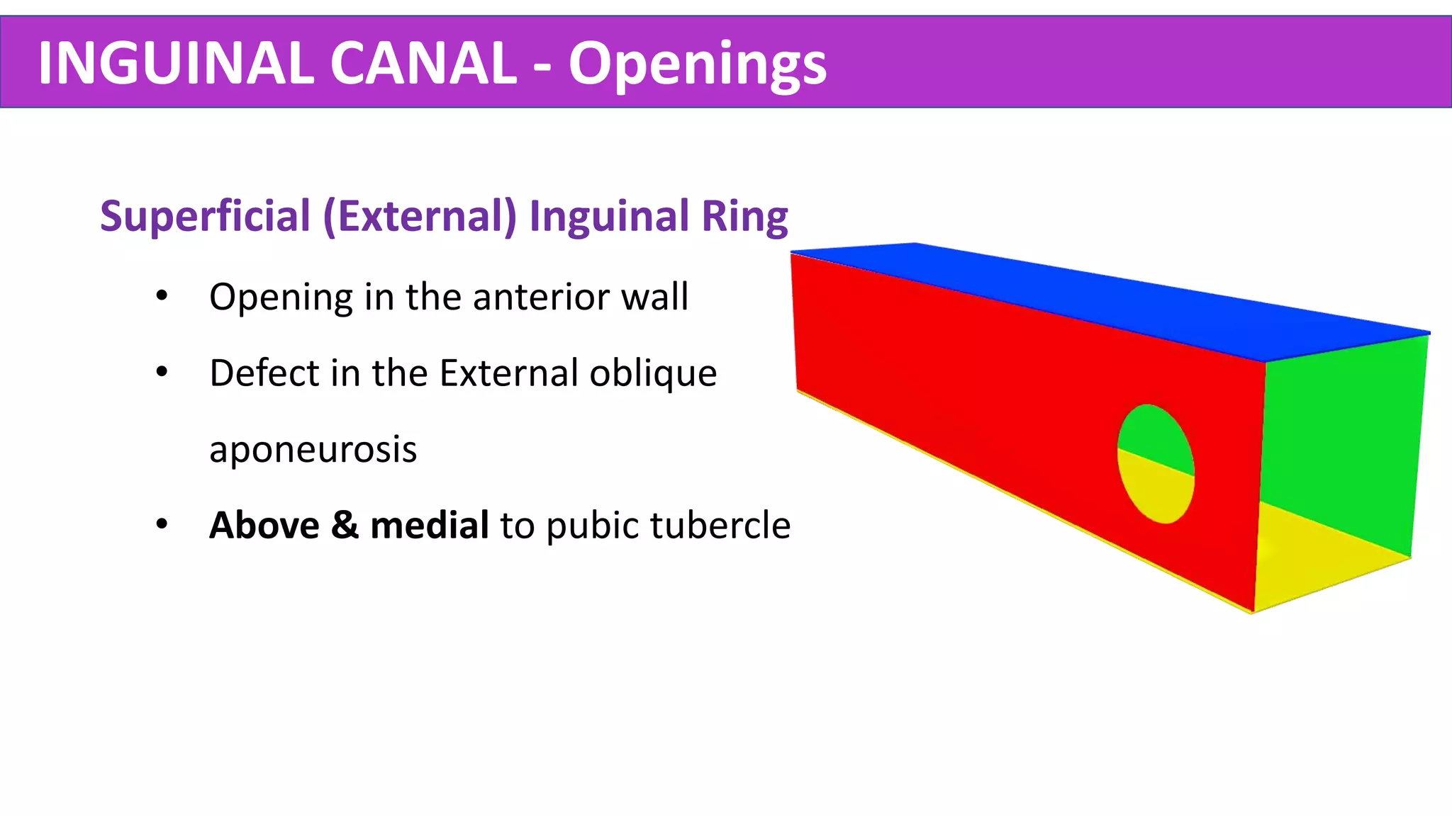
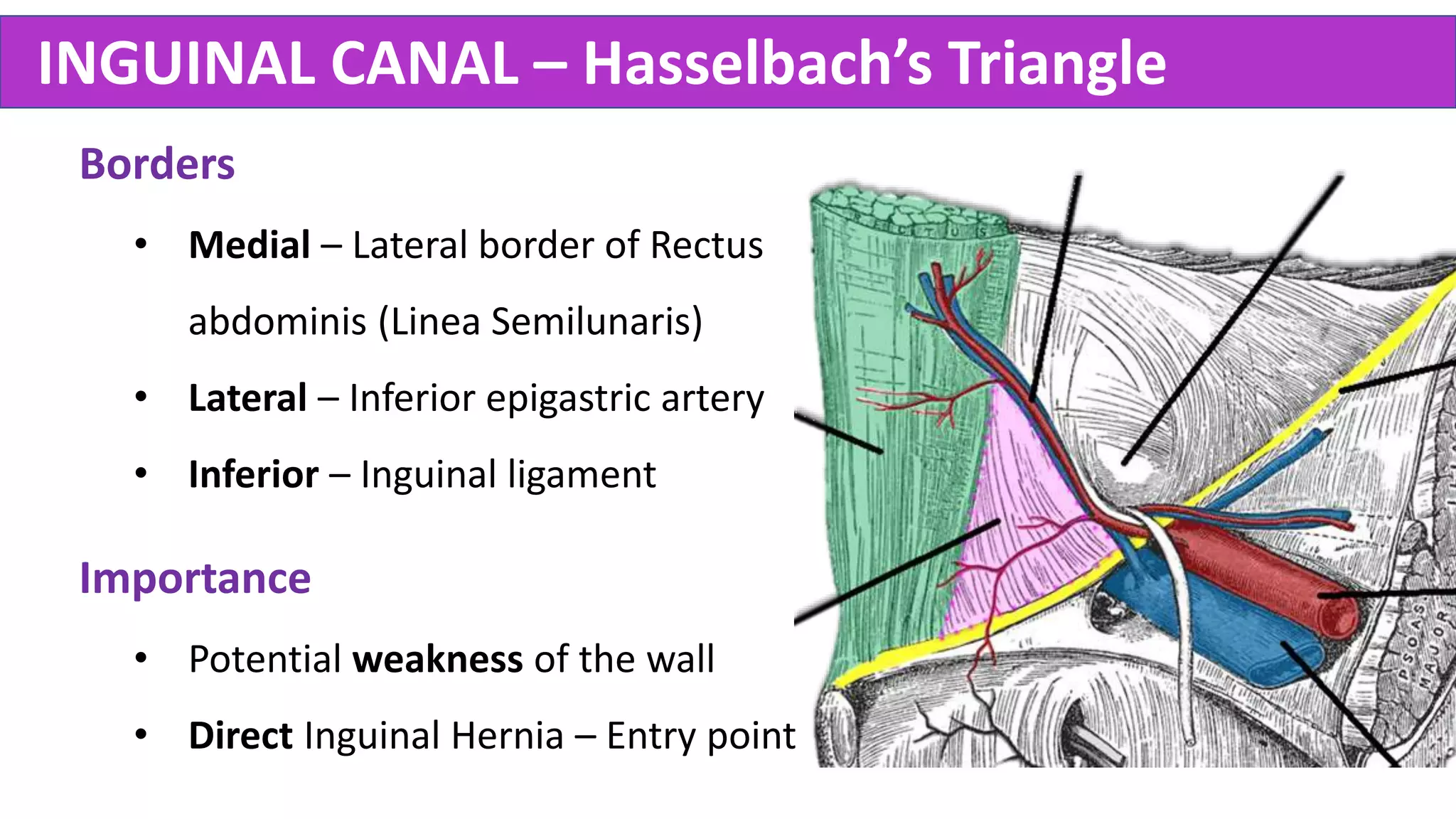
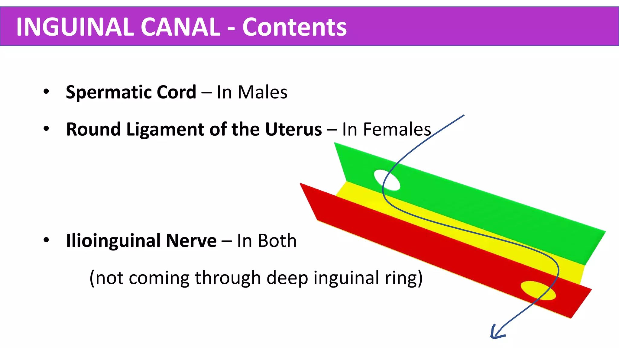
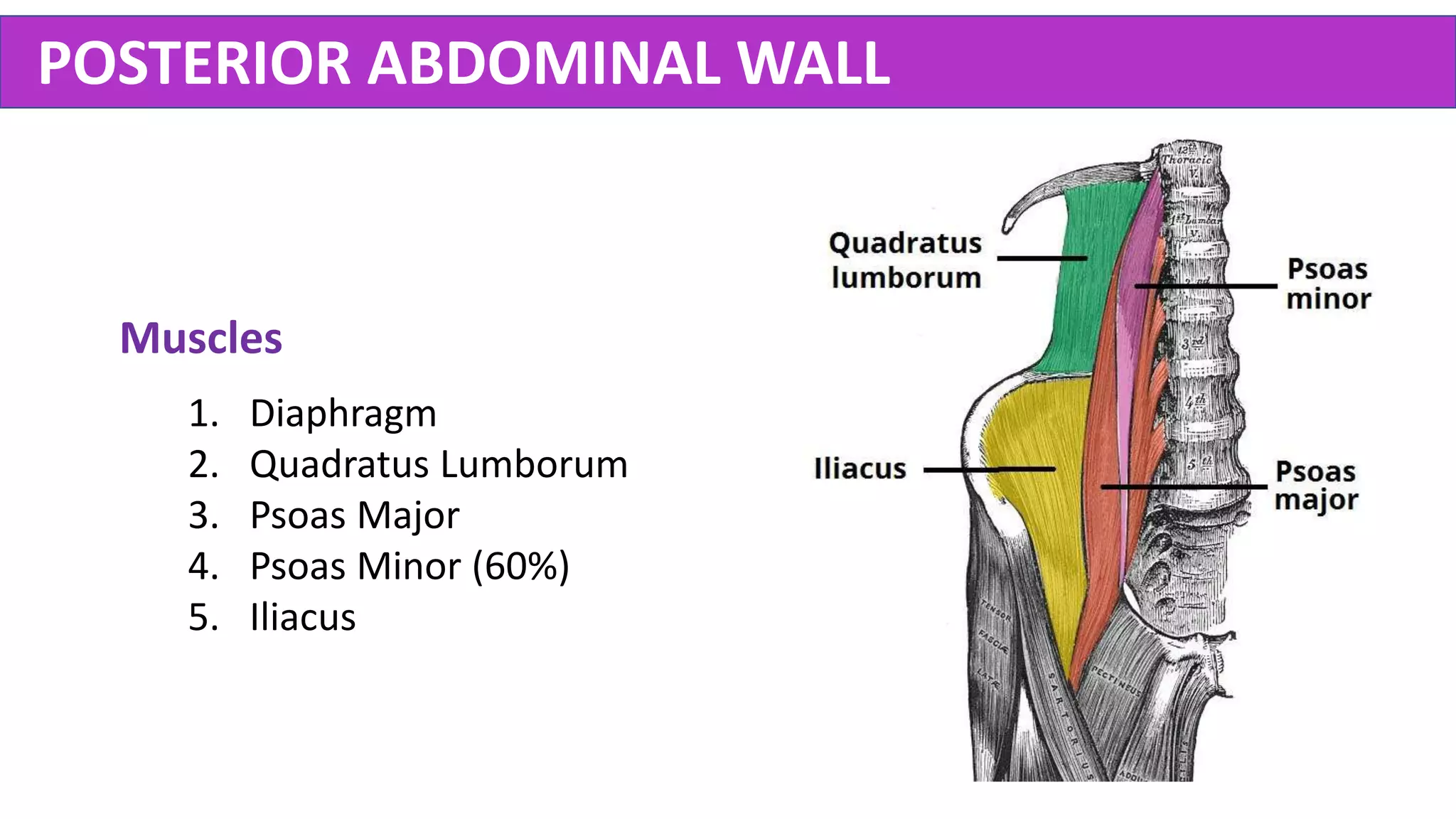
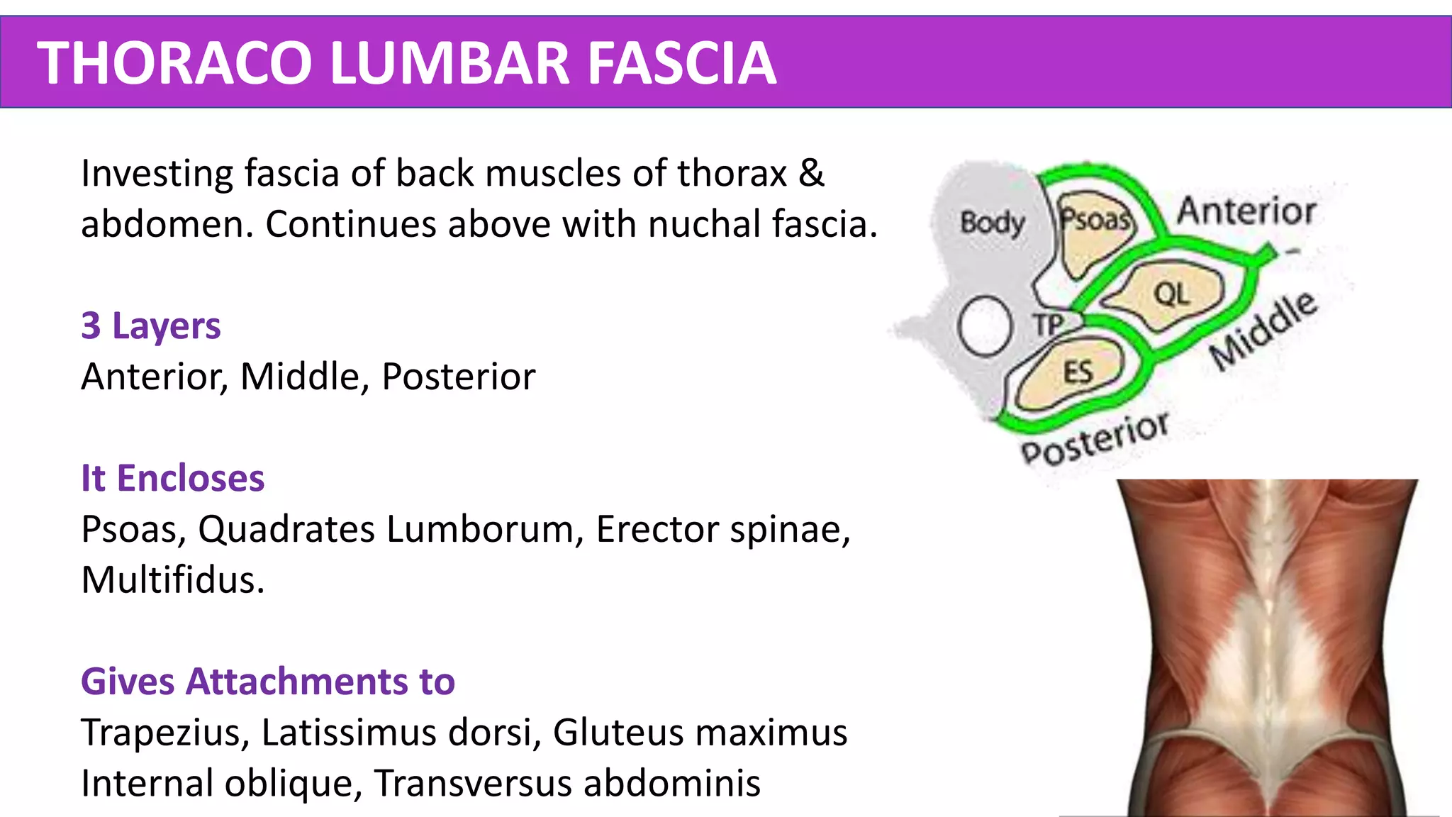
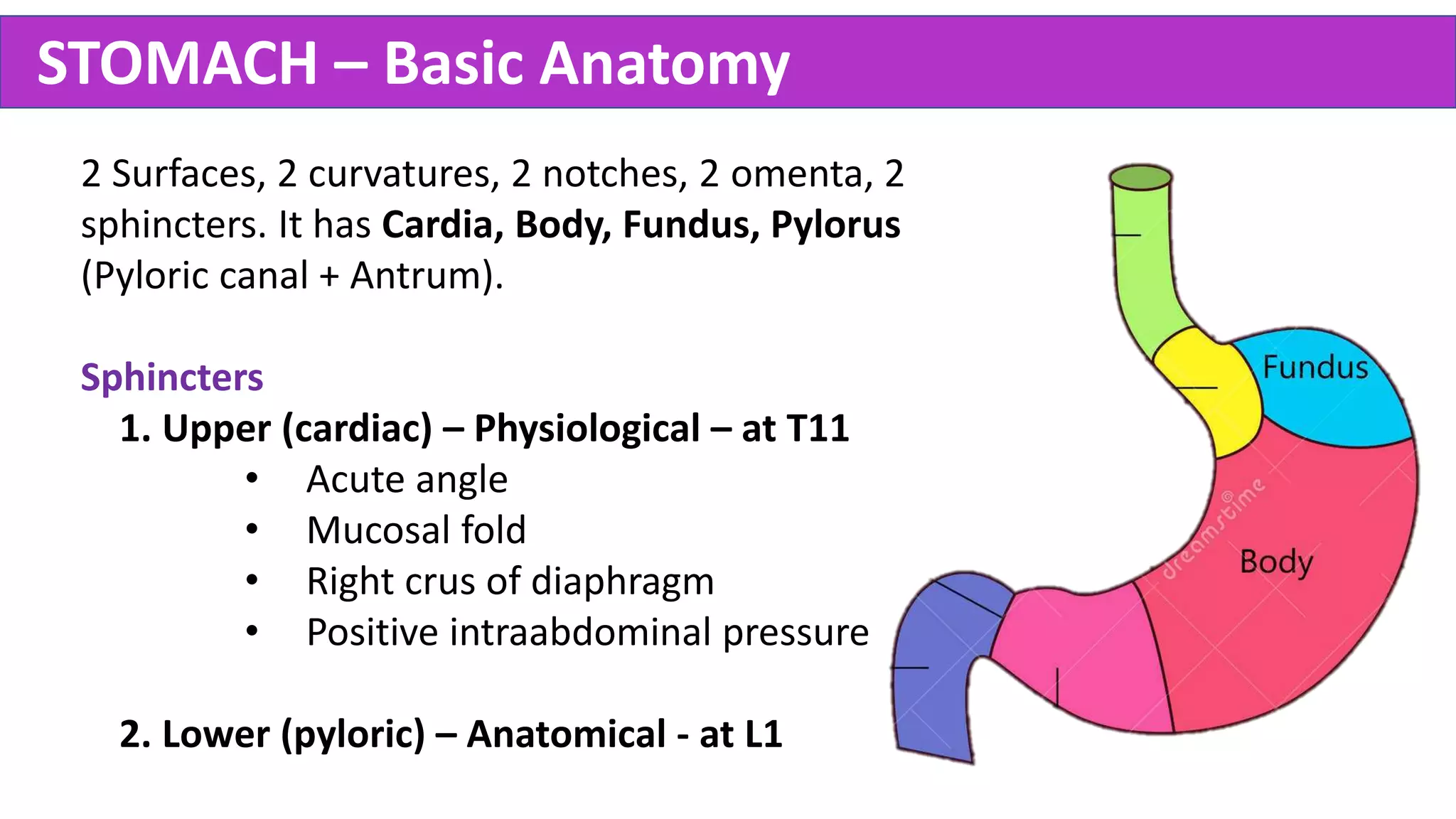
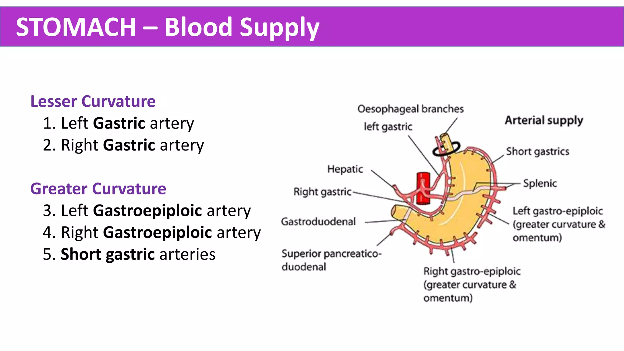
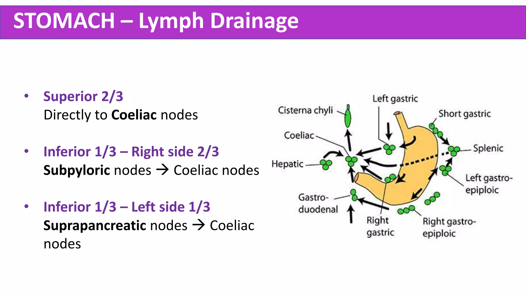
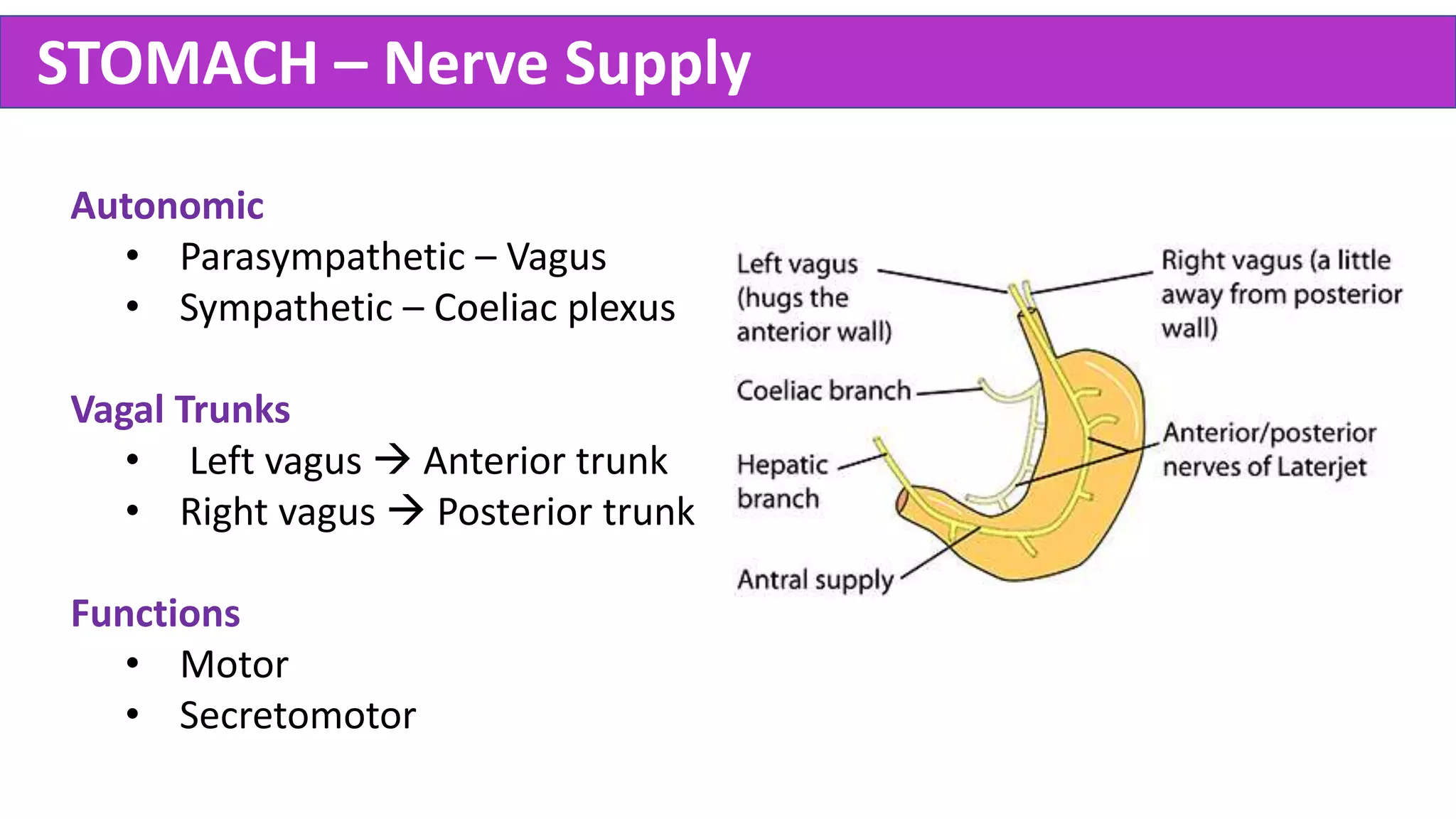
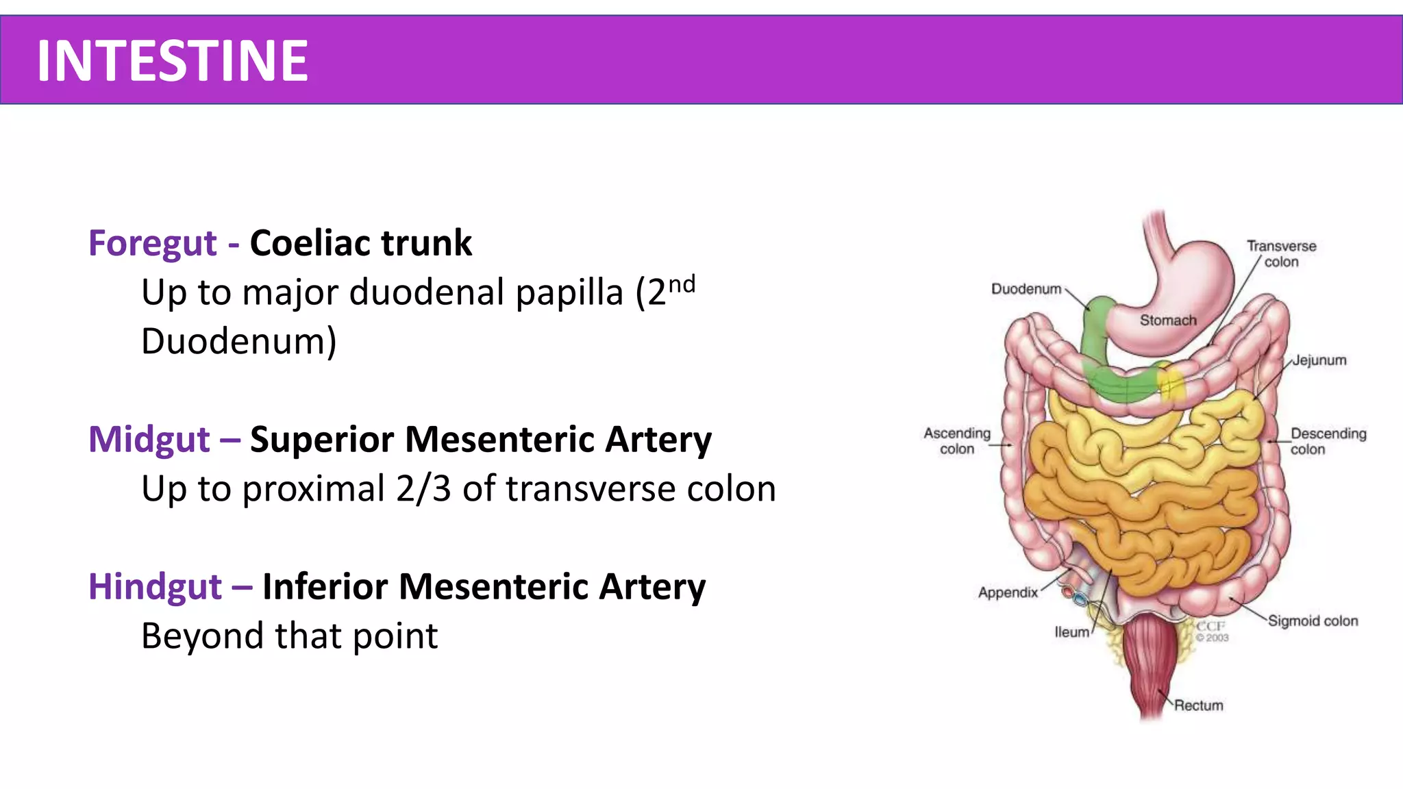
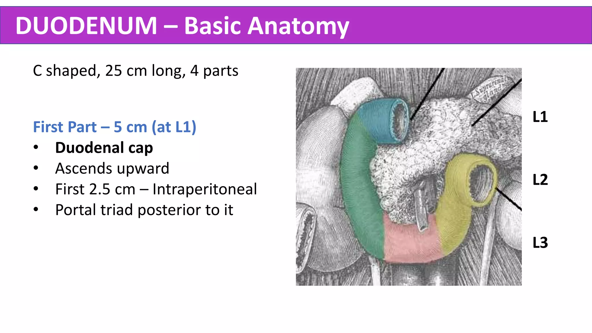
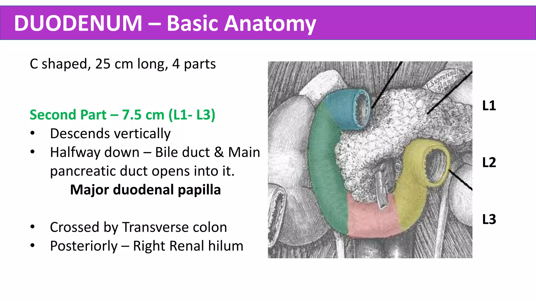
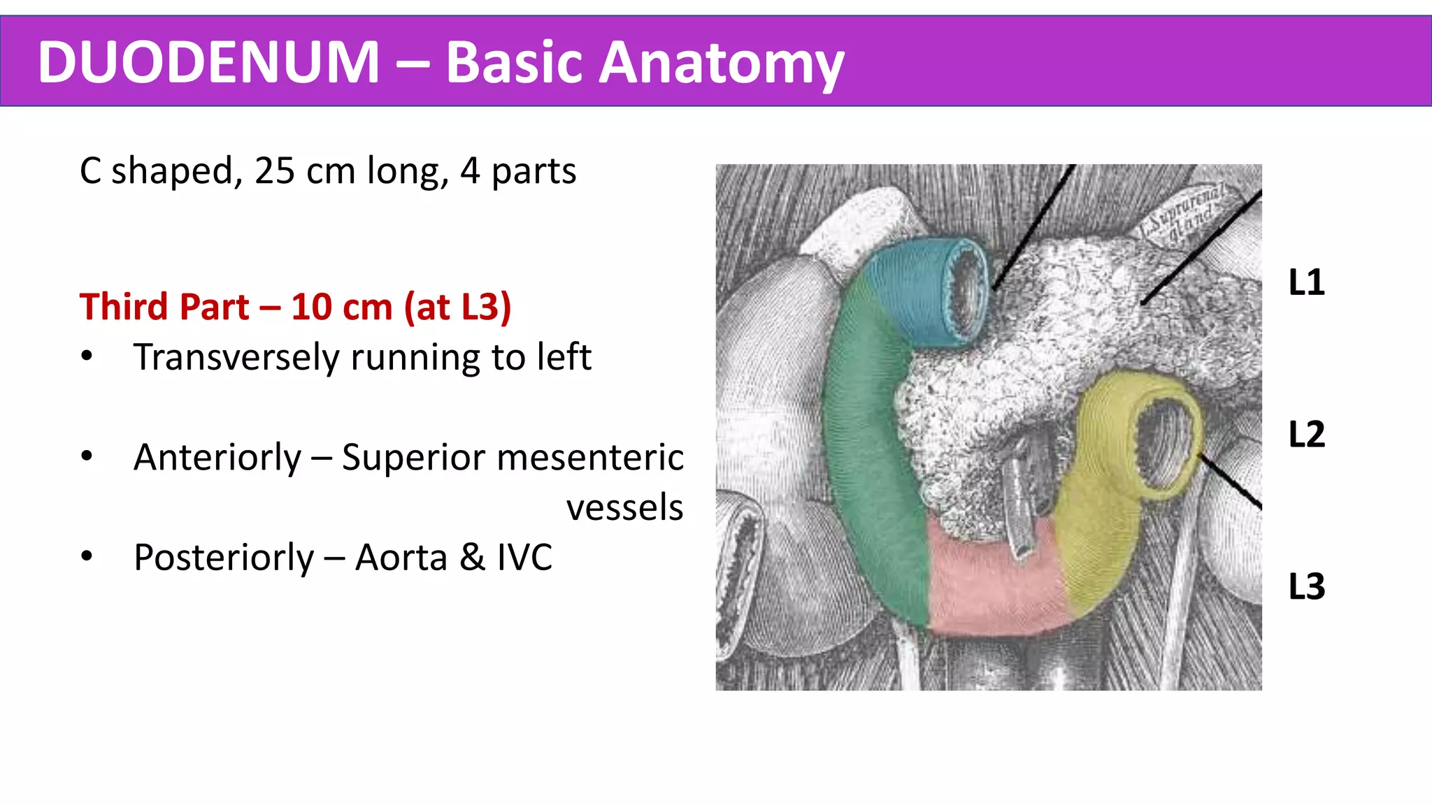
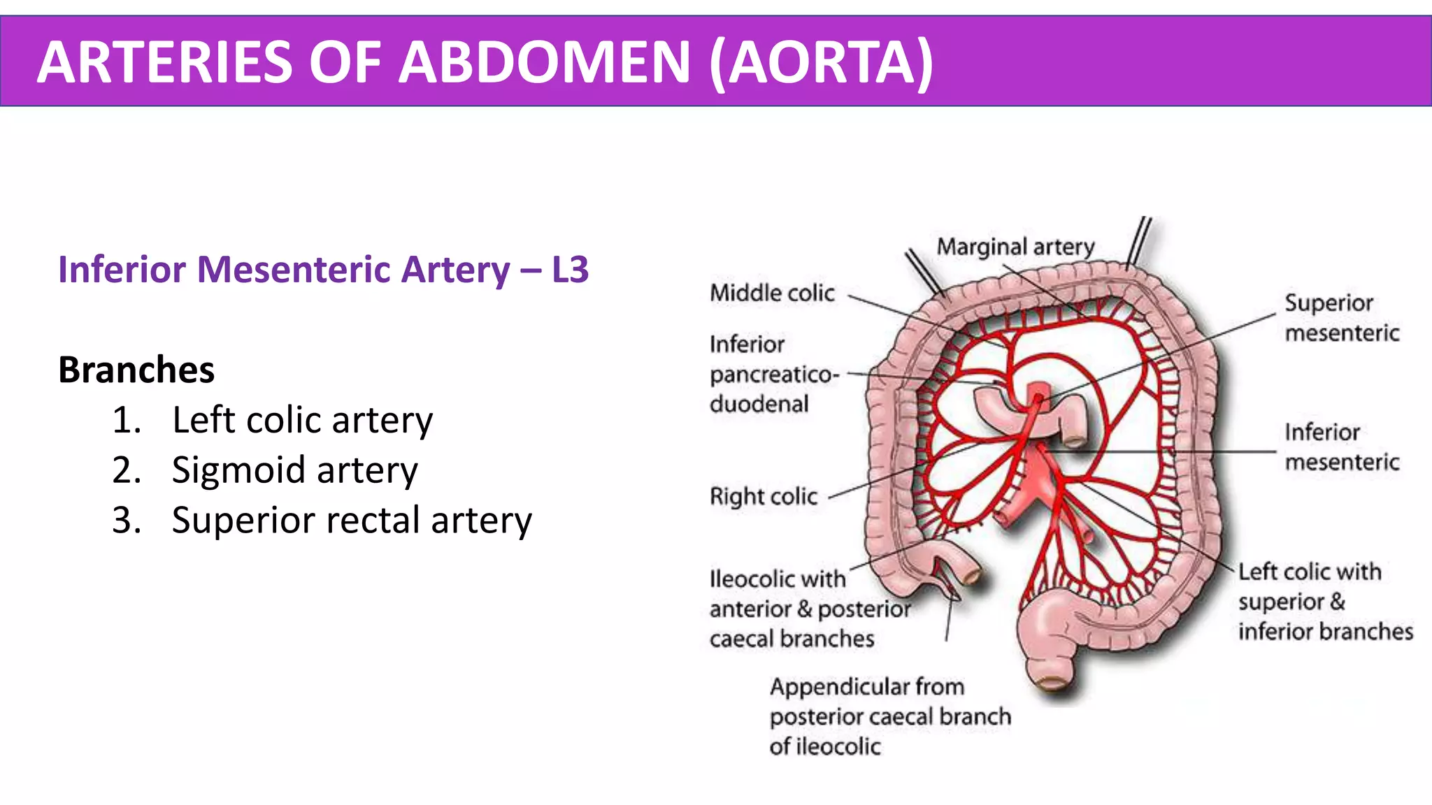
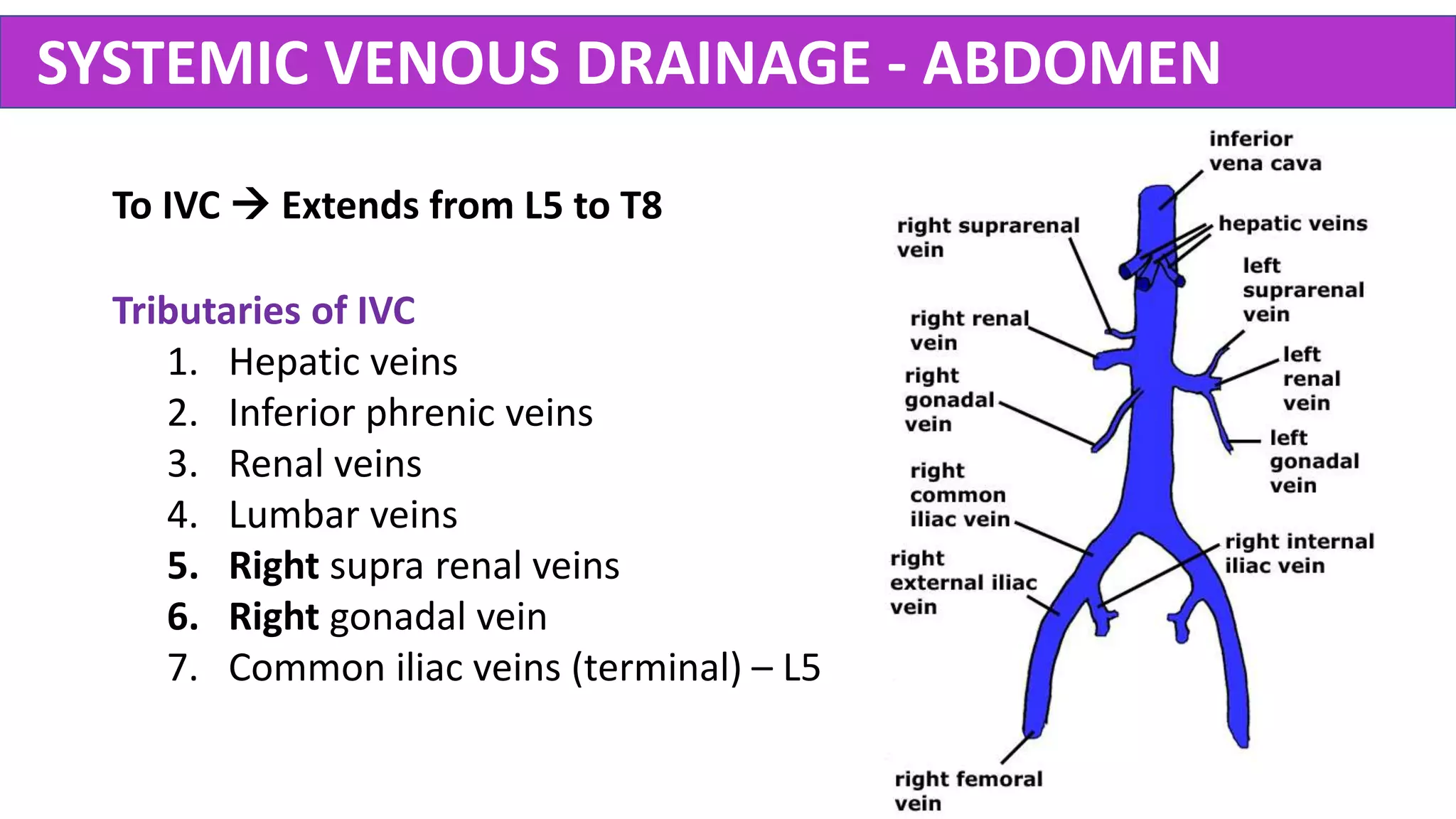
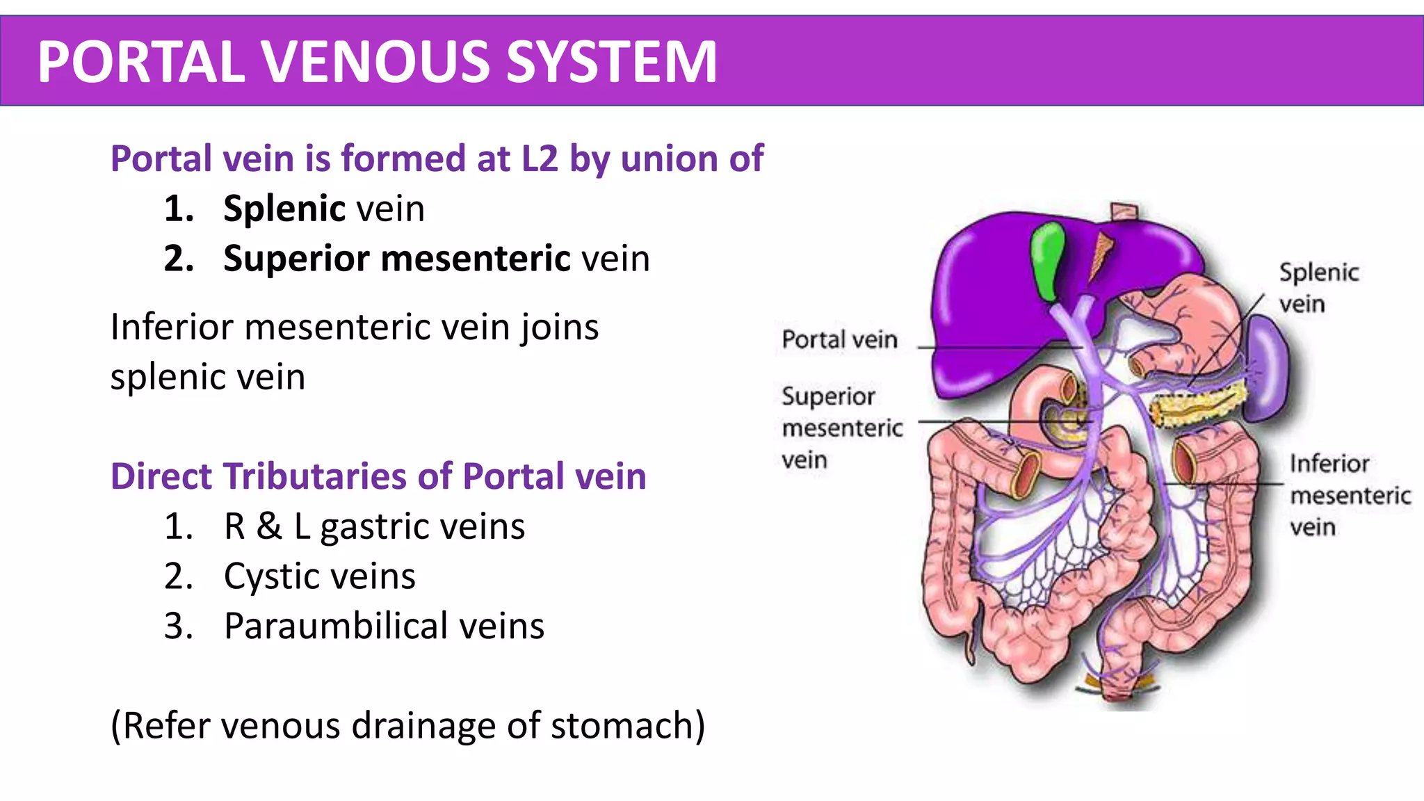
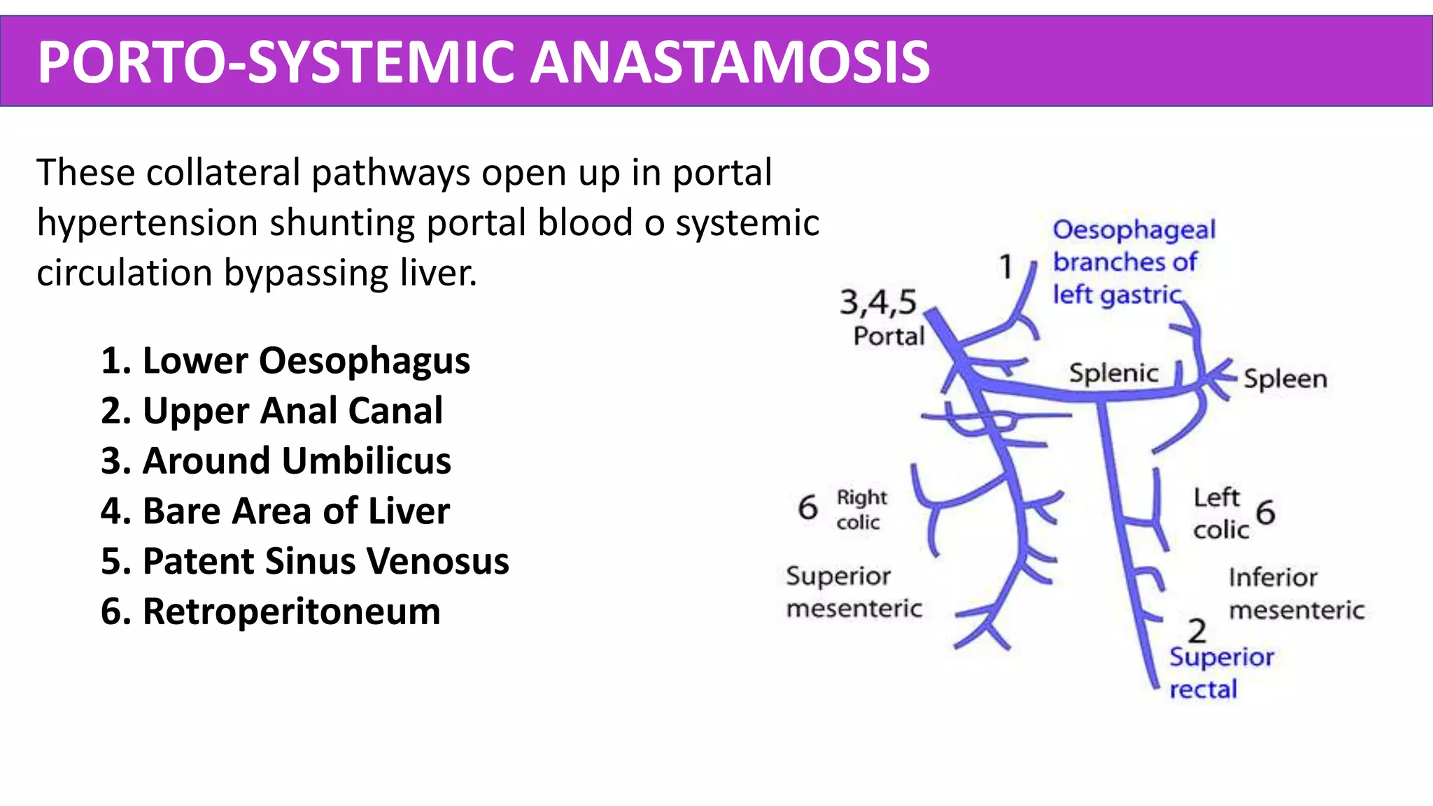
ABDOMEN
ANATOMY OF
Dr. Rajith Eranga
www.rermedapps.com
SURFACE ANATOMY – 9 Regions
A. Midclavicular Plane
Vertical line joining the mid-points of
clavicles to mid-inguinal points.
B. Transpyloric Plane (L1)
Horizontal line joining the mid-point
between jugular notch & pubic symphysis.
C. Intertubercular Plane (L5)
Horizontal line joining the two iliac tubercles.
A
B
C
A
SURFACE ANATOMY - Planes
1. Transpyloric Plane – L1
• Pylorus of Stomach
• Neck of Pancreas
• Fundus of Gallbladder
• Renal Hilum
• Duodenojejunal flexure
• End of Spinal Cord (adult)
2. Subcostal Plane – L3
• Origin of Inferior mesenteric artery
3. Supracristal Plane – L4
• Bifurcation of Aorta
ANTERIOR ABDOMINAL WALL - Layers
1
2
3
4
5
6
7
89
1. External Oblique
2. Internal Oblique
3. Transversus Abdominis
4. Rectus Abdominis
5. Pyramidalis
ANTERIOR ABDOMINAL WALL - Muscles
Rectus Sheath
INGUINAL CANAL – Basic Anatomy
• It is an oblique intermuscular passage in
the lower abdominal wall, located parallel
& just above the medial half of the
inguinal ligament.
• Extends from the deep inguinal ring to
the superficial inguinal ring.
• Directed downwards, forwards &
medially.
• 4 cm in length. Larger in males.
RAJITH ERANGA
Anterior Wall
Full Extent
• Skin
• Superficial fascia
• External Oblique Aponeurosis
Lateral 1/3
• Fibres of Internal oblique muscle M
L
INGUINAL CANAL - Walls
RAJITH ERANGA
Floor
Formed by
1. Medial part of inguinal ligament.
2. Lacunae ligament at the medial
end.
INGUINAL CANAL - Walls
RAJITH ERANGA
Roof
Formed by the arching fibres
of the
1. Internal Oblique
2. Transverse Abdominis
muscles.
INGUINAL CANAL - Walls
RAJITH ERANGA
Posterior Wall
Full Extent
• Fascia Transversalis
• Extraperitoneal fat
• Parietal peritoneum
Medial 2/3
• Conjoint tendon
• Reflected part of the inguinal ligament
M
L
INGUINAL CANAL - Walls
RAJITH ERANGA
Deep (Internal) Inguinal Ring
• Opening in the posterior wall
• Defect in the transversalis fascia
• Demarcated medially by the
inferior epigastric artery
INGUINAL CANAL - Openings
RAJITH ERANGA
Superficial (External) Inguinal Ring
• Opening in the anterior wall
• Defect in the External oblique
aponeurosis
• Above & medial to pubic tubercle
INGUINAL CANAL - Openings
RAJITH ERANGA
INGUINAL CANAL – Hasselbach’s Triangle
Borders
• Medial – Lateral border of Rectus
abdominis (Linea Semilunaris)
• Lateral – Inferior epigastric artery
• Inferior – Inguinal ligament
Importance
• Potential weakness of the wall
• Direct Inguinal Hernia – Entry point
RAJITH ERANGA
• Spermatic Cord – In Males
• Round Ligament of the Uterus – In Females
INGUINAL CANAL - Contents
• Spermatic Cord – In Males
• Round Ligament of the Uterus – In Females
• Ilioinguinal Nerve – In Both
(not coming through deep inguinal ring)
INGUINAL CANAL - Contents
POSTERIOR ABDOMINAL WALL
Muscles
1. Diaphragm
2. Quadratus Lumborum
3. Psoas Major
4. Psoas Minor (60%)
5. Iliacus
THORACO LUMBAR FASCIA
Investing fascia of back muscles of thorax &
abdomen. Continues above with nuchal fascia.
3 Layers
Anterior, Middle, Posterior
It Encloses
Psoas, Quadrates Lumborum, Erector spinae,
Multifidus.
Gives Attachments to
Trapezius, Latissimus dorsi, Gluteus maximus
Internal oblique, Transversus abdominis
PERITONEUM – Basic Anatomy
Serous membrane lining abdominal cavity & viscera.
Layers
1. Parietal – Somatic mesoderm
2. Visceral – Splanchnic mesoderm
Functions
1. Convey neurovascular bundle
2. Lubrication – Prevent friction while moving
3. Maintain position of organs
INTRAPERITONEAL Vs RETROPERITONEAL
Intraperitoneal Organs
Wrapped by visceral peritoneum completely.
Eg: Spleen, Liver, Stomach, Jejunum
Retroperitoneal Organs
Covered with peritoneum only anteriorly.
1. Primarily Retroperitoneal
Kidney, Adrenal
2. Secondarily Retroperitoneal
Ascending & Descending colons
PosteriorAbdominalWall
PERITONEAL REFLECTIONS
1. Mesenteries
Suspends organs to posterior abdominal wall
A. The mesentery (of the small intestine)
B. Transverse Mesocolon
C. Sigmoid Mesocolon
D. Meso-appendix
PERITONEAL REFLECTIONS
2. Omenta
Attach Stomach & proximal duodenum
to other organs.
• Lesser Omentum (2 Layered)
From lesser curvature to liver
• Greater Omentum (4 layered)
From greater curvature to
transverse mesocolon
PERITONEAL REFLECTIONS
3. Ligaments
Attach organs to other organs or wall.
• Phrenicocolic ligament
• Gastrosplenic ligament
• Splenorenal ligament
• Falciform ligament
• Gastrohepatic ligament
• Hepatoduodenal ligament
Lesser
Omentum
PERITONEAL CAVITY - Divisions
1. Greater Sac
1. Supracolic compartment
2. Infracolic compartments (Left & Right)
2. Lesser Sac (Omental Bursa)
• Supracolic & Infracolic compartments are
connected by R & L paracolic gutters.
• Greater & Lesser sacs are connected by
Epiploic foramen (of Winslow).
Potential space between parietal & visceral peritoneum.
EPIPLOIC (OMENTAL) FORAMEN
Borders
• Superior – Caudate lobe
• Inferior – Duodenum (1st Part)
• Anterior – Hepatoduodenal ligament
(containing bile duct, hepatic artery,
portal vein)
• Posterior - IVC
Importance
Pringle’s manoeuvre
Lesser sac communicates with greater sac via this.
PERITONEAL RECESSES
a. Lesser Sac (omental bursa)
b. Subphrenic Spaces
1. Right
2. Left
c. Subhepatic Spaces
1. Right (Morrison’s pouch)
2. Left
d. Paraduodenal fossae - 4
e. Retrocecal fossa
f. Intersigmoid fossa
STOMACH – Basic Anatomy
2 Surfaces, 2 curvatures, 2 notches, 2 omenta, 2
sphincters. It has Cardia, Body, Fundus, Pylorus
(Pyloric canal + Antrum).
Sphincters
1. Upper (cardiac) – Physiological – at T11
• Acute angle
• Mucosal fold
• Right crus of diaphragm
• Positive intraabdominal pressure
2. Lower (pyloric) – Anatomical - at L1
STOMACH BED
1. Lesser sac
2. Left Crus of Diaphragm
3. Pancreas
4. Spleen
5. Left Kidney
6. Left Suprarenal
7. Splenic artery
8. Transverse Colon
9. Transverse Mesocolon
STOMACH – Blood Supply
Lesser Curvature
1. Left Gastric artery
2. Right Gastric artery
Greater Curvature
3. Left Gastroepiploic artery
4. Right Gastroepiploic artery
5. Short gastric arteries
STOMACH – Blood Supply
To Portal Vein
1. Left Gastric vein
2. Right Gastric vein
To Superior Mesenteric Vein
3. Left Gastroepiploic vein
To Splenic Vein
4. Right Gastroepiploic vein
5. Short gastric veins
STOMACH – Lymph Drainage
• Superior 2/3
Directly to Coeliac nodes
• Inferior 1/3 – Right side 2/3
Subpyloric nodes  Coeliac nodes
• Inferior 1/3 – Left side 1/3
Suprapancreatic nodes  Coeliac
nodes
STOMACH – Nerve Supply
Autonomic
• Parasympathetic – Vagus
• Sympathetic – Coeliac plexus
Vagal Trunks
• Left vagus  Anterior trunk
• Right vagus  Posterior trunk
Functions
• Motor
• Secretomotor
INTESTINE
Foregut - Coeliac trunk
Up to major duodenal papilla (2nd
Duodenum)
Midgut – Superior Mesenteric Artery
Up to proximal 2/3 of transverse colon
Hindgut – Inferior Mesenteric Artery
Beyond that point
DUODENUM – Basic Anatomy
C shaped, 25 cm long, 4 parts
First Part – 5 cm (at L1)
• Duodenal cap
• Ascends upward
• First 2.5 cm – Intraperitoneal
• Portal triad posterior to it
L1
L2
L3
DUODENUM – Basic Anatomy
C shaped, 25 cm long, 4 parts
Second Part – 7.5 cm (L1- L3)
• Descends vertically
• Halfway down – Bile duct & Main
pancreatic duct opens into it.
Major duodenal papilla
• Crossed by Transverse colon
• Posteriorly – Right Renal hilum
L1
L2
L3
DUODENUM – Basic Anatomy
C shaped, 25 cm long, 4 parts
Third Part – 10 cm (at L3)
• Transversely running to left
• Anteriorly – Superior mesenteric
vessels
• Posteriorly – Aorta & IVC
L1
L2
L3
DUODENUM – Basic Anatomy
C shaped, 25 cm long, 4 parts
Fourth Part – 2.5 cm (L3 - L2)
• Ascends upward
• Ligament of Trietz
R crus to duodnojejunal junction
L1
L2
L3
DUODENUM – Blood Supply
Site of anastomosis (Watershed)
between foregut & midgut
vessels.
Coeliac trunk 
Superior Pancreatico duodenal
+
Superior Mesenteric Artery 
Inferior Pancreatico duodenal
JEJUNUM & ILEUM
JEJUNUM ILEUM
Diameter Larger Smaller
Wall Thicker Thinner
Region Umbilical Suprapubic
Mesentery Less fat & thin More fat & thicker
Vessels 1 or 2 Arcades Multiple arcades
Lumen Plicae circularis Peyer’s patches
Histology Tall villi, Deep crypts Short villi, shallow crypts
CAECUM
• Below ile-ocaecal valve
• Usually intraperitoneal
• 3 teniae meet at the base of
appendix
• Retrocaecal fossa behind it
APPENDIX
• A diverticulum
• At McBurney’s point
• Meso-appendix – Appendicular
artery (from ileocolic artery)
• End arterial supply – Gangrene!
• Usually retrocaecal.
Appendicectomy
Using Gridiron or Lanz incision
COLON
Length - 150cm
Parts
1. Ascending colon - Retroperitoneal
2. Transverse colon - Intraperitoneal
3. Descending colon - Retroperitoneal
4. Sigmoid colon - Intraperitoneal
Structure
• Omental appendices
• Tinae coli - 3
• Haustrations
SPLEEN
• Lies on 9, 10, 11 ribs
• Becomes palpable when it is twice normal
size
• Thin capsule
• Splenic notch in anterior border
• Related closely to stomach, splenic flexure,
left kidney & tail of pancreas
• Splenorenal ligament - splenic vessels
• Gastrosplenic ligament - short gastric vessels
LIVER – Basic Anatomy
Largest solid organ (1.5Kg)
Location
Right Hypogastrium + Epigastrium + Left
Hypogastrium
Lobes
1. Right lobe (largest)
2. Left lobe
3. Caudate lobe
4. Quadrate lobe
LIVER – Peritoneal Attachments
• Falciform ligament & Ligamentum
teres – to abdominal wall
• 2 Coronary ligaments (Ant & Post
folds) – demarcates bare area
• 2 Triangular ligaments (L & R) –
attach to diaphragm
• Lesser omentum – attach to
stomach
Bare Area of Liver
LIVER – Porta Hepatis
• Area on under surface
• Cross-bar of ‘H’
• Structures enter/leave liver
• Peritoneum is reflected around it
Contents
• Hepatic artery
• Common hepatic duct
• Portal vein
• Lymphatics & LN
• Autonomic nerves
LIVER – Blood Supply
Dual blood supply
1. Hepatic artery (25%)
2. Portal vein (75%)
Venous drainage
Central veins
Collecting veins
Hepatic veins IVC
LIVER – Nerve Supply & Lymph Drainage
Nerve Supply
• Capsule (Glisson’s capsule)
Lower intercostal nerves
• Liver Parenchyma
Hepatic plexus (Celiac plexus + Vagus)
Lymph drainage
• Anterior aspect - Hepatic LN Cysterna Chyli
• Posterior aspect - Posterior mediastinal LN (via bare area)
GALLBLADDER
• Bile reservoir (50ml)
• In GB Fossa (Between Right & quadrate lobes)
• Fundus, Body, Neck, Cystic duct
• Blood supply – Cystic artery & Liver bed
Clinicals
• Murphy’s sign - Read
• Courvoisier's law - Read
• Cholecystitis – Inflammation of GB
• Cholangitis – Inflammation of bile duct
• Charcot’s triad – Fever + Jaundice + RUQ pain
BILLIARY SYSTEM
Calot’s Triangle
• Between Cystic duct,
Common hepatic duct & Liver.
• Cystic artery is located here.
• Important in cholecystectomy.
• R & L hepatic duct  Common Hepatic duct
• Common hepatic duct + Cystic duct  Bile duct
• Bile duct opens to 2nd part of duodenum with
main pancreatic duct at ampulla of Vater
• It is guarded by sphincter of Oddi
PANCREAS
• Retroperitoneal
• Exocrine + Endocrine gland
• Head, Uncinate process, Neck, Body, Tail
Relations & Development (Read)
Ducts
1. Main duct – at Amulla of Vater
2. Accessory duct - Proximally
Clinicals
• Pancreatic CA - Bile duct obstruction
• Annular Pancreas - Duodenal obstruction
• Pancreatic pseudocyst
KIDNEYS – Basic Anatomy
• Retroperitoneal
• From T12 to L3 (Hilum at L1)
• Parenchyma  Cortex & Medulla
• Cortex extends to Medulla = Pyramids
• Apex of Pyramids = Renal Papilla
• Renal papilla  Minor Calyx  Major
Calyx  Renal Pelvis Ureter
KIDNEYS – Renal Fascia & Fat
From deep to superficial
KIDNEYS – Arterial Supply
Renal arteries (from aorta)  Divides at hilum
• Anterior – 75%
• Posterior – 25%
• Five Segmental arteries from them
These arteries are end arteries!
Segmental arteries divides further…
Segmental  Interlobar  Arcuate 
interlobular Afferent arterioles
 Glomerulus
URETERS
• 25 cm long
• Retroperitoneal, adhered to peritoneum
• 3 parts – Abdominal, Pelvic & Intravesical
Sites of Potential Obstruction of Calculi
1. Pelviureteric junction
2. Pelvic Brim
3. Vesicoureteric junction
URETERS - Relations
Posteriorly
• Psoas (medial border)
• Transverse processes of L2-L5
• Sacroiliac joint
• Bifurcation of common iliac Artery
• Genitofemoral nerve
Anteriorly
• 2nd Duodenum
• Gonadal vessels
• Right colic vessels
• Ileocolic vessels
SUPRARENAL GLANDS - Layers
Cortex – Mesoderm
1. Zona Glomerulosa
2. Zona Fasciculata
3. Zona Reticularis
Medulla – Ectoderm (Neural crest cells)
SUPRARENAL GLANDS – Vasculature
Arterial Supply – 3 arteries
1. Superior adrenal artery  Inferior Phrenic
2. Middle adrenal artery  Abdominal Aorta
3. Inferior adrenal artery  Renal Artery
Venous Drainage – Single vein
1. Left side – to Left Renal vein  IVC
2. Right side  IVC
ARTERIES OF ABDOMEN (AORTA)
Branches
Anterior Branches (Unpaired)
1. Coeliac trunk (L1)
2. Superior Mesenteric (L1)
3. Inferior Mesenteric (L3)
Lateral Branches (Paired)
1. Inferior Phrenic Arteries
2. Lumbar Arteries (4 pairs)
3. Renal arteries
4. Supra renal arteries
5. Gonadal arteries
At L4  Bifurcate to two common iliac
arteries + Median sacral artery
ARTERIES OF ABDOMEN (AORTA)
Coeliac Trunk – L1
Branches
1. Left Gastric Artery
2. Splenic Artery
3. Common Hepatic Artery
ARTERIES OF ABDOMEN (AORTA)
Superior Mesenteric Artery – L1
Branches
1. Inferior Pancreaticoduodenal
2. Middle Colic artery
3. Right Colic artery
4. Ileocolic artery
ARTERIES OF ABDOMEN (AORTA)
Inferior Mesenteric Artery – L3
Branches
1. Left colic artery
2. Sigmoid artery
3. Superior rectal artery
SYSTEMIC VENOUS DRAINAGE - ABDOMEN
Tributaries of IVC
1. Hepatic veins
2. Inferior phrenic veins
3. Renal veins
4. Lumbar veins
5. Right supra renal veins
6. Right gonadal vein
7. Common iliac veins (terminal) – L5
To IVC  Extends from L5 to T8
PORTAL VENOUS SYSTEM
Portal vein is formed at L2 by union of
1. Splenic vein
2. Superior mesenteric vein
Inferior mesenteric vein joins
splenic vein
Direct Tributaries of Portal vein
1. R & L gastric veins
2. Cystic veins
3. Paraumbilical veins
(Refer venous drainage of stomach)
PORTO-SYSTEMIC ANASTAMOSIS
These collateral pathways open up in portal
hypertension shunting portal blood o systemic
circulation bypassing liver.
1. Lower Oesophagus
2. Upper Anal Canal
3. Around Umbilicus
4. Bare Area of Liver
5. Patent Sinus Venosus
6. Retroperitoneum
THANK YOU 
END OF THE LECTURE

Abdomen Anatomy - Made Easy

  • 1.
    ABDOMEN ANATOMY OF Dr. RajithEranga www.rermedapps.com
  • 2.
    SURFACE ANATOMY –9 Regions A. Midclavicular Plane Vertical line joining the mid-points of clavicles to mid-inguinal points. B. Transpyloric Plane (L1) Horizontal line joining the mid-point between jugular notch & pubic symphysis. C. Intertubercular Plane (L5) Horizontal line joining the two iliac tubercles. A B C A
  • 3.
    SURFACE ANATOMY -Planes 1. Transpyloric Plane – L1 • Pylorus of Stomach • Neck of Pancreas • Fundus of Gallbladder • Renal Hilum • Duodenojejunal flexure • End of Spinal Cord (adult) 2. Subcostal Plane – L3 • Origin of Inferior mesenteric artery 3. Supracristal Plane – L4 • Bifurcation of Aorta
  • 4.
    ANTERIOR ABDOMINAL WALL- Layers 1 2 3 4 5 6 7 89
  • 5.
    1. External Oblique 2.Internal Oblique 3. Transversus Abdominis 4. Rectus Abdominis 5. Pyramidalis ANTERIOR ABDOMINAL WALL - Muscles Rectus Sheath
  • 6.
    INGUINAL CANAL –Basic Anatomy • It is an oblique intermuscular passage in the lower abdominal wall, located parallel & just above the medial half of the inguinal ligament. • Extends from the deep inguinal ring to the superficial inguinal ring. • Directed downwards, forwards & medially. • 4 cm in length. Larger in males.
  • 7.
    RAJITH ERANGA Anterior Wall FullExtent • Skin • Superficial fascia • External Oblique Aponeurosis Lateral 1/3 • Fibres of Internal oblique muscle M L INGUINAL CANAL - Walls
  • 8.
    RAJITH ERANGA Floor Formed by 1.Medial part of inguinal ligament. 2. Lacunae ligament at the medial end. INGUINAL CANAL - Walls
  • 9.
    RAJITH ERANGA Roof Formed bythe arching fibres of the 1. Internal Oblique 2. Transverse Abdominis muscles. INGUINAL CANAL - Walls
  • 10.
    RAJITH ERANGA Posterior Wall FullExtent • Fascia Transversalis • Extraperitoneal fat • Parietal peritoneum Medial 2/3 • Conjoint tendon • Reflected part of the inguinal ligament M L INGUINAL CANAL - Walls
  • 11.
    RAJITH ERANGA Deep (Internal)Inguinal Ring • Opening in the posterior wall • Defect in the transversalis fascia • Demarcated medially by the inferior epigastric artery INGUINAL CANAL - Openings
  • 12.
    RAJITH ERANGA Superficial (External)Inguinal Ring • Opening in the anterior wall • Defect in the External oblique aponeurosis • Above & medial to pubic tubercle INGUINAL CANAL - Openings
  • 13.
    RAJITH ERANGA INGUINAL CANAL– Hasselbach’s Triangle Borders • Medial – Lateral border of Rectus abdominis (Linea Semilunaris) • Lateral – Inferior epigastric artery • Inferior – Inguinal ligament Importance • Potential weakness of the wall • Direct Inguinal Hernia – Entry point
  • 14.
    RAJITH ERANGA • SpermaticCord – In Males • Round Ligament of the Uterus – In Females INGUINAL CANAL - Contents
  • 15.
    • Spermatic Cord– In Males • Round Ligament of the Uterus – In Females • Ilioinguinal Nerve – In Both (not coming through deep inguinal ring) INGUINAL CANAL - Contents
  • 16.
    POSTERIOR ABDOMINAL WALL Muscles 1.Diaphragm 2. Quadratus Lumborum 3. Psoas Major 4. Psoas Minor (60%) 5. Iliacus
  • 17.
    THORACO LUMBAR FASCIA Investingfascia of back muscles of thorax & abdomen. Continues above with nuchal fascia. 3 Layers Anterior, Middle, Posterior It Encloses Psoas, Quadrates Lumborum, Erector spinae, Multifidus. Gives Attachments to Trapezius, Latissimus dorsi, Gluteus maximus Internal oblique, Transversus abdominis
  • 18.
    PERITONEUM – BasicAnatomy Serous membrane lining abdominal cavity & viscera. Layers 1. Parietal – Somatic mesoderm 2. Visceral – Splanchnic mesoderm Functions 1. Convey neurovascular bundle 2. Lubrication – Prevent friction while moving 3. Maintain position of organs
  • 19.
    INTRAPERITONEAL Vs RETROPERITONEAL IntraperitonealOrgans Wrapped by visceral peritoneum completely. Eg: Spleen, Liver, Stomach, Jejunum Retroperitoneal Organs Covered with peritoneum only anteriorly. 1. Primarily Retroperitoneal Kidney, Adrenal 2. Secondarily Retroperitoneal Ascending & Descending colons PosteriorAbdominalWall
  • 20.
    PERITONEAL REFLECTIONS 1. Mesenteries Suspendsorgans to posterior abdominal wall A. The mesentery (of the small intestine) B. Transverse Mesocolon C. Sigmoid Mesocolon D. Meso-appendix
  • 21.
    PERITONEAL REFLECTIONS 2. Omenta AttachStomach & proximal duodenum to other organs. • Lesser Omentum (2 Layered) From lesser curvature to liver • Greater Omentum (4 layered) From greater curvature to transverse mesocolon
  • 22.
    PERITONEAL REFLECTIONS 3. Ligaments Attachorgans to other organs or wall. • Phrenicocolic ligament • Gastrosplenic ligament • Splenorenal ligament • Falciform ligament • Gastrohepatic ligament • Hepatoduodenal ligament Lesser Omentum
  • 23.
    PERITONEAL CAVITY -Divisions 1. Greater Sac 1. Supracolic compartment 2. Infracolic compartments (Left & Right) 2. Lesser Sac (Omental Bursa) • Supracolic & Infracolic compartments are connected by R & L paracolic gutters. • Greater & Lesser sacs are connected by Epiploic foramen (of Winslow). Potential space between parietal & visceral peritoneum.
  • 24.
    EPIPLOIC (OMENTAL) FORAMEN Borders •Superior – Caudate lobe • Inferior – Duodenum (1st Part) • Anterior – Hepatoduodenal ligament (containing bile duct, hepatic artery, portal vein) • Posterior - IVC Importance Pringle’s manoeuvre Lesser sac communicates with greater sac via this.
  • 25.
    PERITONEAL RECESSES a. LesserSac (omental bursa) b. Subphrenic Spaces 1. Right 2. Left c. Subhepatic Spaces 1. Right (Morrison’s pouch) 2. Left d. Paraduodenal fossae - 4 e. Retrocecal fossa f. Intersigmoid fossa
  • 26.
    STOMACH – BasicAnatomy 2 Surfaces, 2 curvatures, 2 notches, 2 omenta, 2 sphincters. It has Cardia, Body, Fundus, Pylorus (Pyloric canal + Antrum). Sphincters 1. Upper (cardiac) – Physiological – at T11 • Acute angle • Mucosal fold • Right crus of diaphragm • Positive intraabdominal pressure 2. Lower (pyloric) – Anatomical - at L1
  • 27.
    STOMACH BED 1. Lessersac 2. Left Crus of Diaphragm 3. Pancreas 4. Spleen 5. Left Kidney 6. Left Suprarenal 7. Splenic artery 8. Transverse Colon 9. Transverse Mesocolon
  • 28.
    STOMACH – BloodSupply Lesser Curvature 1. Left Gastric artery 2. Right Gastric artery Greater Curvature 3. Left Gastroepiploic artery 4. Right Gastroepiploic artery 5. Short gastric arteries
  • 29.
    STOMACH – BloodSupply To Portal Vein 1. Left Gastric vein 2. Right Gastric vein To Superior Mesenteric Vein 3. Left Gastroepiploic vein To Splenic Vein 4. Right Gastroepiploic vein 5. Short gastric veins
  • 30.
    STOMACH – LymphDrainage • Superior 2/3 Directly to Coeliac nodes • Inferior 1/3 – Right side 2/3 Subpyloric nodes  Coeliac nodes • Inferior 1/3 – Left side 1/3 Suprapancreatic nodes  Coeliac nodes
  • 31.
    STOMACH – NerveSupply Autonomic • Parasympathetic – Vagus • Sympathetic – Coeliac plexus Vagal Trunks • Left vagus  Anterior trunk • Right vagus  Posterior trunk Functions • Motor • Secretomotor
  • 32.
    INTESTINE Foregut - Coeliactrunk Up to major duodenal papilla (2nd Duodenum) Midgut – Superior Mesenteric Artery Up to proximal 2/3 of transverse colon Hindgut – Inferior Mesenteric Artery Beyond that point
  • 33.
    DUODENUM – BasicAnatomy C shaped, 25 cm long, 4 parts First Part – 5 cm (at L1) • Duodenal cap • Ascends upward • First 2.5 cm – Intraperitoneal • Portal triad posterior to it L1 L2 L3
  • 34.
    DUODENUM – BasicAnatomy C shaped, 25 cm long, 4 parts Second Part – 7.5 cm (L1- L3) • Descends vertically • Halfway down – Bile duct & Main pancreatic duct opens into it. Major duodenal papilla • Crossed by Transverse colon • Posteriorly – Right Renal hilum L1 L2 L3
  • 35.
    DUODENUM – BasicAnatomy C shaped, 25 cm long, 4 parts Third Part – 10 cm (at L3) • Transversely running to left • Anteriorly – Superior mesenteric vessels • Posteriorly – Aorta & IVC L1 L2 L3
  • 36.
    DUODENUM – BasicAnatomy C shaped, 25 cm long, 4 parts Fourth Part – 2.5 cm (L3 - L2) • Ascends upward • Ligament of Trietz R crus to duodnojejunal junction L1 L2 L3
  • 37.
    DUODENUM – BloodSupply Site of anastomosis (Watershed) between foregut & midgut vessels. Coeliac trunk  Superior Pancreatico duodenal + Superior Mesenteric Artery  Inferior Pancreatico duodenal
  • 38.
    JEJUNUM & ILEUM JEJUNUMILEUM Diameter Larger Smaller Wall Thicker Thinner Region Umbilical Suprapubic Mesentery Less fat & thin More fat & thicker Vessels 1 or 2 Arcades Multiple arcades Lumen Plicae circularis Peyer’s patches Histology Tall villi, Deep crypts Short villi, shallow crypts
  • 39.
    CAECUM • Below ile-ocaecalvalve • Usually intraperitoneal • 3 teniae meet at the base of appendix • Retrocaecal fossa behind it
  • 40.
    APPENDIX • A diverticulum •At McBurney’s point • Meso-appendix – Appendicular artery (from ileocolic artery) • End arterial supply – Gangrene! • Usually retrocaecal. Appendicectomy Using Gridiron or Lanz incision
  • 41.
    COLON Length - 150cm Parts 1.Ascending colon - Retroperitoneal 2. Transverse colon - Intraperitoneal 3. Descending colon - Retroperitoneal 4. Sigmoid colon - Intraperitoneal Structure • Omental appendices • Tinae coli - 3 • Haustrations
  • 42.
    SPLEEN • Lies on9, 10, 11 ribs • Becomes palpable when it is twice normal size • Thin capsule • Splenic notch in anterior border • Related closely to stomach, splenic flexure, left kidney & tail of pancreas • Splenorenal ligament - splenic vessels • Gastrosplenic ligament - short gastric vessels
  • 43.
    LIVER – BasicAnatomy Largest solid organ (1.5Kg) Location Right Hypogastrium + Epigastrium + Left Hypogastrium Lobes 1. Right lobe (largest) 2. Left lobe 3. Caudate lobe 4. Quadrate lobe
  • 44.
    LIVER – PeritonealAttachments • Falciform ligament & Ligamentum teres – to abdominal wall • 2 Coronary ligaments (Ant & Post folds) – demarcates bare area • 2 Triangular ligaments (L & R) – attach to diaphragm • Lesser omentum – attach to stomach Bare Area of Liver
  • 45.
    LIVER – PortaHepatis • Area on under surface • Cross-bar of ‘H’ • Structures enter/leave liver • Peritoneum is reflected around it Contents • Hepatic artery • Common hepatic duct • Portal vein • Lymphatics & LN • Autonomic nerves
  • 46.
    LIVER – BloodSupply Dual blood supply 1. Hepatic artery (25%) 2. Portal vein (75%) Venous drainage Central veins Collecting veins Hepatic veins IVC
  • 47.
    LIVER – NerveSupply & Lymph Drainage Nerve Supply • Capsule (Glisson’s capsule) Lower intercostal nerves • Liver Parenchyma Hepatic plexus (Celiac plexus + Vagus) Lymph drainage • Anterior aspect - Hepatic LN Cysterna Chyli • Posterior aspect - Posterior mediastinal LN (via bare area)
  • 48.
    GALLBLADDER • Bile reservoir(50ml) • In GB Fossa (Between Right & quadrate lobes) • Fundus, Body, Neck, Cystic duct • Blood supply – Cystic artery & Liver bed Clinicals • Murphy’s sign - Read • Courvoisier's law - Read • Cholecystitis – Inflammation of GB • Cholangitis – Inflammation of bile duct • Charcot’s triad – Fever + Jaundice + RUQ pain
  • 49.
    BILLIARY SYSTEM Calot’s Triangle •Between Cystic duct, Common hepatic duct & Liver. • Cystic artery is located here. • Important in cholecystectomy. • R & L hepatic duct  Common Hepatic duct • Common hepatic duct + Cystic duct  Bile duct • Bile duct opens to 2nd part of duodenum with main pancreatic duct at ampulla of Vater • It is guarded by sphincter of Oddi
  • 50.
    PANCREAS • Retroperitoneal • Exocrine+ Endocrine gland • Head, Uncinate process, Neck, Body, Tail Relations & Development (Read) Ducts 1. Main duct – at Amulla of Vater 2. Accessory duct - Proximally Clinicals • Pancreatic CA - Bile duct obstruction • Annular Pancreas - Duodenal obstruction • Pancreatic pseudocyst
  • 51.
    KIDNEYS – BasicAnatomy • Retroperitoneal • From T12 to L3 (Hilum at L1) • Parenchyma  Cortex & Medulla • Cortex extends to Medulla = Pyramids • Apex of Pyramids = Renal Papilla • Renal papilla  Minor Calyx  Major Calyx  Renal Pelvis Ureter
  • 52.
    KIDNEYS – RenalFascia & Fat From deep to superficial
  • 53.
    KIDNEYS – ArterialSupply Renal arteries (from aorta)  Divides at hilum • Anterior – 75% • Posterior – 25% • Five Segmental arteries from them These arteries are end arteries! Segmental arteries divides further… Segmental  Interlobar  Arcuate  interlobular Afferent arterioles  Glomerulus
  • 54.
    URETERS • 25 cmlong • Retroperitoneal, adhered to peritoneum • 3 parts – Abdominal, Pelvic & Intravesical Sites of Potential Obstruction of Calculi 1. Pelviureteric junction 2. Pelvic Brim 3. Vesicoureteric junction
  • 55.
    URETERS - Relations Posteriorly •Psoas (medial border) • Transverse processes of L2-L5 • Sacroiliac joint • Bifurcation of common iliac Artery • Genitofemoral nerve Anteriorly • 2nd Duodenum • Gonadal vessels • Right colic vessels • Ileocolic vessels
  • 56.
    SUPRARENAL GLANDS -Layers Cortex – Mesoderm 1. Zona Glomerulosa 2. Zona Fasciculata 3. Zona Reticularis Medulla – Ectoderm (Neural crest cells)
  • 57.
    SUPRARENAL GLANDS –Vasculature Arterial Supply – 3 arteries 1. Superior adrenal artery  Inferior Phrenic 2. Middle adrenal artery  Abdominal Aorta 3. Inferior adrenal artery  Renal Artery Venous Drainage – Single vein 1. Left side – to Left Renal vein  IVC 2. Right side  IVC
  • 58.
    ARTERIES OF ABDOMEN(AORTA) Branches Anterior Branches (Unpaired) 1. Coeliac trunk (L1) 2. Superior Mesenteric (L1) 3. Inferior Mesenteric (L3) Lateral Branches (Paired) 1. Inferior Phrenic Arteries 2. Lumbar Arteries (4 pairs) 3. Renal arteries 4. Supra renal arteries 5. Gonadal arteries At L4  Bifurcate to two common iliac arteries + Median sacral artery
  • 59.
    ARTERIES OF ABDOMEN(AORTA) Coeliac Trunk – L1 Branches 1. Left Gastric Artery 2. Splenic Artery 3. Common Hepatic Artery
  • 60.
    ARTERIES OF ABDOMEN(AORTA) Superior Mesenteric Artery – L1 Branches 1. Inferior Pancreaticoduodenal 2. Middle Colic artery 3. Right Colic artery 4. Ileocolic artery
  • 61.
    ARTERIES OF ABDOMEN(AORTA) Inferior Mesenteric Artery – L3 Branches 1. Left colic artery 2. Sigmoid artery 3. Superior rectal artery
  • 62.
    SYSTEMIC VENOUS DRAINAGE- ABDOMEN Tributaries of IVC 1. Hepatic veins 2. Inferior phrenic veins 3. Renal veins 4. Lumbar veins 5. Right supra renal veins 6. Right gonadal vein 7. Common iliac veins (terminal) – L5 To IVC  Extends from L5 to T8
  • 63.
    PORTAL VENOUS SYSTEM Portalvein is formed at L2 by union of 1. Splenic vein 2. Superior mesenteric vein Inferior mesenteric vein joins splenic vein Direct Tributaries of Portal vein 1. R & L gastric veins 2. Cystic veins 3. Paraumbilical veins (Refer venous drainage of stomach)
  • 64.
    PORTO-SYSTEMIC ANASTAMOSIS These collateralpathways open up in portal hypertension shunting portal blood o systemic circulation bypassing liver. 1. Lower Oesophagus 2. Upper Anal Canal 3. Around Umbilicus 4. Bare Area of Liver 5. Patent Sinus Venosus 6. Retroperitoneum
  • 65.
    THANK YOU  ENDOF THE LECTURE